Download presentation
Presentation is loading. Please wait.

1
Appendicular Skeleton: Bones of The Upper Limb

2
Shoulder: Arm: Forearm: Hand: (27 bones) Clavicle Scapula Humerus
Ulna (M) Radius (L) Hand: (27 bones) Carpal bones (8) Metacarpals (5) Phalanges (14)
 Radius (L) Hand: (27 bones) Carpal bones (8) Metacarpals (5) Phalanges (14)")
3
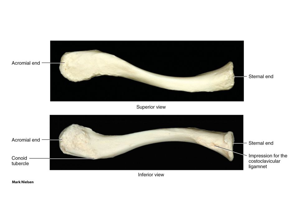
Clavicle “S” shaped bone (resilience)
medial 2/3 convex anteriorly , lateral 1/3 concave Sternal end (rounded) manubrium & 1st rib Acromial end (flat) acromion
 manubrium & 1st rib. Acromial end (flat) acromion.")
4
importance of the clavicle:
Connects upper limb to axial skeleton (strut) Protects neurovascular bundle that supply the upper limb Transmits shocks from upper limb to axial skeleton
 Protects neurovascular bundle that supply the upper limb. Transmits shocks from upper limb to axial skeleton.")
6
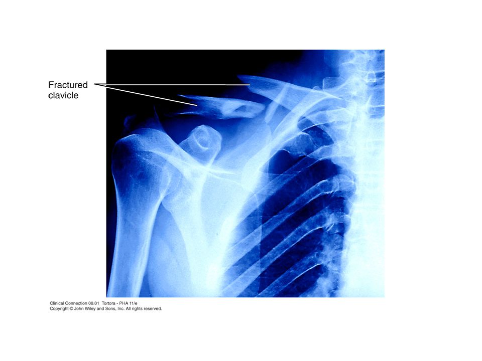
Fractures of Clavicle Common indirect impact to upper limb
Between middle & lateral thirds Medial part / lateral Shoulder drop Medial rotation of humerus

8
Scapula (shoulder blade)
Triangular flat bone Anterior (costal) surface 2nd – 7th ribs Posterior (spinous) surface
 surface. 2nd – 7th ribs. Posterior (spinous) surface.")
9
Left Scapula (Posterior Aspect)
1.Coracoid Process 3. Superior border 4. Supraspinous Fossa 6. Scapular Spine 7.Medial border 8.Infraspinous Fossa 9.Inferior Angle 10.Lateral border 11.Glenoid Cavity Margin 12.Acromion Process

10
Clinical: Winged Scapula
Medial border of scapula hangs away from thoracic wall 1. Damage to serratus anterior m. 2. Injury to long thoracic n. During fights

11
Humerus (brachial bone)
The arm bone Articulates with: Scapula (shoulder joint) Radius & Ulna (elbow joint)
 Radius & Ulna (elbow joint)")
12
Upper part Head (1/3 of sphere) Anatomical Neck Lesser Tubercle Intertubercular Groove Greater Tubercle Surgical Neck Deltoid Tuberosity

13
Right Humerus, Lower part
1. Radial Fossa 2. Lateral Epicondyle 3. Capitulum 4. Trochlea 5. Medial Epicondyle 6. Coronoid Fossa 7. Olecranon Fossa

14
Humerus Fractures 1. Fracture of the surgical neck:
Common in elderly (osteoporosis) due to falls down
 due to falls down.")
15
2. Transverse fracture of humerus shaft:
direct blow to the arm displacement depends on relation to deltoid insertion below deltoid tuberosity: upper laterally (deltoid) lower superiorly (biceps) Above deltoid tuberosity: ??
 lower superiorly (biceps) Above deltoid tuberosity:")
16
3. Supracondylar fracture:
fractures of the distal part of humerus 4. Fracture of medial epicondyle: pulls the epicondyle inferiorly weakness in fingers/hand flexion injury to ulnar n.

17
Bones of The Forearm Radius: - Lateral bone of the forearm
- Articulations: Humerus & Ulna Scaphoid & Lunate - Shaft is convex lat. & wider below than above

18
Right Radius Head of Radius Neck of Radius Radial Tuberosity
Anterior Posterior Radius Ulna Head of Radius Neck of Radius Radial Tuberosity Radius (Shaft) Styloid Process
 Styloid Process.")
19
Stabilizing bone of forearm
Bones of The Forearm Ulna: - Medial bone of the forearm - Articulations: Humerus & Radius Radius - Longer than Radius Stabilizing bone of forearm

20
Right Ulna Olecranon Process Trochlear notch Coronoid Process
Anterior Posterior Radius Ulna Olecranon Process Trochlear notch Coronoid Process Ulnar Tuberosity Styloid Process

21
“dinner-fork deformity”
Fractures of Radius Colles’ Fracture: Fracture of distal end of the radius, where the distal fragment moves posteriorly & superiorly due to falls on open hands “dinner-fork deformity” Smith’s Fracture: Reversed ??

22
Proximal, middle & distal
Bones of The Hand Carpal bones (8): Proximal row (4) & distal row (4) Metacarpals (5): Base, shaft & head Phalanges: Proximal, middle & distal Thumb (Exception)
: Proximal row (4) & distal row (4) Metacarpals (5): Base, shaft & head. Phalanges: Proximal, middle & distal. Thumb (Exception)")
23
Carpals of proximal row: Scaphoid (boat) 2. Lunate (moon)
3. Triquetrum (3 cornered) Pisiform (pea)
 4. Pisiform (pea)")
24
1. Trapezium (4-sided) 2. Trapezoid (wedge)
Carpals of distal row: 1. Trapezium (4-sided) 2. Trapezoid (wedge) 3. Capitate (large, with rounded head) 4. Hamate (hummer, hook process)
 2. Trapezoid (wedge) 3. Capitate (large, with rounded head) 4. Hamate (hummer, hook process)")
25
Stop Letting Those People Touch The Cadaver’s Hand

26
Lower Limb Divided into 4 regions: Gluteal Region: hip bone
Thigh: femur Leg: tibia & fibula Foot: tarsals, metatarsals & phalanges

27
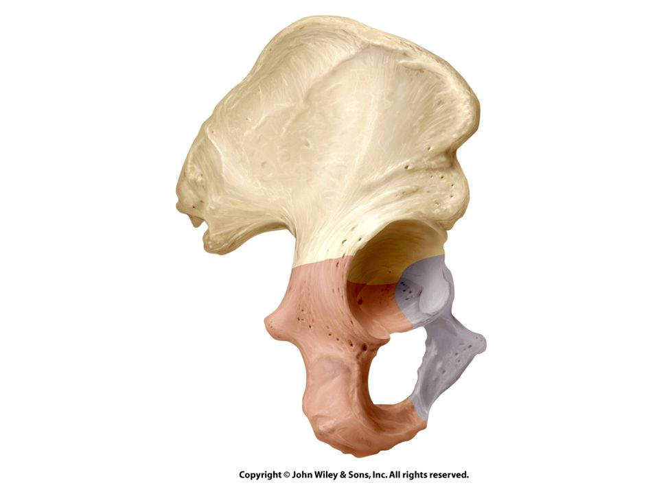
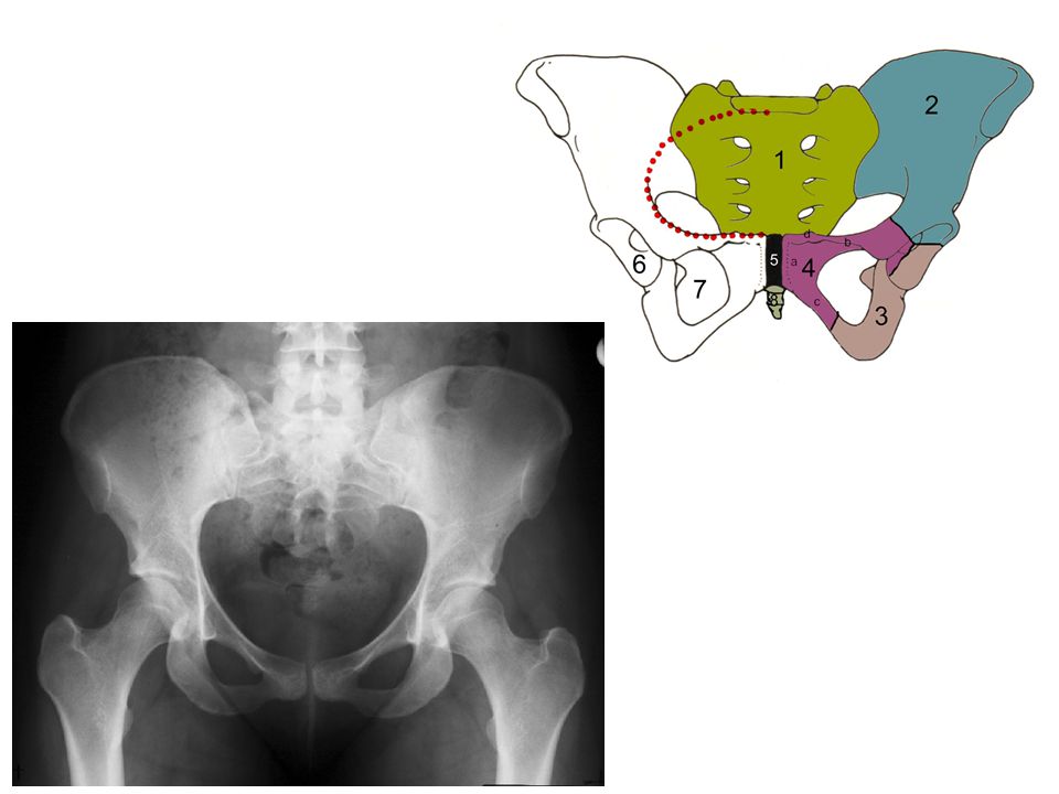
Hip Bone Made up of 3 bones: ilium (flat) ischium (L) pubis (V)
Meet at the Acetabulum: a socket where the femur head articulates to form the hip joint Beneath the acetabulum is an opening: “Obturator Foramen” Posterior: 2 notches (greater & lesser sciatic notches)
")
29
ilium (L, flank= خاصرة) The largest part of hip bone
Consist of: body & wing (ala) Has 4 spines (?) The sup. Border of the wing: Iliac crest (palpable) Iliac crest extension?
 Has 4 spines ( ) The sup. Border of the wing: Iliac crest (palpable) Iliac crest extension")
30
Ischium (L-shaped) (G, socket) Forms inferoposterior part of hip bone
Consists of: Body (joins?) Ramus (joins?) Spine tuberosity
 Ramus (joins ) Spine. tuberosity.")
31
Pubis (V-shaped) Forms the ant. part of hip bone Consists of:
Body (articulates?) Sup. & Inf. rami Tubercle
 Sup. & Inf. rami. Tubercle.")
33
Comparison of Female & Male Pelves
Pelvic Inlet ?? Pubic Arch 90o Vs. 90o Pelvic Outlet wide Vs. Narrow Sacrum concavity

34
Femur longest, heaviest, and strongest bone in the body
Proximally: articulates with the acetabulum of the hip bone forming the hip joint Distally: articulate with the condyles of the tibia forming the knee joint

35
Surface Features of Femur
Head Neck Lesser trochanter Greater trochanter Shaft Linea Aspera Femoral Condyles

37
weight-bearing bone of the leg
Tibia Tibia: L, flute Located medially weight-bearing bone of the leg Surface features: Tibial condyles Tibial tuberosity Medial malleolus

38
Fibula Slender bone, smaller than tibia Located laterally Features:
Head Lateral malleolus Fibula is a common source of bone for grafting

39
Tarsal Bones 7 bones Talus (ankle= كاحل) Calcaneus (heel= كعب)
Navicular (little boat) Medial Cuneiform (wedge-shape) Intermediate Cuneiform Lateral Cuneiform Cuboid
 Medial Cuneiform (wedge-shape) Intermediate Cuneiform. Lateral Cuneiform. Cuboid.")
Similar presentations