Download presentation
Presentation is loading. Please wait.

1
The Appendicular Skeleton, Articulations and Movement
Ex. 11 & 13 The Appendicular Skeleton, Articulations and Movement

2
Important figures & quiz knowledge
All figures and tables are important to know & understand Don’t forget: next week is the practical exam Practical review: Friday, 10/2 SC 115 2-5 PM 3 classes overlapping (I will be going back and forth)
")
3
Appendicular skeleton: 126 bones
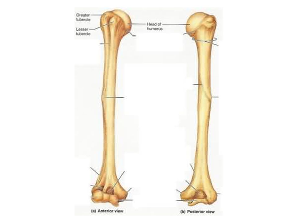
64 bones in upper limbs and pectoral girdle Shoulder girdle functional aspects Attachment of the upper limbs to the axial skeleton Attachment points for many trunk and neck muscles Clavicle/ collarbone – sternal end attaches to sternal manubrium. (Figure 11.2, p147) Acromial end - articulates with scapula & holds arm away from the top of the thorax Scapulae/ shoulder blades – have no directs attachment to the axial skeleton but is loosely held in place by the trunk muscles. (Figure 11.2, p147) Spine - deltoid muscle attachment Acromion process – connects with the clavicle Coracoid process – serves as attachment point for some of the upper limb muscles Glenoid cavity – a shallow socket that receives the head of the arm bone – humerus The Arm Humerus – long bone. (Figure 11.3, p148) Head – fits into the shallow glenoid cavity of the scapula Greater and lesser tubercles - attachemnt for biceps muscles The Forearm (Figure 11.4, page 149) Radius - lateral bone of the forearm Ulna - medial bone of the forearm Radial notch – articulates with the head of the radius The Hand – manus (Figure 11.5, page 150) Three groups of bones: Carpus – wrist. 8 carpal bones Metacarpals – palm: numbered 1 to 5 from the thumb. Phalanges – fingers: numbered 1 to 5 from the thumb 14 bones Each finger contains three phalanges except for the thumb which has only two
 Acromial end - articulates with scapula & holds arm away from the top of the thorax. Scapulae/ shoulder blades – have no directs attachment to the axial skeleton but is loosely held in place by the trunk muscles. (Figure 11.2, p147) Spine - deltoid muscle attachment. Acromion process – connects with the clavicle. Coracoid process – serves as attachment point for some of the upper limb muscles. Glenoid cavity – a shallow socket that receives the head of the arm bone – humerus. The Arm. Humerus – long bone. (Figure 11.3, p148) Head – fits into the shallow glenoid cavity of the scapula. Greater and lesser tubercles - attachemnt for biceps muscles. The Forearm (Figure 11.4, page 149) Radius - lateral bone of the forearm. Ulna - medial bone of the forearm. Radial notch – articulates with the head of the radius. The Hand – manus (Figure 11.5, page 150) Three groups of bones: Carpus – wrist. 8 carpal bones. Metacarpals – palm: numbered 1 to 5 from the thumb. Phalanges – fingers: numbered 1 to 5 from the thumb. 14 bones. Each finger contains three phalanges except for the thumb which has only two.")
10
Appendicular skeleton: 126 bones
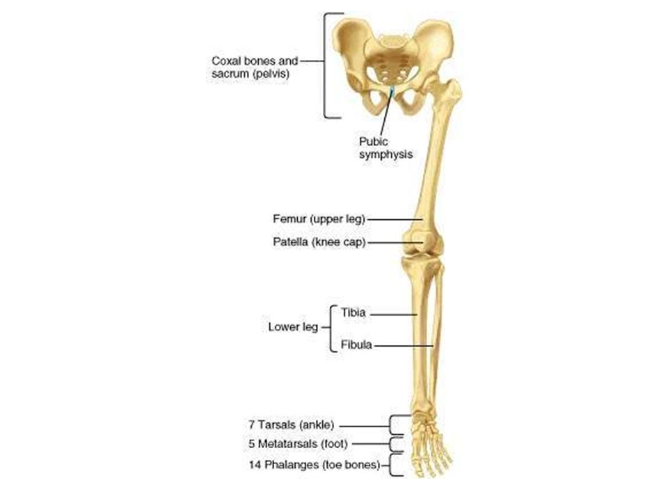
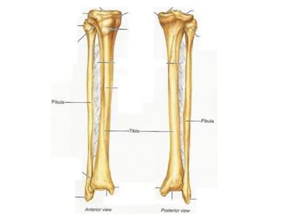
62 bones in lower limbs and pelvic girdle The Pelvic (Hip) Girdle (Figure 11.6, pages 151-2) Formed by two coxal bones (2 fused bones) Bones are heavy and massive, and attach securely to the axial skeleton Sockets for the heads of the femurs (thigh bones) are deep and heavily reinforced by ligaments to ensure a stable, strong attachment Ability to bear weight is more important than mobility and flexibility Combined weight of the upper body rests on the pelvis Each coxal bone is a result of the fusion of three bones: Ilium – large flaring bone Ischium – “sit – down” bone Pubis - anterior poertion of the coxal bone All three bones fuse at the deep hemispherical socket – acetabulum, which receives the head of the thigh bone Comparison of the Male and Female Pelves (Table 11.1, page 153) Bones of males: usually larger, heavier, and have more prominent bone markings Female pelvis reflects modifications for childbearing Wider, shallower, lighter, and rounder The Leg (Figure 11.8, page 155) Tibia - larger & medial bone Fibula - parallel to the tibia The Foot (Figure 11.9, page 156) 7 tarsal bones, 5 metatarsals (instep), 14 phalanges (toes) Body weight is concentrated on two largest tarsals Calcaneus - heel bone Talus – b/n tibia and calcaneus Each toe has three phalanges except the great toe, which has two The Arches The foot has two important functions: weight bearing and propulsion. These functions require a high degree of stability and flexibility The foot has three arches Medial longitudinal arch is the highest and most important of the three arches. It is composed of the calcaneus, talus, navicular, cuneiforms, and the first three metatarsals Lateral longitudinal arch is lower and flatter than the medial arch. It is composed of the calcaneus, cuboid, and the fourth and fifth metatarsal Transverse arch is composed of the cuneiforms, the cuboid, and the five metatarsal bases Arches are maintained by the shapes of the bones as well as by ligaments and tendons
 Girdle (Figure 11.6, pages 151-2) Formed by two coxal bones (2 fused bones) Bones are heavy and massive, and attach securely to the axial skeleton. Sockets for the heads of the femurs (thigh bones) are deep and heavily reinforced by ligaments to ensure a stable, strong attachment. Ability to bear weight is more important than mobility and flexibility. Combined weight of the upper body rests on the pelvis. Each coxal bone is a result of the fusion of three bones: Ilium – large flaring bone. Ischium – sit – down bone. Pubis - anterior poertion of the coxal bone. All three bones fuse at the deep hemispherical socket – acetabulum, which receives the head of the thigh bone. Comparison of the Male and Female Pelves (Table 11.1, page 153) Bones of males: usually larger, heavier, and have more prominent bone markings. Female pelvis reflects modifications for childbearing. Wider, shallower, lighter, and rounder. The Leg (Figure 11.8, page 155) Tibia - larger & medial bone. Fibula - parallel to the tibia. The Foot (Figure 11.9, page 156) 7 tarsal bones, 5 metatarsals (instep), 14 phalanges (toes) Body weight is concentrated on two largest tarsals. Calcaneus - heel bone. Talus – b/n tibia and calcaneus. Each toe has three phalanges except the great toe, which has two. The Arches. The foot has two important functions: weight bearing and propulsion. These functions require a high degree of stability and flexibility. The foot has three arches. Medial longitudinal arch is the highest and most important of the three arches. It is composed of the calcaneus, talus, navicular, cuneiforms, and the first three metatarsals. Lateral longitudinal arch is lower and flatter than the medial arch. It is composed of the calcaneus, cuboid, and the fourth and fifth metatarsal. Transverse arch is composed of the cuneiforms, the cuboid, and the five metatarsal bases. Arches are maintained by the shapes of the bones as well as by ligaments and tendons.")
18
Articulations Articulations – joints, bone – bone contact Function:
Hold bones together Allow flexibility so that body movements can occur Functional classification (based on amount of movement) (p. 170, Fig 13.1) Synarthroses – immovable joints, ex.: sutures in the axial skeleton Amphiarthroses – slightly movable joints, ex.: vertebral disc, in the axial skeleton Diarthroses – freely movable joints, ex.: synovial joints in the limbs
 (p. 170, Fig 13.1) Synarthroses – immovable joints, ex.: sutures in the axial skeleton. Amphiarthroses – slightly movable joints, ex.: vertebral disc, in the axial skeleton. Diarthroses – freely movable joints, ex.: synovial joints in the limbs.")
19
Structural classifications (based connective tissue type
Fibrous joint – joined by fibrous c.t., little or no movement Suture – edges of bone interlock (ex.: skull) Syndesmoses – bones connected by short ligaments (ex.: between distal ends of tibia and fibula) Cartilaginous joint – articulating bone ends joined by cartilage plate or pad, slightly movable Symphysis – fibrocartilage (ex.: pubic symphysis) Synchondrosis – hyaline cartilage (ex.: epiphyseal plate) Synovial joint – joined by joint cavity containing synovial fluid, freely movable (p. 171, Fig 13.2)
 Syndesmoses – bones connected by short ligaments (ex.: between distal ends of tibia and fibula) Cartilaginous joint – articulating bone ends joined by cartilage plate or pad, slightly movable. Symphysis – fibrocartilage (ex.: pubic symphysis) Synchondrosis – hyaline cartilage (ex.: epiphyseal plate) Synovial joint – joined by joint cavity containing synovial fluid, freely movable (p. 171, Fig 13.2)")
21
Subclassifications of synovial joints
Based on # of axes, joint shape, & allowable motion type Classification of synovial joints by shape of joint, and type of motion allowed (p.172; 13.3): Plane/gliding joint: the wrist Bone surfaces slide across each other, allowing a wide range of movements Hinge joint: elbow and ankles Allow for flexion and extension Pivot joint: the skull on its spinal axis Movement is limited to rotation Condyloid/ Ellipsoid joint: structurally similar to a ball and socket joint but without rotation Saddle joint: the thumb Bone surfaces are concave, allowing movement in all direction but only limited rotation Ball and Socket joint: the rounded head of one bone fits into a socket-like cavity of another, such as the hip and shoulder joints Allow free rotation Classification of synovial joints by the number of axes Nonaxial joint - movement tends to be linear rather than angular Joint surfaces flat and glide over one another instead of around ex.: carpal bones Uniaxial joint - movement in one plane around one axis Move like a door hinge ex.: elbow joint and interphalangeal joints of hand and foot Pivot joint - bone pivots around another bone ex.: radius pivots around stationary ulnar, also atlantoaxial joint C1 (atlas) pivots around stationary C2(axis) on the odontoid process Biaxial joint - motion occurs in two different axis in two planes Condyloid (ellipsoidal) –metacarpophalangeal joints of fingers or toes Saddle – carpometacarpal joints at thumb Triaxial/ multiaxial joint - motion in all three axes ex.: ball and socket - hip and shoulder More motion than any other type of joint
: Plane/gliding joint: the wrist. Bone surfaces slide across each other, allowing a wide range of movements. Hinge joint: elbow and ankles. Allow for flexion and extension. Pivot joint: the skull on its spinal axis. Movement is limited to rotation. Condyloid/ Ellipsoid joint: structurally similar to a ball and socket joint but without rotation. Saddle joint: the thumb. Bone surfaces are concave, allowing movement in all direction but only limited rotation. Ball and Socket joint: the rounded head of one bone fits into a socket-like cavity of another, such as the hip and shoulder joints. Allow free rotation. Classification of synovial joints by the number of axes. Nonaxial joint - movement tends to be linear rather than angular. Joint surfaces flat and glide over one another instead of around. ex.: carpal bones. Uniaxial joint - movement in one plane around one axis. Move like a door hinge. ex.: elbow joint and interphalangeal joints of hand and foot. Pivot joint - bone pivots around another bone. ex.: radius pivots around stationary ulnar, also atlantoaxial joint C1 (atlas) pivots around stationary C2(axis) on the odontoid process. Biaxial joint - motion occurs in two different axis in two planes. Condyloid (ellipsoidal) –metacarpophalangeal joints of fingers or toes. Saddle – carpometacarpal joints at thumb. Triaxial/ multiaxial joint - motion in all three axes. ex.: ball and socket - hip and shoulder. More motion than any other type of joint.")
24
Movements of synovial joints
Flexion Sagittal plane Decreases joint angle & distance betw. articulated bones Extension Opp. flexion Increases joint angle & distance betw. bones Abduction Frontal plane Movement of limb away from midline or median plane Adduction Opp. abduction Movement toward midline or median plane Rotation Movement of bone around its own logitudinal axis w/o lateral or medial displacement Circumduction: combination of previously listed movements (except rotation) Pronation: movement of palm from anterior or superior position to a posterior or inferior position Supination: opp. of pronation Inversion: medial turning of sole of foot Eversion: opp. of inversion Dorsiflextion: movement of ankle joint dorsally Plantar flexion: movement of ankle joint ventrally
 Pronation: movement of palm from anterior or superior position to a posterior or inferior position. Supination: opp. of pronation. Inversion: medial turning of sole of foot. Eversion: opp. of inversion. Dorsiflextion: movement of ankle joint dorsally. Plantar flexion: movement of ankle joint ventrally.")
Similar presentations


 and type of substance.>")

 Function (range of motion)>")


 Weakest parts of the skeleton Weakest parts of the skeleton Articulation – site where two or more bones meet Articulation – site.>")

 Articulations – wherever 2 bones meet Classified by function –Synarthrosis (Immovable) –Amphiarthrosis (slightly.>")


