Download presentation
Presentation is loading. Please wait.

1
Overview of Structure of the Adult Skull
Biology 323 Human Anatomy for Biology Majors Lecture 16 Dr. Stuart S. Sumida Overview of Structure of the Adult Skull

4
The developing skull has three component origins:
Condrocranium (base of skull / braincase) Dermatocranium (flat bones of skull) Splanchnocranium (bones derived from gill arch elements)
 Dermatocranium (flat bones of skull) Splanchnocranium (bones derived from gill arch elements)")
5
Condrocranium Endochondral Mesoderm Dermatocranium Dermal Neural Crest
Mode of Germ Layer Formation Origin Condrocranium Endochondral Mesoderm Dermatocranium Dermal Neural Crest Splanchnocranium Endochondral Neural Crest

6
CHONDROCRANIUM: Bones of the base of the skull.
Most major cranial nerves escape the skull through these. Endochondral Mostly Mesodermal Include: ethmoid, sphenoid (part), occipital (part) right and left temporal (parts).
, occipital (part) right and left temporal (parts).")
8
orbit II/ Ethmoid Sphenoid Petrous temporal Basioccipital

11
Flat bones of skull: DERMATOCRANIUM
(These and others.)
")
18
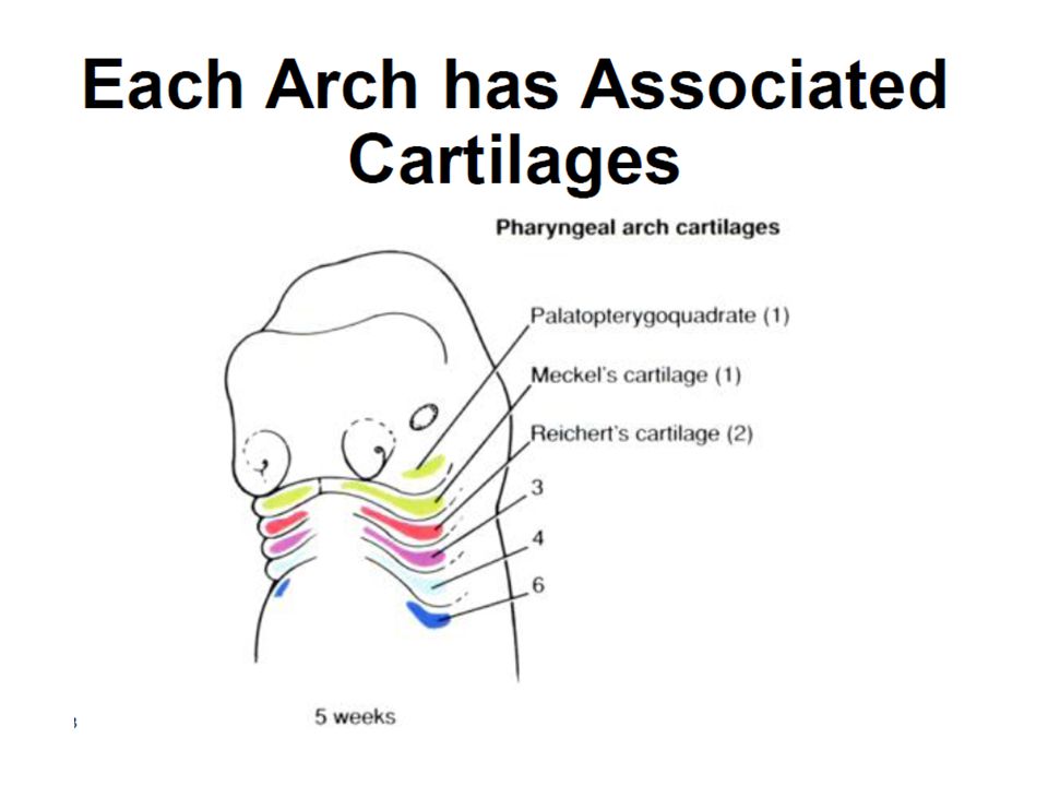
Gill Pouch Bones: become SPLANCHNOCRANIUM

21
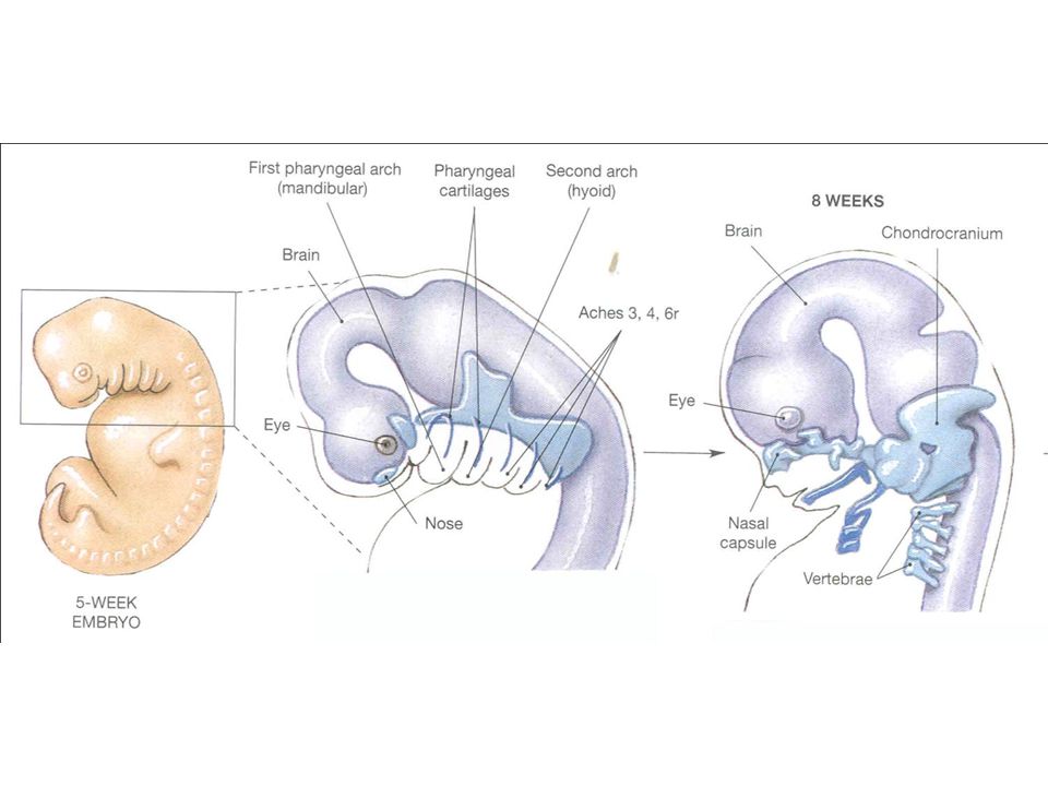
This lecture will revolve around the early embryology of the vertebrate skull. One of the landmark achievments in this was the summary of that topic – based initially the developing embryonic shark head – by Edwin S. Goodrich. Thus it has come to be known as the “Goodrich Diagram”.

22
From Studies on the structure and development of vertebrates by Edwin S. Goodrich, 1958.

23
If you’re working with the PowerPoint files, save space in your notes to draw here.

29
Mandible (Lower Jaw)
")
30
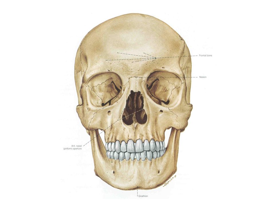
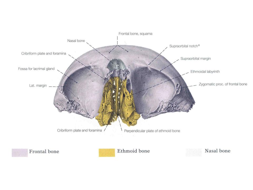
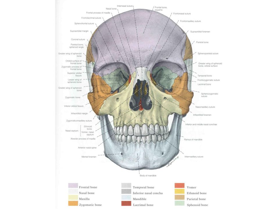
Skull – Anterior View

33
Anterior Inferior Frontal

34
Ethmoid superior lateral

36
Bones of the Orbit

37
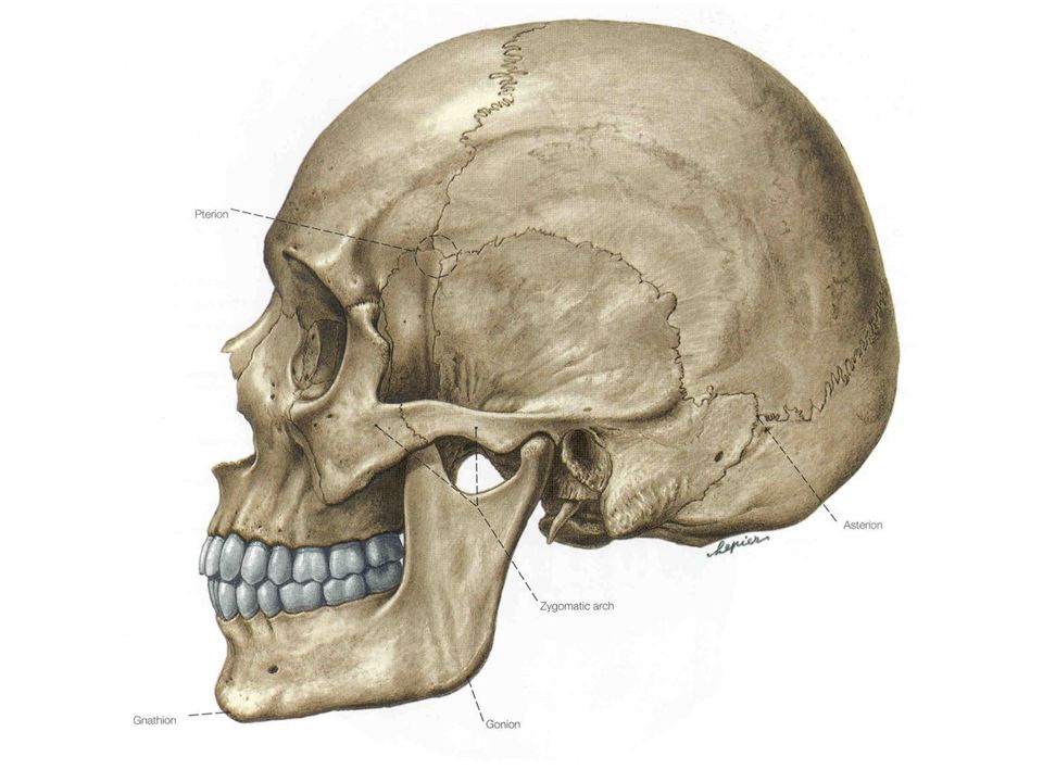
Skull – Lateral View

40
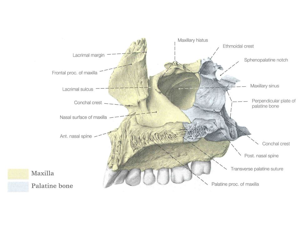
Maxilla: Lateral View

41
External Internal Parietal

42
Temporal Internal External

43
Neonatal Temporal Bone

44
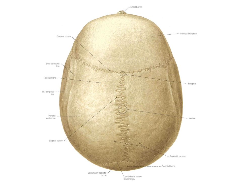
Skull – Superior View

46
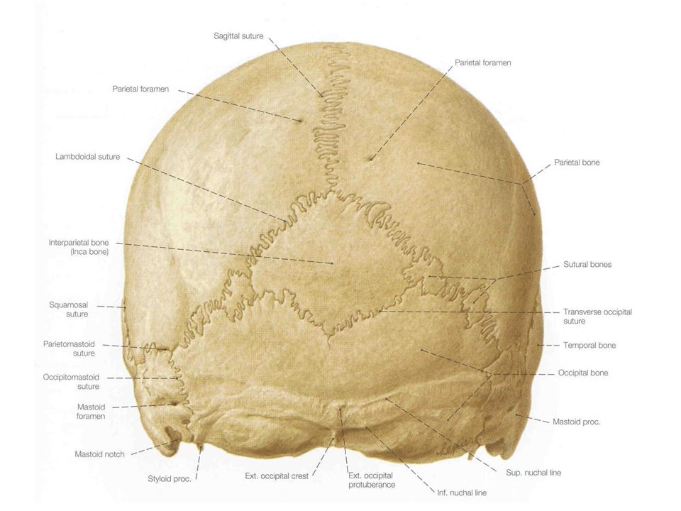
Skull – Posterior View

48
Inferior Internal Occipital

49
Skull – Inferior View

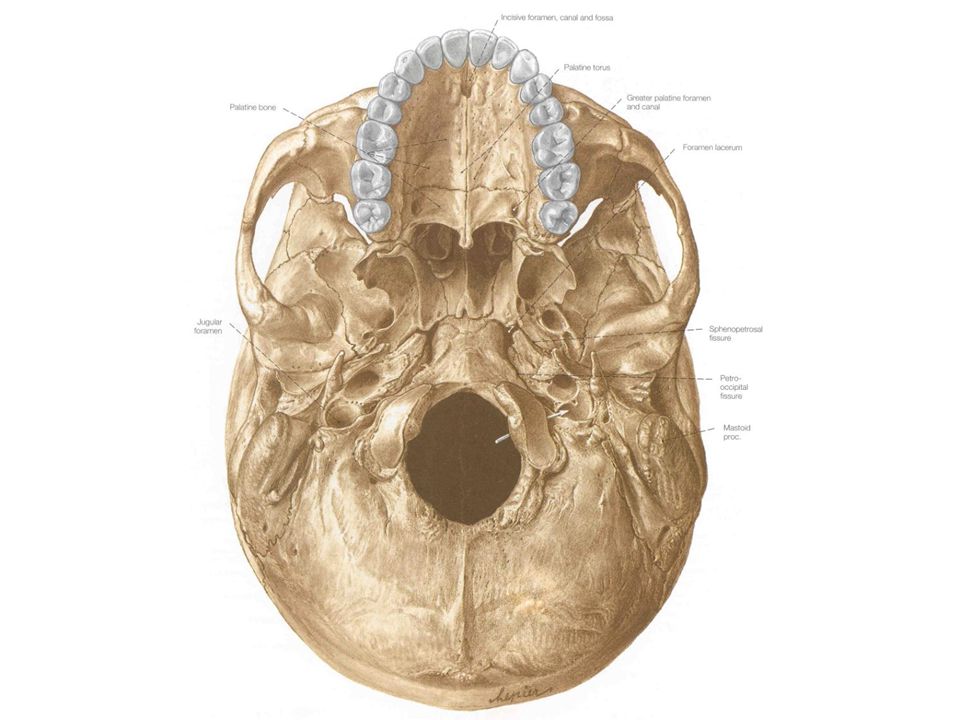
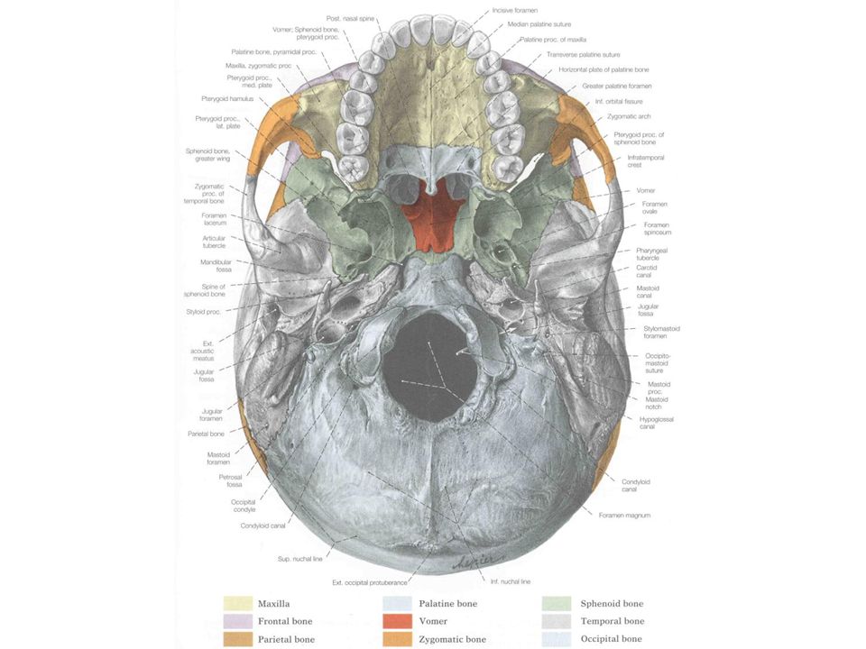
52
Hard Palate

53
Infratemporal Region

54
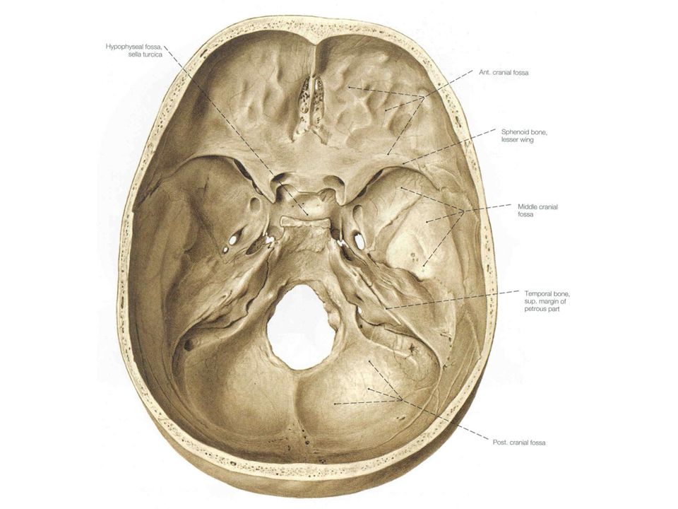
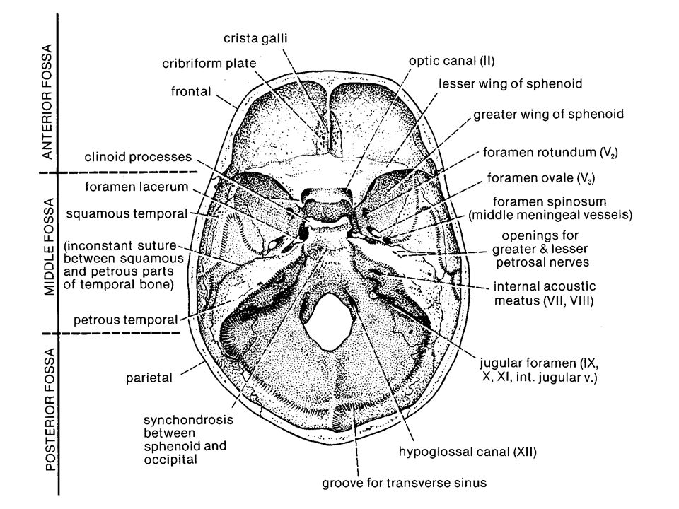
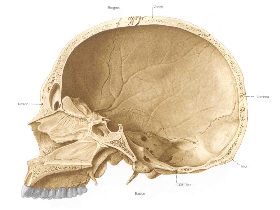
Skull – Internal View

60
Sphenoid Occipital

61
Major Ligaments Near Jaw Joint

62
Major Ligaments Near Jaw Joint:
Stylomandibular Sphenomandibular

64
Bones of the Basicranium
Ethmoid Sphenoid Temporal Occipital

65
Ethmoid superior lateral

66
Anterior Posterior Sphenoid

67
Temporal Internal External

68
Neonatal Temporal Bone

69
Sphenoid Occipital

71
Dermal Roofing Bones Nasals Frontal Parietals

73
Anterior Inferior Frontal

75
External Internal Parietal

76
Dermal Facial Bones Maxilla Zygomatic Lacrimal

78
Maxilla: Lateral View

80
Bones of the Orbit

81
Dermal Palatal Bones Maxilla Palatine Vomer

82
Hard Palate

84
Bones of Splanchnopleure Sphenoid Greater Wing
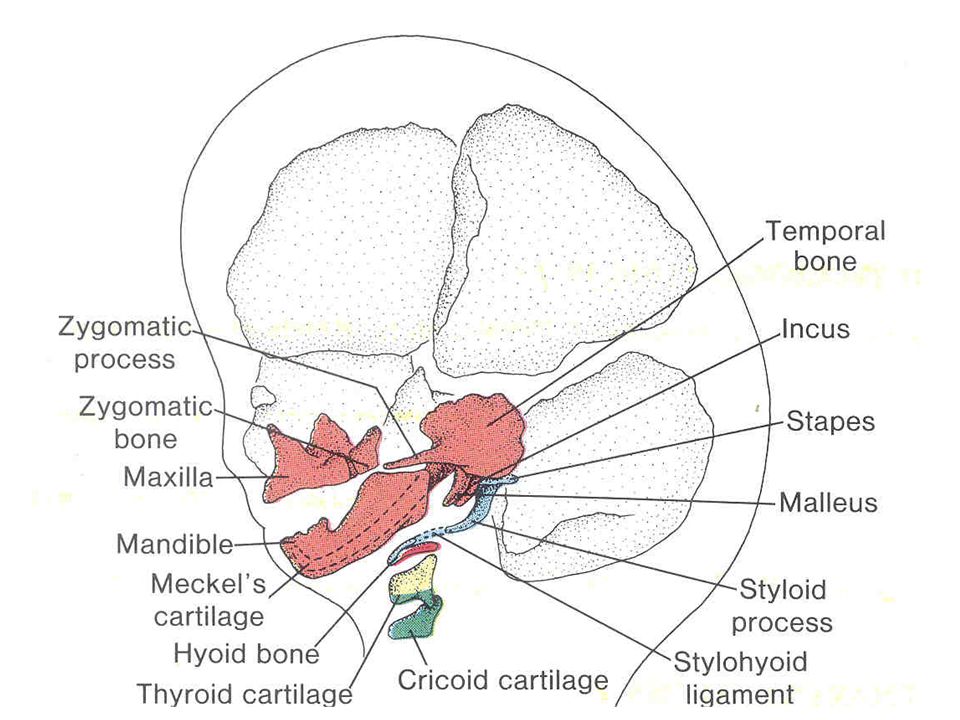
Temporal Styloid Process Middle Ear Ossicles

85
Anterior Posterior Sphenoid

86
Sphenoid/ anterior view of orbital surface and foramina with associated cranial nerves indicated
II V1 III Opth. a. IV VI Opthalmic v. V2 Nerve of pterygoid canal ALSO CALLED VIDIAN NERVE (VII parasympathetics + sympathetics) V3
 V3.")
87
Neonatal Temporal Bone

88
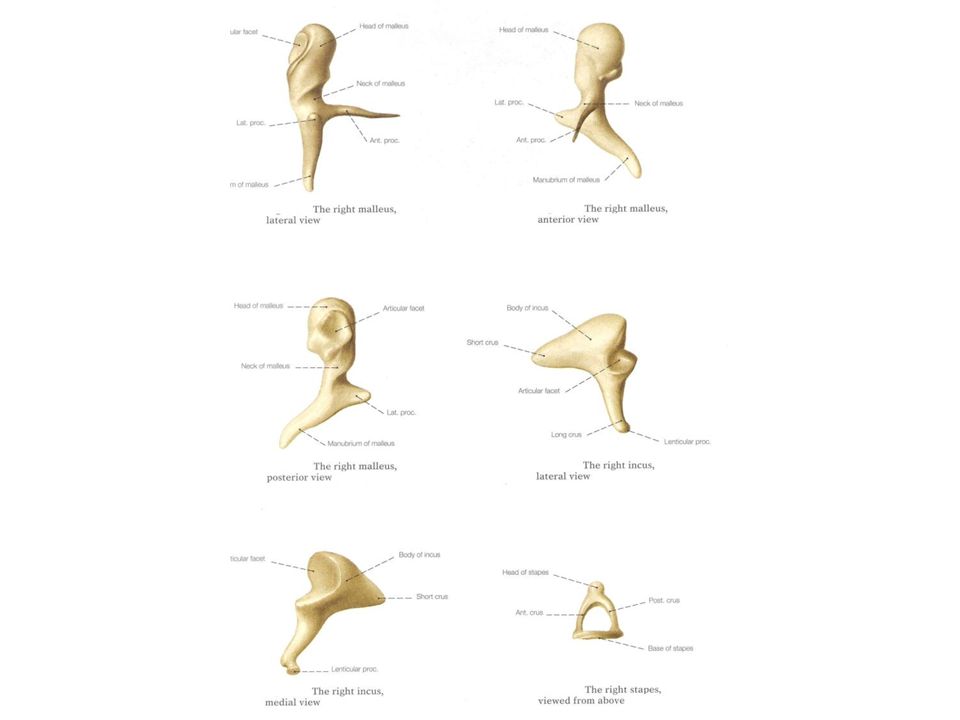
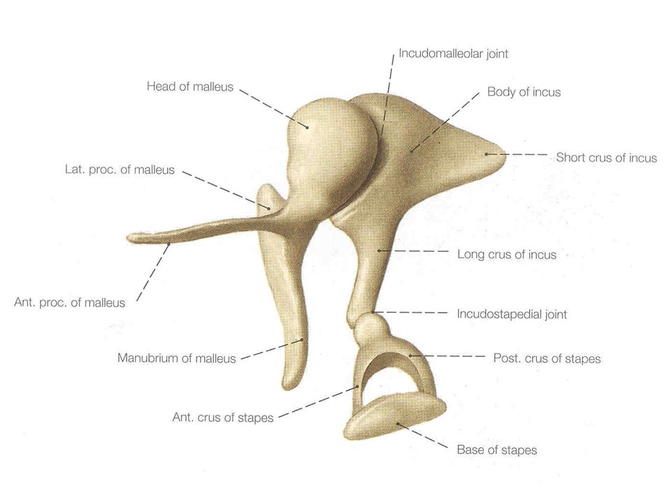
Middle Ear Ossicles Malleus Incus Stapes

Similar presentations


 Process = Prominence or extension.>")


