Download presentation
Presentation is loading. Please wait.

1
Respiration and Breathing http://www.youtube.com/watch?v=Kl4cU9sG_ 08&list=PLJicmE8fK0Ehrg3meytY7DT8LJiwu U3Th&index=121

2
The air we breathe consists of nitrogen, carbon dioxide, trace gases and valuable oxygen vital for life. In humans, the only means we have of obtaining oxygen is through the lungs. Absence of oxygen for more than just a few minutes can result in death.

3
Breathing is not: YawningHiccuping - Stretching the facial- Spasm of the muscles diaphragm

4
Respiration involves all processes related to the exchange of oxygen and carbon dioxide, including breathing, gas exchange and cellular respiration. –External respiration – involves the exchange of oxygen and carbon dioxide between the air and the cells of the lungs. –Internal respiration – involves the exchange of oxygen and carbon dioxide between the blood and the tissue fluids.

5
Respiration Cellular respiration – involves the combustion of oxygen to produce ATP and carbon dioxide in the cells.

6
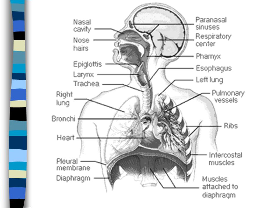
Anatomy Know the pathway for inhaled and exhaled air in the respiratory system Know terms such as nasal cavity, oral cavity, pharynx, epiglottis, larynx, trachea, lung, bronchi, bronchioles, intercostal muscles, diaphragm, alveoli https://www.youtube.com/watch?v=Cy1lfZAIojs How Do Your Lungs Work?

7
Chandler's project https://www.youtube.com/watch?v=8rm ZkaloqxE&feature=youtu.be https://www.youtube.com/watch?v=8rm ZkaloqxE&feature=youtu.be How your lungs work http://www.nhlbi.nih.gov/health/health- topics/topics/hlw/whathappens.html http://www.nhlbi.nih.gov/health/health- topics/topics/hlw/whathappens.html Inflating a cow’s lungs http://www.sciencechannel.com/tv-shows/outrageous-acts-of- science/videos/how-lungs- work.htm?utm_source=Outbrain&utm_medium=Referral&utm_c ampaign=OutbrainOrganicTraffic=obnetwork http://www.sciencechannel.com/tv-shows/outrageous-acts-of- science/videos/how-lungs- work.htm?utm_source=Outbrain&utm_medium=Referral&utm_c ampaign=OutbrainOrganicTraffic=obnetwork

9
Anatomy Nasal cavity – filters the air with the help of cilia and mucous. It also moistens and warms the air before it goes to the lungs. Pharynx – opens from the nasal cavity and branches into two structures: Esophagus – muscular tube that moves food to the stomach. Trachea – the windpipe which is supported by C-shaped cartilaginous rings and covered with ciliated cells and mucous that act as a secondary filter.

10
Epiglottis – a flap of skin that covers the opening of the trachea to prevent food from entering the lungs while swallowing. Larynx – voice box, composed of 2 thin sheets of elastic ligaments that vibrate as air is forced out of them. These are also called the vocal cords and are protected by a thick band of cartilage called the Adam’s apple. Bronchi- are two tubes that branch from the trachea and carry air to the left and right lungs.

11
Bronchioles – smaller branches off the bronchi that become progressively smaller until they reach the alveoli. Alveoli – air sacs in the lung where gas exchange occurs. Alveoli are covered with a slippery lipoprotein film called a surfactant that prevents the sacs from collapsing and sticking together. Gases diffuse in and out of the alveoli according to concentration gradients.

12
Pleural membranes – thin membranes surrounding the outer surface of the lungs. They are filled with fluid to reduce friction between the lungs and the chest cavity during inhalation. Diaphragm – a large sheet of muscle that separates the organs of the thoracic cavity from those of the abdominal cavity. As the muscle contracts, the diaphragm flattens decreasing pressure inside the chest cavity, drawing air into the lungs. Relaxed, the muscle is dome-shaped.

13
Inter-costal muscles – found between the ribs. As they contract, the ribs are pulled outward and upward, increasing the chest volume and contribute to inspiration. http://sprojects.mmi.mcgill.ca/resp/anato my.swf http://sprojects.mmi.mcgill.ca/resp/anato my.swf

14
Breathing Movements A pressure difference between the atmosphere and the chest cavity determine the movement of gases into and out of the lungs. Gases, like other substances, move from higher to lower concentration or from high pressure to low pressure.

15
Breathing Movements Know what happens to the diaphragm and the internal and external intercostal muscles when inhaling and exhaling –Understand the pressure of the chest cavity and how it facilitates the moving of air in and out http://www.youtube.com/watch?v=858cJYK2pXU&feature=related http://www.youtube.com/watch?v=hp-gCvW8PRY&feature=related http://www.youtube.com/watch?v=0YRicc-73FQ&feature=related http://www.youtube.com/watch?v=RmMcqnXqrvA&NR=1

16
collapsed lung http://www.youtube.com/watch?v=R-l1nzOezIw http://www.youtube.com/watch?v=R-l1nzOezIw

18
http://highered.mcgraw- hill.com/sites/0072437316/student_view0/chapter44/animatio ns.html#

19
Circulatory & Respiratory Systems - CrashCourse Biology #27 http://www.youtube.com/watch?v=9fxm 85Fy4sQ http://www.youtube.com/watch?v=9fxm 85Fy4sQ

20
Regulation of Breathing Breathing movements are controlled by the medulla oblongata in the brain information about the amount of CO 2 and O 2 is directed by chemoreceptors which send a message to the medulla –there are separate receptors for CO 2 (more sensitive) and O 2
 and O 2")
22
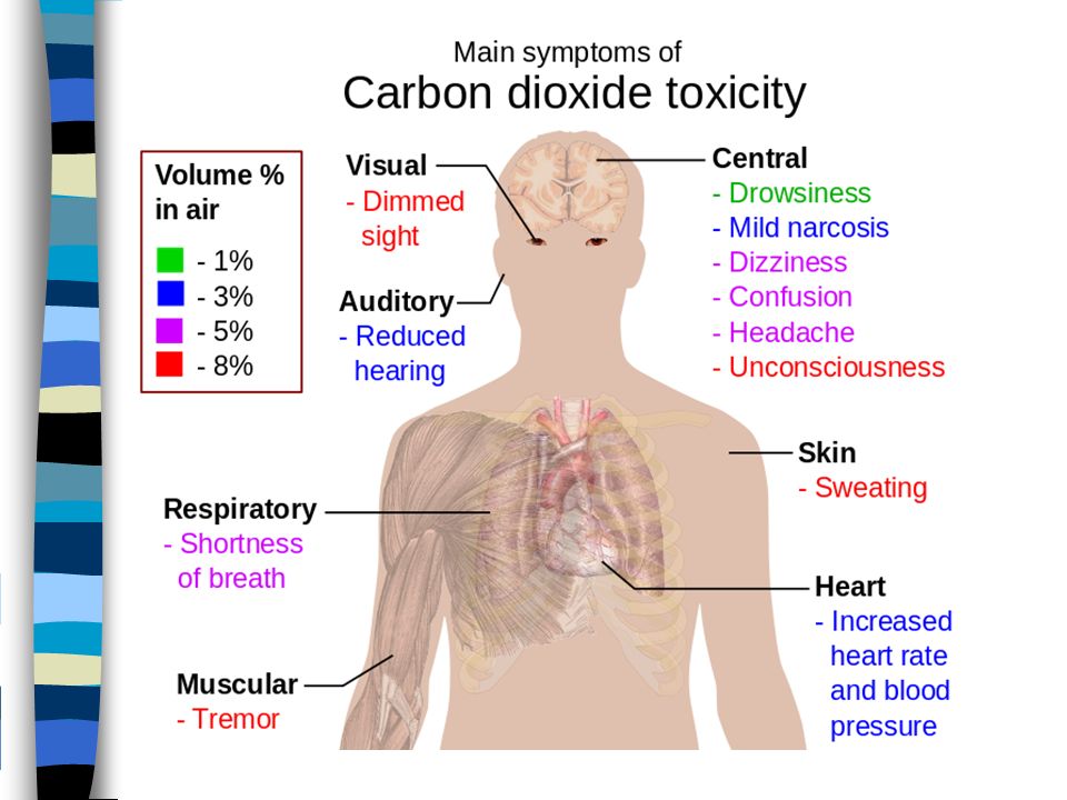
What Happens when CO 2 levels increase? chemoreceptors sense increase in CO 2 the diaphragm and intercostal muscles’ activity increases (stimulated by medulla oblongata) this increases breathing movements and therefore increases the amount of CO 2 being exhaled when CO 2 levels fall, the chemoreceptors become inactive and breathing rates return to normal
 this increases breathing movements and therefore increases the amount of CO 2 being exhaled when CO 2 levels fall, the chemoreceptors become inactive and breathing rates return to normal.")
23
What Happens when CO 2 levels increase? Drugs like morphine and barbiturates (aka. Downers/depressants) can make the medulla less sensitive to CO 2 levels and as a result, breathing rate decreases which could eventually cause death Why can’t you hold your breath forever? Why do people breathe into paper bags when having an anxiety attack?
 can make the medulla less sensitive to CO 2 levels and as a result, breathing rate decreases which could eventually cause death Why can’t you hold your breath forever. Why do people breathe into paper bags when having an anxiety attack .")
24
How a feedback loop works

25
Feedback Loop for CO 2 High CO 2 Chemoreceptors Medulla DiaphragmIntercostals Breathing Rate Increases More O 2 absorbed by blood inactivates chemoreceptors

27
What Happens When O 2 Levels Are Low? oxygen chemoreceptors called the carotid and aortic bodies detect when oxygen levels are low and become stimulated a message is sent to the medulla the medulla sends nerve impulses to the diaphragm and the ribs begin breathing movements this will increase the amount of oxygen in the blood the O 2 receptors are only called into action when O 2 levels fall and CO 2 levels remain in the normal range

28
What Happens When O 2 Levels Are Low? Some examples – when you hold your breath, your O 2 levels drop while the CO 2 levels increase and the high CO 2 levels will initiate breathing movements –in high altitudes where there is less O 2 present, the opposite will happen. Low levels of O 2 is not accompanied by high CO 2 levels, the oxygen chemoreceptors initiate breathing movements –when carbon monoxide poisoning occurs, CO (carbon monoxide) competes with O 2 on the binding sites of the hemoglobin molecules in the blood. This reduces the O 2 levels in the blood, stimulating the oxygen chemoreceptors to initiate breathing movements
 competes with O 2 on the binding sites of the hemoglobin molecules in the blood. This reduces the O 2 levels in the blood, stimulating the oxygen chemoreceptors to initiate breathing movements.")
29
Feedback Loop Low Blood O 2 Chemoreceptors Medulla DiaphragmIntercostals Breathing Rate Increases

30
Breathing Graphs

31
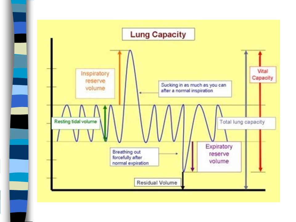
Breathing Graphs www.videosurf.com/video/pulmonary-function-test-pft-120987100 www.videosurf.com/video/pulmonary-function-test-pft-120987100 Tidal Volume (TV) – volume of air being inhaled and exhaled during normal breathing. Inspiratory Reserve (IR) – maximum amount of air that is inhaled above tidal volume. Expiratory Reserve (ER) – amount of air that can be exhaled after normal exhalation/ Residual Volume – volume of air that always stays in the lungs Vital Capacity (VC) – total volume of air that the lungs can inhale and exhale. Vital Capacity = Tidal Volume + Inspiratory Reserve + Expiratory Reserve VC = TV + IR + ER Total capacity = vital capacity + residual volume
 – maximum amount of air that is inhaled above tidal volume. Expiratory Reserve (ER) – amount of air that can be exhaled after normal exhalation/ Residual Volume – volume of air that always stays in the lungs Vital Capacity (VC) – total volume of air that the lungs can inhale and exhale. Vital Capacity = Tidal Volume + Inspiratory Reserve + Expiratory Reserve VC = TV + IR + ER Total capacity = vital capacity + residual volume.")
34
Respiration and Blood

35
Oxygen Transport –O 2 in the alveoli diffuses into the fluid around the cells surrounding the capillary bed –O 2 then diffuses through the capillary walls and into the blood plasma and the oxygen binds on the hemoglobin molecules in the red blood cell http://www.youtube.com/watch?v=WXOBJEXxNEo&feature=related http://media.pearsoncmg.com/bc/bc_campbell_biology_7/media/interactivemedia/activities/ load.html?42&E

36
Respiration and Blood Carbon Dioxide Transport –23% of CO 2 is carried on hemoglobin –7% is carried in plasma –70% of CO 2 (from cellular respiration) enters the red blood cells and in order to maintain blood pH, is chemically converted to carbonic acid (H 2 CO 3 ) in a reaction that is catalyzed by carbonic anhydrase (an enzyme) carbonic acid molecules dissociate forming bicarbonate ions and hydrogen ions bicarbonate diffuses out or RBC into the plasma
 enters the red blood cells and in order to maintain blood pH, is chemically converted to carbonic acid (H 2 CO 3 ) in a reaction that is catalyzed by carbonic anhydrase (an enzyme) carbonic acid molecules dissociate forming bicarbonate ions and hydrogen ions bicarbonate diffuses out or RBC into the plasma")
37
Respiration and Blood Carbon Dioxide Transport –when blood rich in CO 2 reaches the lungs, bicarbonate ions combine with hydrogen ions, reforming carbonic acid carbonic acid molecules dissociate forming bicarbonate ions and hydrogen ions carbonic acid dissociates, forming water and CO 2 which diffuses out of the blood and into the alveoli when present in normal amounts, the ratio of carbonic acid to bicarbonate creates an acid-base balance in the blood, helping to keep the pH at a level where the body's cellular functions are most efficient

38
Respiration and Blood Carbon Dioxide Transport –CO 2 travels from the capillaries to the alveoli, this is driven by concentration differences (the concentration of CO 2 in the capillaries is slightly higher than in the alveoli) – CO 2 is then expelled out of the lungs in exhalation
 – CO 2 is then expelled out of the lungs in exhalation")
39
Respiration and Blood http://www.dnatube.com/video/372/Maintena nce-of-pH-Buffer http://www.dnatube.com/video/372/Maintena nce-of-pH-Buffer Reaction Summary CO 2 + H 2 O H 2 CO 3 (carbonic acid)In RBC carbonic anhydrase H 2 CO 3 HCO 3 - + H + In RBC then to plasma (bicarbonate) HCO 3 - + H + H 2 CO 3 In RBC H 2 CO 3 CO 2 In RBC H 2 O
In RBC carbonic anhydrase H 2 CO 3 HCO H + In RBC then to plasma (bicarbonate) HCO H + H 2 CO 3 In RBC H 2 CO 3 CO 2 In RBC H 2 O")
40
https://www.youtube.com/watch? v=qDrV33rZlyA https://www.youtube.com/watch? v=qDrV33rZlyA Respiration Gas Exchange summary (13 min)
.")
41
The Function of the Hydrogen Ions the H + ions help to dislodge O 2 from the hemoglobin causing O 2 to diffuse into the tissues by removing H + ions from the plasma, the hemoglobin acts as a buffer when the deoxygenated blood from the veins reaches the lungs, O 2 dislodges the H + from the hemoglobin the free H + combines with bicarbonate to eventually form CO 2 and H 2 O this is called blood buffering, it helps to maintain blood pH

42
Respiratory System Disorders Laryngitis – swelling of the larynx which leads to temporary voice loss Respiratory Distress Syndrome – newborn babies lack the lipoprotein coating the alveoli. Extreme force is required by the baby to breathe. May result in death Pleuracy – inflammation of the pleural membranes (a thin membrane that covers the outer surface of the lung) caused by rubbing together. This results in a fluid buildup in the chest. Exhaling becomes more difficult.
 caused by rubbing together. This results in a fluid buildup in the chest. Exhaling becomes more difficult..")
43
Respiratory System Disorders Bronchitis – caused by a bacterial or viral infection. It causes mucus cells of the respiratory pathway to secrete more mucus. Tissue swelling occurs and air passages narrow, restricting breathing in and out Asthma – sufferers require extreme force to exhale, as a result, more air comes in than goes out. Caused by allergies (causing tissues to swell), or muscle spasms on the surface of the bronchioles Sinusitis – inflammation of the sinuses, mucus discharge and blockage of the nasal passages. Leads to headaches.
, or muscle spasms on the surface of the bronchioles Sinusitis – inflammation of the sinuses, mucus discharge and blockage of the nasal passages. Leads to headaches..")
44
Respiratory System Disorders Emphysema – inhalation is easier than exhalation. The buildup of pressure on the alveoli causes them to rupture, thus reducing the surface area for gas exchange. Breathing rate will increase Pneumonia – inflammation of the lungs caused by bacteria, viruses, or inhalation of irritating gases. Leads to cough and fever, shortness of breath, chills, sweating, blood in mucus Lung Cancer – cancer cells destroy healthy lung tissue Cystic Fibrosis – the mucus coating on the insides of the lungs becomes very sticky leading to breathing problems (genetic condition)
.")
45
VIDEO CLIPS ON DISORDERS http://videos.howstuffworks.com/discovery-health/14058-body-invaders-pneumonia-video.htm PNEUMONIA http://videos.howstuffworks.com/discovery-health/14058-body-invaders-pneumonia-video.htm http://videos.howstuffworks.com/discovery-health/13720-keeping-active-with-asthma- video.htm\ ASTHMA http://videos.howstuffworks.com/discovery-health/13720-keeping-active-with-asthma- video.htm\ http://videos.howstuffworks.com/hsw/26274-smoking-health-video.htm http://videos.howstuffworks.com/hsw/26273-smoking-addiction-video.htm http://www.youtube.com/watch?v=2ZUPNv_D_nc COPD https://www.youtube.com/watch?v=sCgah1rO2ik http://conditions.healthguru.com/video/what-is-emphysema http://www.videosurf.com/video/emphysema-54012146 EMPHYSEMA http://www.videosurf.com/video/new-hope-for-cystic-fibrosis-52952900 CYSTIC FIBROSIS http://www.youtube.com/watch?v=tThouV9edvc http://www.youtube.com/watch?v=xsbM1HYOfzM&feature=related CHRONIC ASTHMA http://www.youtube.com/watch?v=4YhhZISW54Q

46
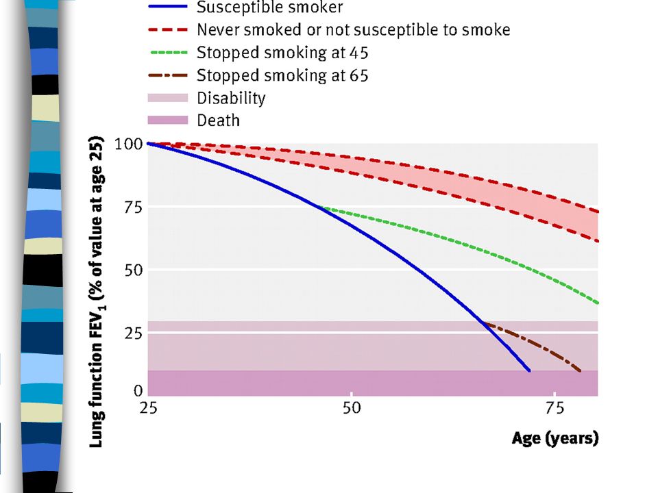
Smoking

Similar presentations


>")


 To improve gas absorption Increase surface.>")


