Download presentation
Presentation is loading. Please wait.

1
The Respiratory System Chapter 10

2
Organs of the Respiratory System Nose Pharynx Larynx Trachea Bronchi Lungs

3
Accessory Structures Thorax Diaphragm

4
The Nose

5
The Nose (naso or rhino) Acts as entrance for air and exit for carbon dioxide Lined with a ciliated epithelial mucus membrane Filters dust and foreign material Warms & moistens entering air Has olfactory receptors
 Acts as entrance for air and exit for carbon dioxide Lined with a ciliated epithelial mucus membrane Filters dust and foreign material Warms & moistens entering air Has olfactory receptors")
6
Nostrils (Nares) Paired openings that vary in pliability and expandability to accommodate an increased need for oxygen. The horse has pliable and expandable nostrils because mouth breathing is not characteristic.

9
Pharynx (Throat) A musculomembranous saclike structure Upper portion is attached to the base of the skull and the lower portion with the esophagus Connects with the nasal chambers, mouth, larynx and eustachian tubes
 A musculomembranous saclike structure Upper portion is attached to the base of the skull and the lower portion with the esophagus Connects with the nasal chambers, mouth, larynx and eustachian tubes")
10
The pharynx is divided into three parts: Nasopharynx – opens into the Eustachian tubes Oropharynx – opens into the mouth Laryngopharynx – opens into the larynx & esophagus

12
The pharynx is used as passageway for both air and food. Depending on the species it may also have a role in vocalization.

13
Larynx (Voice Box) Located just below the pharynx Serves as a passage for air The epiglottis is a lid-like structure that prevents food from entering the airway during swallowing
 Located just below the pharynx Serves as a passage for air The epiglottis is a lid-like structure that prevents food from entering the airway during swallowing")
16
The larynx plays an important role in creating sound. Air passes through the glottis during expiration causing a vibration, and producing a sound.

18
Trachea (Windpipe) A tube formed of smooth muscle with numerous C shaped rings of cartilage embedded in the tissue These rings prevent collapse of the structure.
 A tube formed of smooth muscle with numerous C shaped rings of cartilage embedded in the tissue These rings prevent collapse of the structure.")
21
The end of the trachea separates into smaller airways called the primary bronchi. The Bronchi

23
Lined with a ciliated mucous membrane & C-shaped cartilage rings, which become complete as the bronchi enter the lungs They then divide into smaller secondary branches, then bronchioles, then alveolar ducts The Bronchi

26
The alveolar ducts terminate into alveolar sacs: Small, squamous epithelium lined spaces that allow the lungs to achieve the primary function of oxygen and carbon dioxide exchange.

27
As the bronchioles get smaller, the cartilage rings begin to disappear. There are no rings in the alveolar ducts, sacs or alveoli.

29
The Lungs The primary structures of the respiratory system Occupy almost the entire thoracic space Each contains millions of alveoli and capillaries

31
Membranes in the Lungs Visceral pleura - Serous membrane that encases the lungs Parietal pleura – Lines the thoracic cavity These membranes reduce friction during respiration.

33
The space between these membranes is called the pleural cavity or potential space.

35
Thorax (Chest Cavity) Lined with a membrane similar to the covering of the lungs, that lubricates both surfaces during respiration
 Lined with a membrane similar to the covering of the lungs, that lubricates both surfaces during respiration")
36
The pleural membrane divides the chest into 3 parts: Right pleural cavity Left pleural cavity Mediastinum

37
The mediastinum contains the heart, thymus, esophagus, trachea, bronchi, nerves, arteries, veins and lymphatic vessels.

38
A dome-shaped musculomembranous partition separating the thoracic and abdominal cavity Attaches to the lumbar and abdominal cavities at the lumbar vertebra, lower ribs and sternum The Diaphragm

39
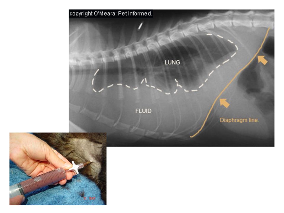
Normal feline chest Diaphragmatic Hernia

40
The diaphragm is the primary muscle of respiration. Inspiration: Contracts, flattens and lowers, increasing thoracic capacity. Expiration: Relaxes and returns to its normal position

42
Respiratory Process The respiratory cycle is divided into three parts: Inspiration Expiration Rest

43
Respiration Involves the exchange of oxygen and carbon dioxide wastes Tissue does not store oxygen and takes in only the oxygen it needs.

44
During exercise the oxygen requirement can more than double. Flow of air in and out of the lungs depends on changes in the thoracic cavity. Inspiration and expiration are in accordance with pressure differences between the atmosphere and air in the lungs

45
Tidal volume (TV): The amount of air inhaled during ordinary respiration Inspiratory reserve volume (IRV): The amount of air that can be forcibly inhaled beyond the normal amount
: The amount of air inhaled during ordinary respiration Inspiratory reserve volume (IRV): The amount of air that can be forcibly inhaled beyond the normal amount")
46
Expiratory reserve volume (ERV): The volume of air that can be forcibly expelled beyond normal expiration Some air will always be trapped in the alveoli no matter how forcibly an animal exhales due to intrathoracic pressure.
: The volume of air that can be forcibly expelled beyond normal expiration Some air will always be trapped in the alveoli no matter how forcibly an animal exhales due to intrathoracic pressure.")
47
Residual Volume (RV): Air remaining in the lungs after forced expiration Minimal volume: Air remaining in the alveoli of a collapsed lung
: Air remaining in the lungs after forced expiration Minimal volume: Air remaining in the alveoli of a collapsed lung")
48
Vital capacity (VC): The largest amount of air that can be moved in and out of the lungs. The sum of the total of inspiratory and expiratory reserve volumes plus tidal volume

50
There are several nerves from the brain that pass down the chest wall and diaphragm to control respiration…

51
Vagus nerve: Originates in the brain and sends branches to the larynx, heart, bronchi, esophagus, stomach, liver and abdomen.

53
Phrenic nerve: Originates in the cervical spine and passes to the diaphragm

54
Thoracic nerve: Originates in the thoracic spinal cord, these are the nerves of the muscles of the thorax

55
Next up… The GI Tract

Similar presentations


. 2.Production of sound (vocal cords). 3.Pulmonary ventilation. 4. Inspiration (intercostals muscles lift.>")

 Anatomy 2) mechanics of breathing 3) Chemistry of respiration.>")


