Download presentation
Presentation is loading. Please wait.

1
Introduction to tissue biomechanics for clinical practice Bone

2
Outcomes At the end of this section of study you will be able to Identify on the skeleton the major landmarks of the bones of the upper and lower limb Have had an introduction to the skill of palpation and practiced identifying bony points on a ‘model’ Describe how a bone grows and responds to stress

3
Getting to know bone Bone is – Metabolically active – Growth is essential for normal skeletal development – Has an excellent capacity for repair – Has the capacity to alter its properties and configurations in relation to mechanical demand

4
Bone Features stiffness and strength Composite tissue - strong collagenous tissue enmeshing hard mineral material Contains blood vessels fat and nerves

5
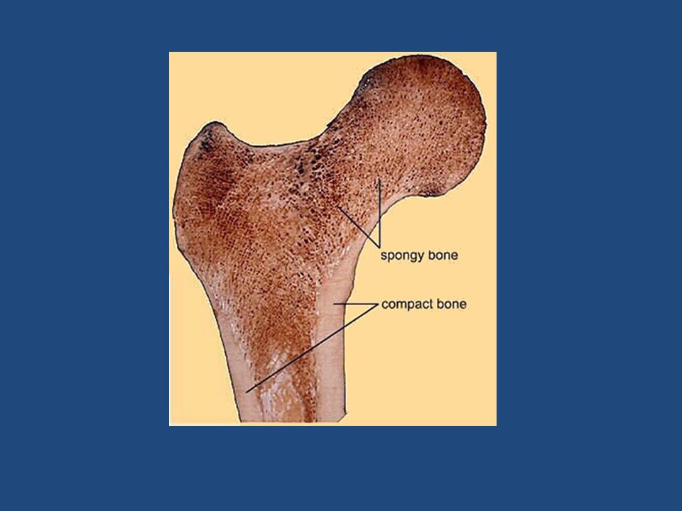
Structure Related to Function Dense cortical bone developed where stress is greatest Alignment of the “trabeculae”in cancellous bone reflects the patterns of habitual stress

6
Expanded distal end of the femur designed for both load bearing and shock absorption Complex configuration of the calcaneum (heel bone) which needs to absorb load and impact from the ankle and lower limb above and transmit force forward to the forefoot
 which needs to absorb load and impact from the ankle and lower limb above and transmit force forward to the forefoot")
7
Cortical bone (dense) Surface of the bones Shaft of the long bones Stiffer than cancellous (spongy) bone
 Surface of the bones Shaft of the long bones Stiffer than cancellous (spongy) bone")
8
Cancellous (spongy)Bone Found in the body of most bones The expanded ends of long bones More porous therefore will store more energy (absorb impact of load bearing)
Bone Found in the body of most bones The expanded ends of long bones More porous therefore will store more energy (absorb impact of load bearing)")
10
Why do we develop lumps and bumps? Muscles attach to the skeleton in a variety of ways – Tendons – localised points of attachment – Aponeurosis – broad sheets of attachment Directly to the periosteum (outer bone lining tissue) Into other sheets of fascia (more about this later) – These attachment points result in raised lines and tubercle/tuberosities being formed as the working muscle applies traction to the developing bone
 Into other sheets of fascia (more about this later) – These attachment points result in raised lines and tubercle/tuberosities being formed as the working muscle applies traction to the developing bone.")
11
Bone adaptation Bone obeys Wolff’s Law “bone is laid down where it is needed and reabsorbed where it is not” This important for us to remember in relation to bone health in response to training/activity altered movement pattern prolonged immobility

12
Bones are shaped by design and function Growth Mechanical demand – weight bearing Muscle attachment – traction from working muscles Post fracture repair Many of the bones in the body have distinguishing marks and features that are clinically significant

13
Lesser trochanter Head of the femur Greater trochanter

14
New terms to learn Tubercle / tuberosity Trochanter Condyle Epicondyle Fossa Foramen Epiphysis Apophysis Sesamoid

15
Things you should know about the pelvic ring 3 Bones - 2 innominate bones and 1 sacrum Innominate (sometimes just called the pelvic bone) is made up of three bones fused together – Ilium – Ischium – Pubis The pelvic ring is formed by 3 joints – 1 symphysis pubis joining the pelvis anteriorly and 2 sacroiliac joints posteriorly
 is made up of three bones fused together – Ilium – Ischium – Pubis The pelvic ring is formed by 3 joints – 1 symphysis pubis joining the pelvis anteriorly and 2 sacroiliac joints posteriorly")
16
More about the pelvis Provides attachment for muscles of the trunk Provides attachment for muscles of the lower limb Provides the acetabulum (socket) for the hip joint Is wider in women than men
 for the hip joint Is wider in women than men")
17
a a b b c c d d e e f f g g

18
Features of the femur Head (ball of the hip joint) – has a fossa in the middle (fovea capitis) Neck – Angle to increase R.O.M. – More acute in women – Common site of osteoporosis Greater trochanter Lesser trochanter Inter-trochanteric line (anterior) Inter-trochanteric crest (posterior)
 Inter-trochanteric crest (posterior).")
19
a a b b c c e e f f g g

20
Features of the femur Gluteal tuberosity Pectineal line Linea aspera Supra condylar lines Lateral and medial epicondyles Lateral and medial condyles Adductor tubercle Inter condylar notch

21
h h i i J J K K l l m m n n

22
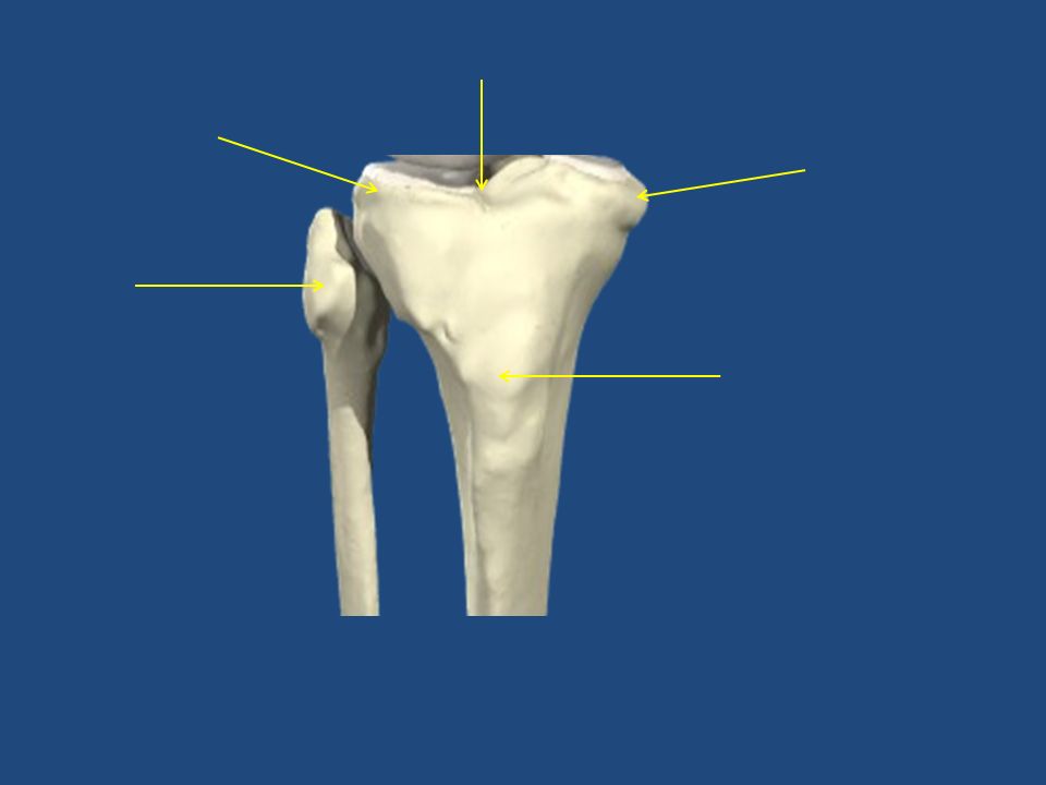
Features of Tibia and Fibula The tibia is a weight bearing bone proximally and distally Superior surface is divided into two by the inter - condylar ridge Tibia is most commonly # long bone Distal end forms the medial malleolus Articulates proximally and distally with the fibula Distal fibula forms the lateral malleolus

24
Features of the foot Talus – is the only bone of the foot to articulate with the leg – Divided in to 3 areas the head neck and body Calcaneus – transmits weight to the ground Navicular – medial Cuboid – lateral 3 cuneiform bones

25
E D C B F G H

26
More on the foot 5 metatarsals – numbered from the medial (big toe) side to the lateral NB base is proximal head is distal 14 phalanges – big toe has only 2 Transverse arch Lateral and medial longitudinal arches
 side to the lateral NB base is proximal head is distal 14 phalanges – big toe has only 2 Transverse arch Lateral and medial longitudinal arches")
28
a b c d e f g

29
a b c d ef

Similar presentations


 (Lower) Leg Foot The lower appendages are attached to the axial skeleton via the pelvic girdle.>")


 Coxae have 3 distinct parts: – Ilium – Ischium – Pubis.>")

 Girdle 2 coxal (hip) bones Ilium Ischium Pubis Sacroiliac Joint Pubic Symphysis Function: Support for vertebral.>")


