Download presentation
Presentation is loading. Please wait.

1
Respiratory System Anatomy and Physiology

2
Functions Delivering air to the lungs Bringing oxygen into the body –Diffuses O 2 from lungs into blood –Circulatory system takes over to deliver O 2 to cells –Why do we need O 2 ? To maintain our energy levels/make ATP Expelling the carbon dioxide back into the air to help maintain blood pH –Diffuses CO 2 from blood into lungs CO 2 and H 2 O when mixed makes carbonic acid and lowers pH of blood.

3
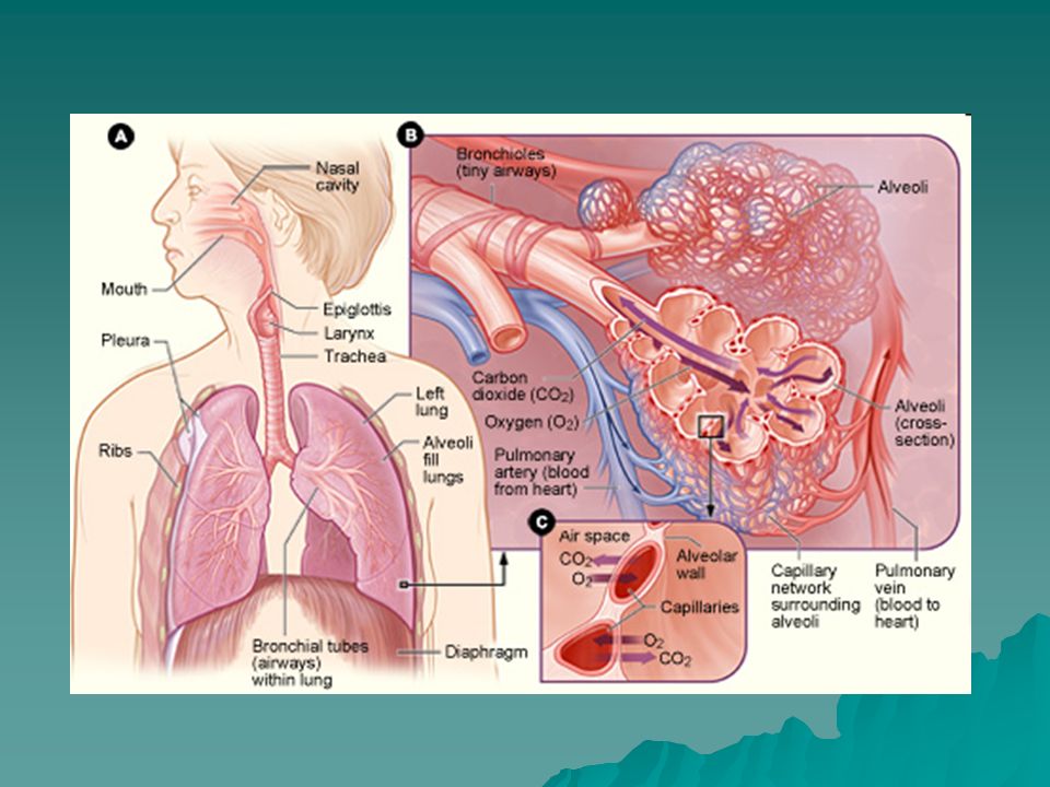
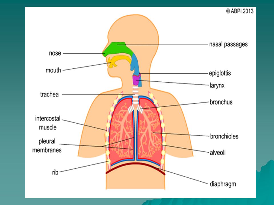
Parts of the Respiratory System Nose – provide opening for air to enter/exit nasal cavity Nasal Cavity – hollow space behind the nose, conducts air to pharynx, sense of smell –Hair, mucus, blood capillaries, and ciliated epithelium that line the nasal cavity filter, moisten, warm, and eliminate debris from the passing air (prevents respiratory infections). Ethmoid bone

4
Parts Continued Pharynx – throat, posterior to nasal cavity -passageway of food moving from oral cavity to esophagus -passageway of air moving between nasal cavity and larynx

5
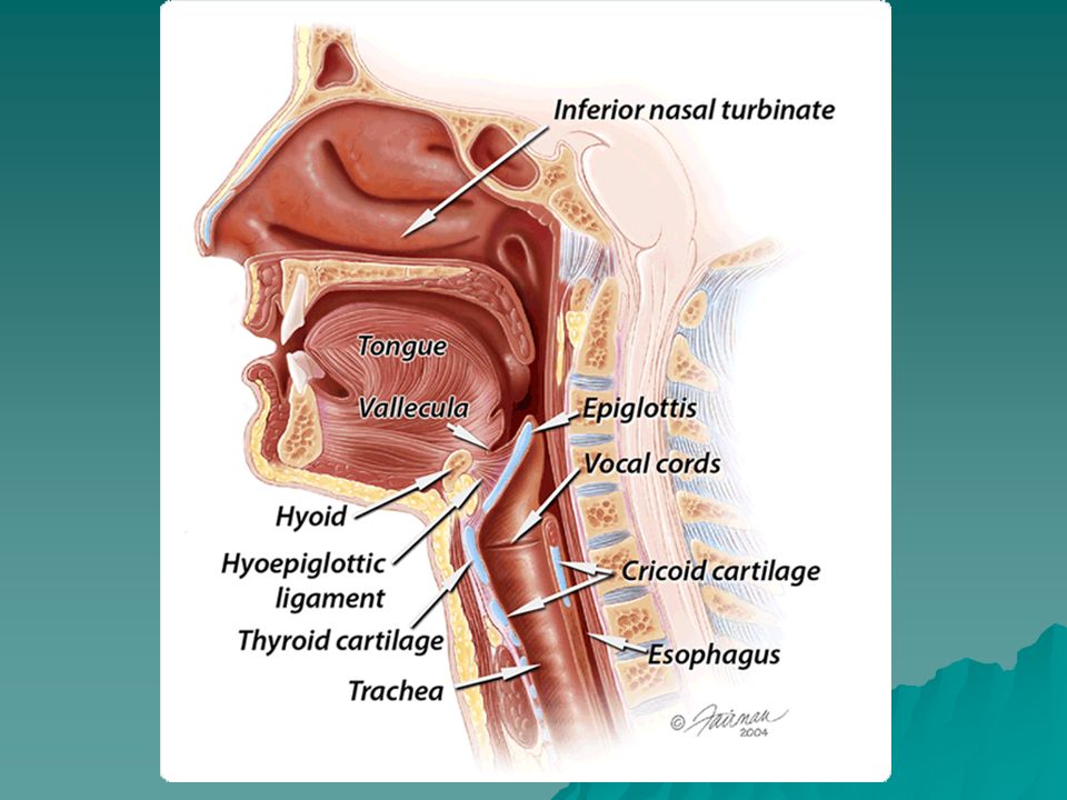
Parts Continued Larynx – passageway for air moving in and out of trachea (superior to trachea), prevents foreign objects from entering trachea –8 rings of cartilage and the mucous membrane has 2 pairs of folds –Houses vocal cords “voice box” – vibrate for speech
, prevents foreign objects from entering trachea –8 rings of cartilage and the mucous membrane has 2 pairs of folds –Houses vocal cords voice box – vibrate for speech")
6
Laryngitis - vocal cords swell, they vibrate differently, leading to hoarseness. Nodules - benign non-cancerous growths on the vocal cords are most often caused by voice misuse or overuse leading to hoarseness. Acid reflux – inflammation, redness, can lead to a sore or even laryngeal cancer if untreated.

7
Parts Continued Thyroid cartilage – big piece of shield-shaped cartilage in front of the larynx (Adam’s apple) – males only Epiglottis – flap of cartilage that closes off the larynx when swallowing so that food goes down the esophagus and not to the lungs (if anything but air enters the larynx – it causes us to cough)
 – males only Epiglottis – flap of cartilage that closes off the larynx when swallowing so that food goes down the esophagus and not to the lungs (if anything but air enters the larynx – it causes us to cough)")
8
Parts Continued Trachea – windpipe (1” wide & 4” long), splits into L & R bronchi – lined with ciliated mucous membrane to sweep debris away from lungs towards pharynx, reinforced with cartilage rings to keep it open (snaps shut when hit)
, splits into L & R bronchi – lined with ciliated mucous membrane to sweep debris away from lungs towards pharynx, reinforced with cartilage rings to keep it open (snaps shut when hit)")
9
Trachea blockage of trachea –heimlich maneuver –blockage causes asphyxiation in minutes tracheostomy

10
Parts Continued Bronchi – trachea divides into 2 to go to each lung Bronchioles or bronchial tree – smaller and smaller branches that end with alveoli –Branching increases surface area to absorb more O2 (lungs are small but each have surface area the size of a tennis court).
.")
11
Asthma

12
Parts Continued Alveoli – air sacs lined with epithelium Lungs – most of the lungs are made of the alveoli (300 million) held together with connective tissue –Left lung – 2 lobes/ Right lung – 3 lobes –Covered by serous membrane (pleura) – keep the lungs from rubbing against the thoracic cavity and adhere lungs to the chest wall for inflation during breathing
 held together with connective tissue –Left lung – 2 lobes/ Right lung – 3 lobes –Covered by serous membrane (pleura) – keep the lungs from rubbing against the thoracic cavity and adhere lungs to the chest wall for inflation during breathing")
13
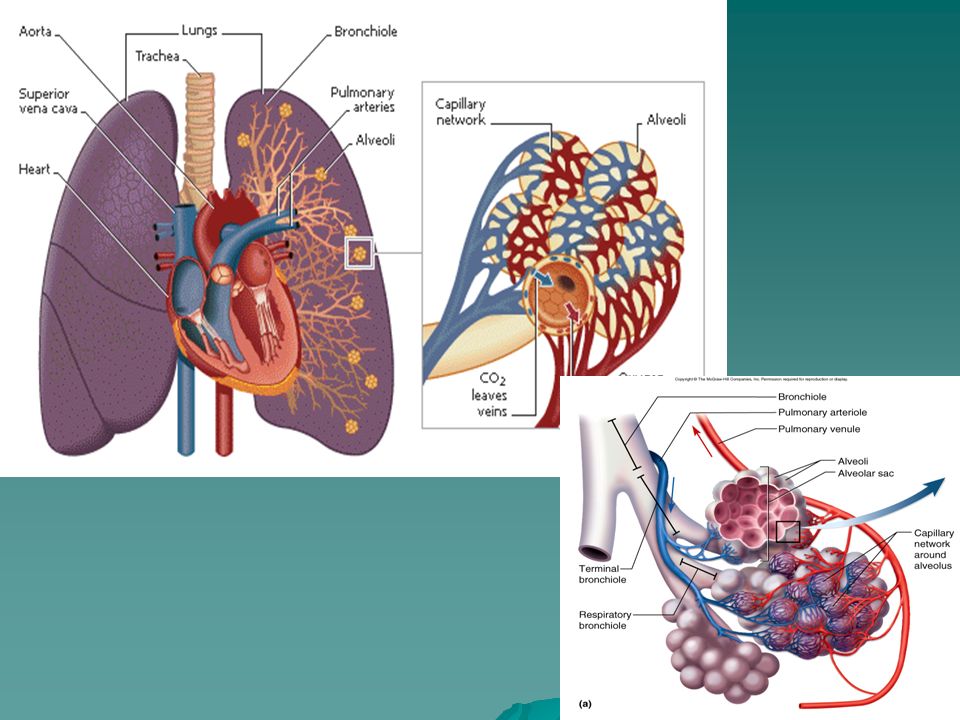
Alveoli Movement of material happens from the alveoli to the capillaries by simple diffusion (oxygen enters the capillaries and carbon dioxide leaves them and enters the alveoli) Macrophages move in and out and destroy bacteria that get in –Air is one way in – debris, smoke, pollutants, etc. needs to be eliminated Embedded in the epithelial cells are cells that make lung surfactant – lipid coating that keeps alveoli from collapsing –“Wind knocked out of you” Surface area for gas exchange of each lung (300 million alveoli) is about the size of 1 tennis court
 is about the size of 1 tennis court.")
14
Alveoli

17
Diaphragm Diaphragm controls breathing, not lungs –Muscle controlled by brain (passive process when sitting) When relaxed, diaphragm is curved upward and when contracted, it flattens out.
 When relaxed, diaphragm is curved upward and when contracted, it flattens out.")
20
Diaphragm and Breathing Lungs respond to air pressure differences within thoracic cavity Inhalation: –Diaphragm contracts (flattens), the volume in the thoracic cavity increases so the pressure decreases –Atmospheric pressure is higher than the pressure in the thoracic cavity so it automatically flows in –Intercostal muscles also expand the ribs aiding in lung expansion and the decrease in pressure (lungs adhere to the thoracic wall by the pleural membrane)
, the volume in the thoracic cavity increases so the pressure decreases –Atmospheric pressure is higher than the pressure in the thoracic cavity so it automatically flows in –Intercostal muscles also expand the ribs aiding in lung expansion and the decrease in pressure (lungs adhere to the thoracic wall by the pleural membrane)")
21
Diaphragm and Breathing Lungs respond to air pressure differences within thoracic cavity Exhalation: –Diaphragm relaxes (moves upward), the volume in the thoracic cavity decreases so the pressure increases –Atmospheric pressure is lower than the pressure in the thoracic cavity so it automatically flows out
, the volume in the thoracic cavity decreases so the pressure increases –Atmospheric pressure is lower than the pressure in the thoracic cavity so it automatically flows out")
22
Volume inc, pressure dec, air entersVolume dec, pressure inc, air exits

23
Breathing Continued Tidal Volume – volume of air inhaled and exhaled (500 mls/1pt.) Inspiratory Reserve Volume – amount that can forcibly be taken in over tidal volume (2000- 3000 mls) Vital capacity – Maximum air the lungs can hold (4500 mls) Expiratory Reserve Volume – amount that can be forcibly exhaled after tidal expiration (about 1000 mls) Residual Volume – amount of air left in the lungs even after forced expiration (about 1000 mls) – allow gas exchange between breaths and keeps alveoli inflated –Lose this when getting the “wind knocked out of you” Sneezing and coughing clear the air passages of debris and excess mucous
 Inspiratory Reserve Volume – amount that can forcibly be taken in over tidal volume ( mls) Vital capacity – Maximum air the lungs can hold (4500 mls) Expiratory Reserve Volume – amount that can be forcibly exhaled after tidal expiration (about 1000 mls) Residual Volume – amount of air left in the lungs even after forced expiration (about 1000 mls) – allow gas exchange between breaths and keeps alveoli inflated –Lose this when getting the wind knocked out of you Sneezing and coughing clear the air passages of debris and excess mucous")
24
Gas Exchange Oxygen diffuses from inside the alveoli (20%) to the capillaries of the lungs (~0%) Most of it is picked up by hemoglobin After the blood is pumped to the body and reaches the capillaries of the body’s tissues – the oxygen is dumped off by hemoglobin and diffuses out of the RBC into the interstitial fluid surrounding the cells It then diffuses into the cells
 to the capillaries of the lungs (~0%) Most of it is picked up by hemoglobin After the blood is pumped to the body and reaches the capillaries of the body’s tissues – the oxygen is dumped off by hemoglobin and diffuses out of the RBC into the interstitial fluid surrounding the cells It then diffuses into the cells")
25
How does more Oxygen Get to the Cells that Need it the Most? i.e. cells doing the most work 1. Blood is diverted to the most active areas (precap. sphincters shut off blood to certain areas not in use) -upper body workout vs. lower body -don’t swim for an hour after you eat 2. Hemoglobin drops off O 2 quicker in areas of low O 2 and high CO 2 (higher acidity) and holds onto it in reverse situations. 3. The higher the concentration difference of oxygen between blood and cells, the faster the rate of diffusion
 -upper body workout vs. lower body -don’t swim for an hour after you eat 2. Hemoglobin drops off O 2 quicker in areas of low O 2 and high CO 2 (higher acidity) and holds onto it in reverse situations. 3. The higher the concentration difference of oxygen between blood and cells, the faster the rate of diffusion.")
26
Control of Respiration Controlled by the autonomic nervous system Normally breath 12-15 times/minute Do have some conscious control over breathing but autonomic centers will ignore information if ph of blood is low (can’t hold your breath until you die) Mostly controlled by level of carbon dioxide in the blood (pH) – act on the medulla oblongata in the brain Oxygen concentration is also measured in the aorta and carotid arteries which send signals to the medulla – only when oxygen is dangerously low Hyperventilating – breathing real fast – lose too much carbon dioxide too fast, blood becomes a little basic and breathing may stop – breath into a bag to increase carbon dioxide levels
 Mostly controlled by level of carbon dioxide in the blood (pH) – act on the medulla oblongata in the brain Oxygen concentration is also measured in the aorta and carotid arteries which send signals to the medulla – only when oxygen is dangerously low Hyperventilating – breathing real fast – lose too much carbon dioxide too fast, blood becomes a little basic and breathing may stop – breath into a bag to increase carbon dioxide levels")
27
Effects of Cigarette Smoke Healthy resp syst is continuously cleansed by mucus trapping dirt and pathogens, cilia then sweeps towards mouth With the first inhalation of smoke, cilia slows. Eventually become paralyzed and disappear – leads to “smokers cough” –Lethal chain reaction – cough chronic bronchitis inc. mucus production and bronchial thickening bronchioles lose elasticity inc. air pressure in alveoli and can rupture = smoking induced emphysema –Cellular changes can occur as well leading to lung cancer. 80% of lung cancer cases are due to cigarette smoking.

28
COPD stands for chronic obstructive pulmonary disease and it is a serious lung disease that makes it harder and harder to breathe. Both chronic bronchitis and emphysema are considered to be COPD. Picture of Alveoli ruptured as a result of emphysema

29
References http://www.cliffsnotes.com/study_gu ide/Function-of-the-Respiratory- System.topicArticleId- 277792,articleId-277729.html

Similar presentations
>")


. 2.Production of sound (vocal cords). 3.Pulmonary ventilation. 4. Inspiration (intercostals muscles lift.>")


>")


 into the atmosphere Filter, moisten,>")


