Download presentation
Presentation is loading. Please wait.

1
Facial Bone Anatomy & Positioning
RTEC 233

2
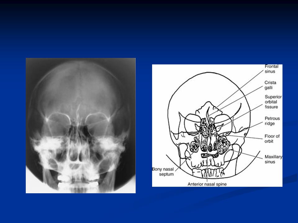
Anterior Aspect of Facial Bones
2 Maxillae 2 Zygomatic bones 2 Lacrimal bones 2 Nasal bones 2 Inferior nasal conchae 2 Palatine bones (not visualized 1 vomer 1 mandible There are 14 bones that make up the facial bones. 6 of which are paired and 2 single bones: mandible and vomer. The facial bones contribute to the shape and form of a person’s face. They also serve as protective housing for the eyes. There are cavities of the orbits, nose and mouth that are largely made up of the facial bones.

3
The 2 maxillae are the largest immovable bones of the face
The 2 maxillae are the largest immovable bones of the face. The maxillae and united at the midline below the nasal septum. Each maxillae assists is forming three cavities: mouth, nasal cavity, and one orbit. Each maxillae consists of 4 processes: 1) frontal process 2) zygomatic process 3)Alveolar process 4) Palatine processes 1) Frontal process projects superiorly along the lateral border of the nose toward the frontal bone. 2) Zygomatic process extends laterally toward the zygoma on each sided. 3) Alveolar process is the inferior aspect. Along the inferior margin of each alveolar process are 8 upper teeth. 4) Palatine process can only be demonstrated inferiorly and form the anterior portion of the mouth called the hard, or bony palate. This bone also contains and important landmark for radiography of the skull and facial bones: acanthion. The acanthion is at the base of the anterior nasal spine at a point where the nose and upper lip meets. The maxillae contain pyramidal shaped cavities called the maxillary sinuses. The infraorbital foramen is located under each orbit on each maxillae which serves as a passage through which infraorbital nerve and artery reach the nose. The maxillae articulate with 2 cranial bones: frontal and ethmoid. Also articulates with all facial bones with the exception of the mandible.
 frontal process 2) zygomatic process 3)Alveolar process 4) Palatine processes. 1) Frontal process projects superiorly along the lateral border of the nose toward the frontal bone. 2) Zygomatic process extends laterally toward the zygoma on each sided. 3) Alveolar process is the inferior aspect. Along the inferior margin of each alveolar process are 8 upper teeth. 4) Palatine process can only be demonstrated inferiorly and form the anterior portion of the mouth called the hard, or bony palate. This bone also contains and important landmark for radiography of the skull and facial bones: acanthion. The acanthion is at the base of the anterior nasal spine at a point where the nose and upper lip meets. The maxillae contain pyramidal shaped cavities called the maxillary sinuses. The infraorbital foramen is located under each orbit on each maxillae which serves as a passage through which infraorbital nerve and artery reach the nose. The maxillae articulate with 2 cranial bones: frontal and ethmoid. Also articulates with all facial bones with the exception of the mandible.")
4
Palatine Bones L-shaped bones
Horizontal portion forms posterior hard palate Vertical portion extends between 1maxillae and 1 pterygoid plate of sphenoid bone Articulates with 2 cranial bones and 4 facial bones Very difficult to visualize of a dry skull because they are internally located. Articulates with 2 cranial bones: 1) Sphenoid 2) Ethmoid Articulates with 4 facial bones: 1) Maxilla 2) Inferior nasal conchae 3) Vomer 4) Adjacent palatine
 Sphenoid. 2) Ethmoid. Articulates with 4 facial bones: 1) Maxilla. 2) Inferior nasal conchae. 3) Vomer. 4) Adjacent palatine.")
5
Zygomatic Bones Forms cheeks Forms lower outer margin of orbits
Articulates with 3 cranial bones Frontal Sphenoid Temporal Articulates with maxillae Sometimes called malar bones. Projecting posteriorly from the zygoma is a thin process connecting to the zygomatic process of the temporal bone. The zygomatic prominence is a landmark on the zygoma and refers to the prominent portion of the zygoma.

6
Inferior Nasal Cochae The only pair of conchae that are separate facial bones Articulates with 1 cranial bone and 3 facial bones Covered with mucous membranes to warm, moisten and cleanse inhaled air The superior and inferior nasal conchae are processes of the ethmoid bone, while the inferior nasal conchae are separate facial bones. The inferior nasal conchae are thin, narrow and extremely thin bones which curl laterally and look like miniature scrolls. The inferior nasal conchae articulate with the ethmoid bone and the maxilla, lacrimal and palatine bones.

7
Lacrimal Bones About the size & shape of a fingernail
Lacrimal foramen for tear duct Lie anteriorly on the medial side of orbit Can be seen on PA and lateral projections Articulates with 2 cranial bones and 2 facial bones Lacrimal means “tear” and is appropriate because the lacrimal bones along with the maxilla form the lacrimal fossae which accommodates the lacrimal sacs. The lacrimal bones articulate with the frontal and ethmoid bones. They also articulate with the maxilla and inferior nasal conchae.

8
Nasal Bones Fused and form bridge of nose Vary in size considerably
The point of junction with the frontal bone is the nasion Articulates with 2 cranial and 2 facial bones

9
Vomer Forms inferosuperior part of nasal septum Deviated nasal septum
Depressions for blood vessels Articulates with 2 cranial bones & 4 facial bones Together the perpendicular plate of the ethmoid bone and the vomer form the bony nasal septum. Anteriorly the septal septum is cartilaginous and is called the septal cartilage. A deviated nasal septum describers the clinical condition where the nasal septum is displaced laterally from the midline of the nose. This deviation occurs at the site of the septal cartilage and the vomer. If there is a severe deviation the nasal passageway can be blocked not allowing the patient to breathe from their nose. The vomer has small furrow-like depressions for blood vessels. In a trauma this can be a source for nosebleed. Articulates with the sphenoid and ethmoid bones of the cranium. Articulates with the R/L palatine bones and the R/L maxillae.

10
Mandible Only movable bone in the skull Densest & largest facial bone
2 bones at birth Contains mental foramina The mandible is the largest and densest bone of the face. It is also the only movable bone in the adult skull. At birth the mandible consists of bilateral pieces held together by a fibrous symphysis that ossifies during the first year of life. There are 2 holes of each side for the transmission of nerves and blood vessels. These openings are called the mental foramina.

11
Pathologic Indications for Facial Radiography
Fractures Blowout Tripod LeFort Coutrecoup Foreign Body Osteomyelitis Neoplasms Secondary Osteomyelitis TMJ Syndrome Fractures- a break in the structure of a bone caused by some sort of trauma. Blowout fracture- is a fracture of the floor of the orbit, caused by a direct blow to the orbital region. The floor ruptures and the inferior rectus muscle is forced through the fracture into the maxillary sinuses. This often causes diplopia (perception of two images), double vision. Tripod- is a fracture caused by a blow to the zygoma. It causes a break in 3 places- orbital process, maxillary process, and the arch. This causes a free floating zygomatic bone. LeFort- is a fracture that is bilateral horizontal fractures of the maxillae. Results in an unstable detached fragment. Countrecoup- is an fracture on one side of a structure caused by a blow from the other side. Foreign body of the eye- metal or other fragments in the eye. This usually occurs in an industrial setting. Neoplasms- new and abnormal growth (tumor). Osteomyelitis- localized infection of the bone/bone marrow. This is usually a result of a trauma, fracture or postoperative bacteria. It also can be spread by blood from a distant site. Secondary Osteomyelitis- an infection of the bone and marrow secondary to sinusitis, results in erosion of the bony margins of the sinus. TMJ syndrome- set of symptoms that includes pain and clicking that indicates dysfunction of the TMJ’s. It can be caused by malocclusion, stress, muscle spasm, or inflammation.
, double vision. Tripod- is a fracture caused by a blow to the zygoma. It causes a break in 3 places- orbital process, maxillary process, and the arch. This causes a free floating zygomatic bone. LeFort- is a fracture that is bilateral horizontal fractures of the maxillae. Results in an unstable detached fragment. Countrecoup- is an fracture on one side of a structure caused by a blow from the other side. Foreign body of the eye- metal or other fragments in the eye. This usually occurs in an industrial setting. Neoplasms- new and abnormal growth (tumor). Osteomyelitis- localized infection of the bone/bone marrow. This is usually a result of a trauma, fracture or postoperative bacteria. It also can be spread by blood from a distant site. Secondary Osteomyelitis- an infection of the bone and marrow secondary to sinusitis, results in erosion of the bony margins of the sinus. TMJ syndrome- set of symptoms that includes pain and clicking that indicates dysfunction of the TMJ’s. It can be caused by malocclusion, stress, muscle spasm, or inflammation.")
12
Tri-pod Fracture

13
Blow out fracture

14
LeFort Fractures

15
FIG 3 - LeFort lines used for classifying fractures of the middle third of the face.
Hodgkinson, D W et al. BMJ 1994;308:46-50 Copyright ©1994 BMJ Publishing Group Ltd.

16
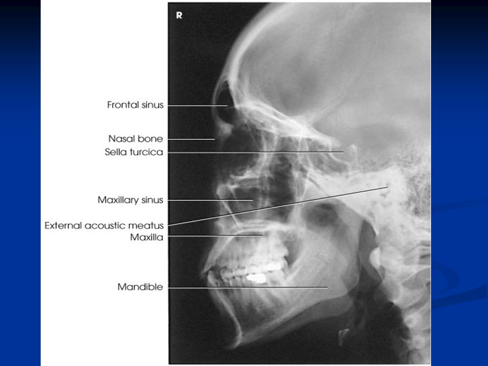
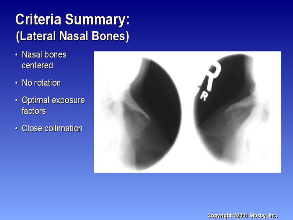
Positioning: Lateral Facial bones
Semiprone or seated MSP parallel IPL perpendicular Suspend respiration CR is perp and enters lateral zygomatic bone ½ way between outer canthus and EAM.

17
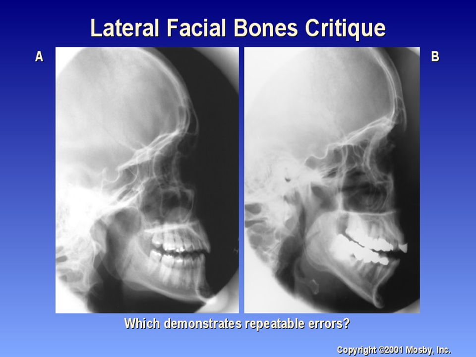
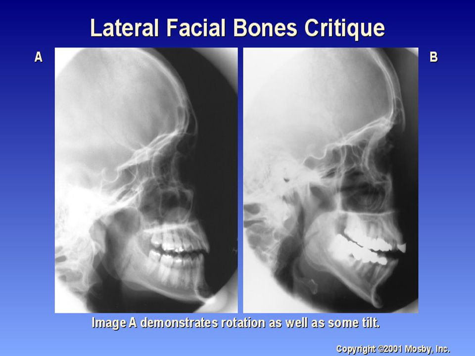
Lateral Facial Radiograph
All facial bones in with zygomatic bone in center Almost SI mandibular rami SI orbital roofs (no tilt) No rotation of sella turcica
 No rotation of sella turcica.")
23
Anatomy Identity

24
Radiograph Anatomy E) Anterior nasal spine
P) Condyle or neck of mandible Q) EAM R) Temporalmandibular fossa of temporal bone S) Greater wings of sphenoid T) Lesser wings of sphenoid U) Ethmoid sinuses between orbits V) Body of maxilla containing maxillary sinuses E) Anterior nasal spine F) Alveolar process of maxilla G) Alveolar process of mandible H) Mentum J) Body of mandible K) Angle of mandib le L) Ramus of mandible M) Coronoid process O) Neck of mandibular condyle
 Condyle or neck of mandible. Q) EAM. R) Temporalmandibular fossa of temporal bone. S) Greater wings of sphenoid. T) Lesser wings of sphenoid. U) Ethmoid sinuses between orbits. V) Body of maxilla containing maxillary sinuses. E) Anterior nasal spine. F) Alveolar process of maxilla. G) Alveolar process of mandible. H) Mentum. J) Body of mandible. K) Angle of mandib le. L) Ramus of mandible. M) Coronoid process. O) Neck of mandibular condyle.")
25
Anatomy Identity

26
Lateral Skull Anatomy A) zygomatic arch J) Body of mandible
B) RT zygomatic bone C) RT nasal bone D) Frontal process of maxilla E) Anterior nasal spine F) Alveolar process of maxilla G) Alveolar process of mandible H) Mentum I) Mental foramen J) Body of mandible K) Angle (gonion) L) Ramus of mandible M) Coronoid process N) Mandibular notch O) Neck of mandibular condyle P) Condyle or head of mandible Q) EAM
 RT zygomatic bone. C) RT nasal bone. D) Frontal process of maxilla. E) Anterior nasal spine. F) Alveolar process of maxilla. G) Alveolar process of mandible. H) Mentum. I) Mental foramen. J) Body of mandible. K) Angle (gonion) L) Ramus of mandible. M) Coronoid process. N) Mandibular notch. O) Neck of mandibular condyle. P) Condyle or head of mandible. Q) EAM.")
27
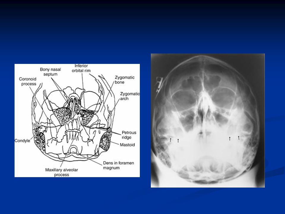
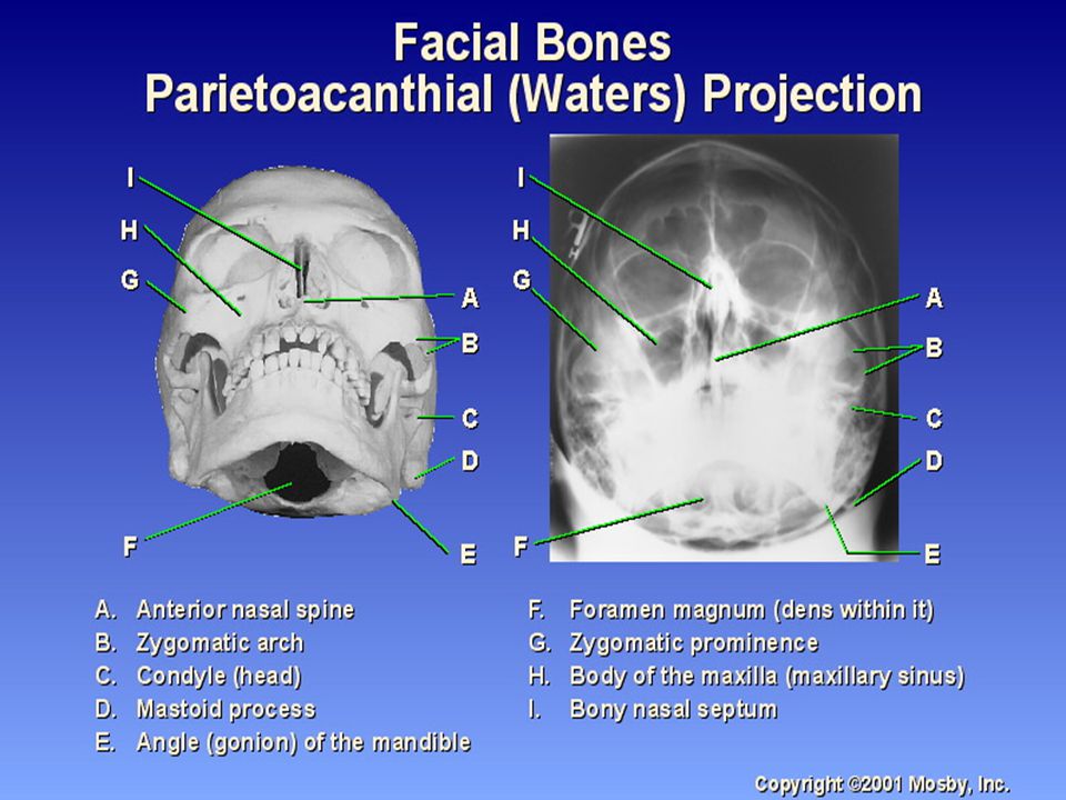
Positioning: Waters Prone or seated upright
Chin on bucky -OML 37 angle with plane of cassette MML & MSP perp Nose 3/4 inch from IR Suspend respiration CR perpendicular to exit acanthion

28
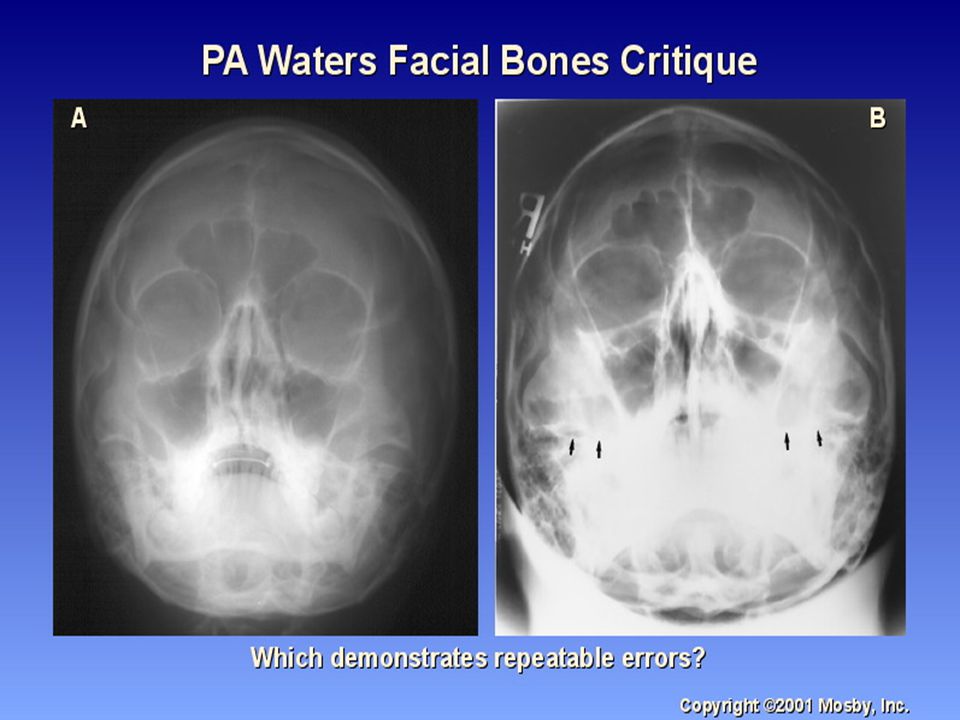
Waters Radiograph Distance from lateral border of skull and orbit equal on each side Petrous ridges projected immediately below maxillary sinuses

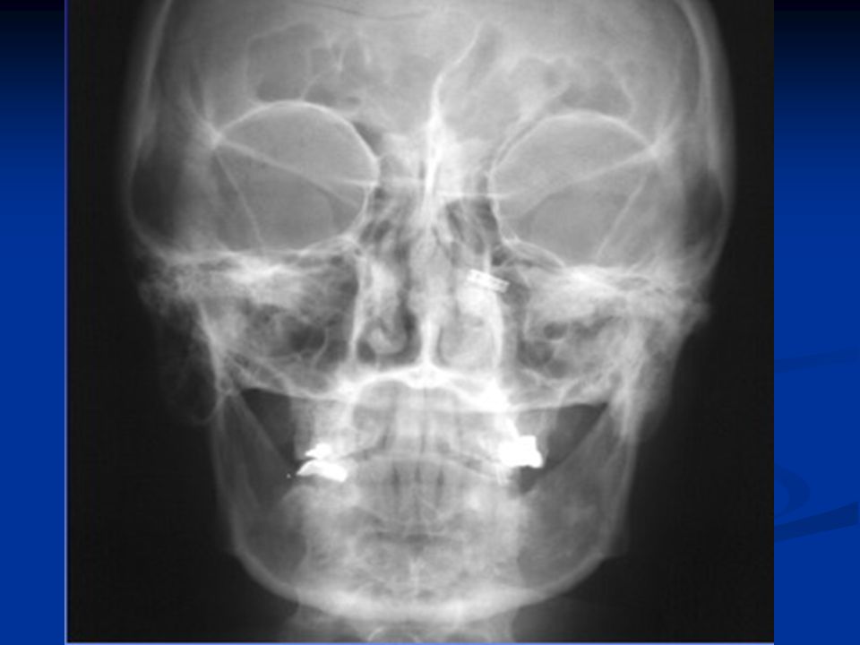
33
Trauma

34
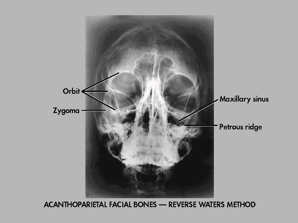
Reverse Waters Supine Extend neck so OML is 37 degree with plane of IR
MML and MSP perp Suspend respiration CR perpendicular and enters acanthion

35
Reverse Waters Radiograph
Distance from lateral border of skull and orbit equal on each side Petrous ridges projected immediately below maxillary sinuses

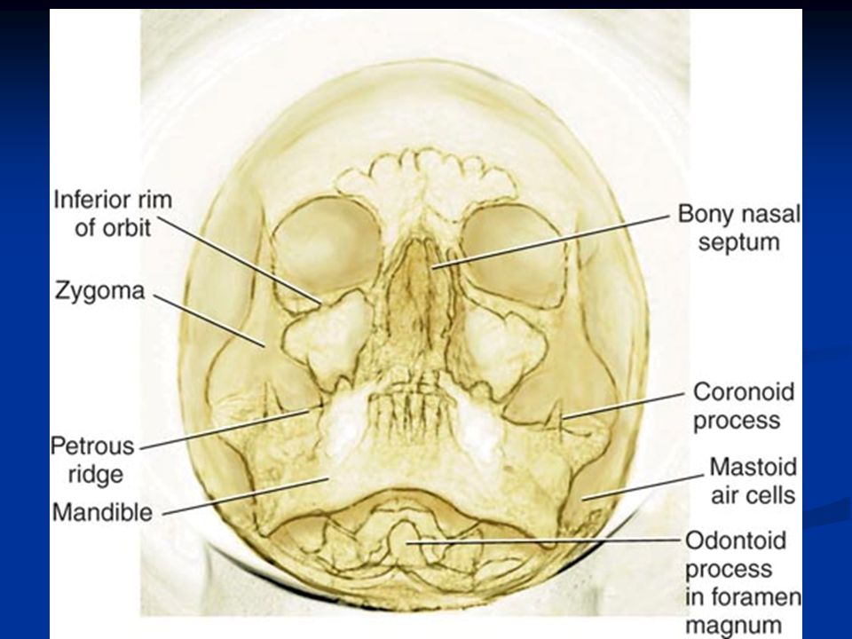
38
Modified Waters OML 55 degree angle from plane of IR MSP perp
CR perpendicular and exits acanthion

39
Modified Waters Radiograph
Petrous ridges projected immediately below the inferior border of the orbits Equal distance from lateral orbit to lateral skull on both sides

43
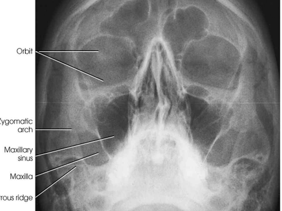
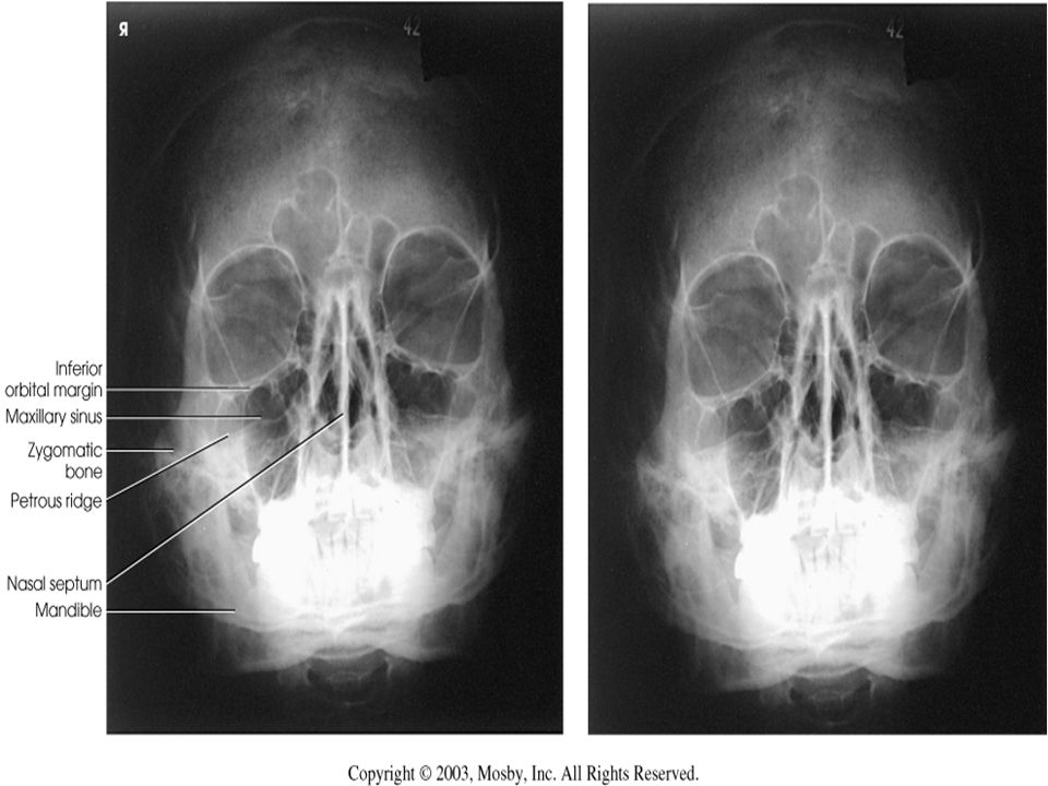
PA Axial - Caldwell Prone or seated upright
Forehead & nose against grid device OML perpendicular CR 15 caudal to exit nasion Suspend respiration

44
PA Axial- Caldwell Radiograph
Equal distance from lat skull to lat orbit Symmetric petrous ridges in lower 1/3 orbit Penetration of frontal bone without excessive density at lateral borders of skull.

48
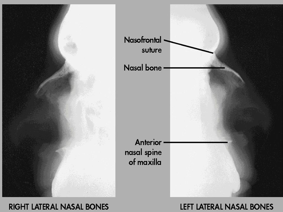
Lateral Nasal Bones Semiprone MSP & IOML parallel IPL perpendicular
CR perpendicular to the bridge of nose at a point 1” distal to the nasion

49
Lateral Nasal bones Radiograph
No rotation of nasal bone and soft tissue Anterior nasal spine and frontonasal suture evident Close collimation

Similar presentations