Download presentation
Presentation is loading. Please wait.

1
Bone Tissue get Vitamin D handout Quiz #1 opens today and is
available until Monday 4/9 at 11pm

2
Quiz information 6 quizzes during the quarter
5 questions worth 2 points each = 10 points total Available Tuesday’s at 3pm until the following Monday at 11pm No make ups!! Clinical cases with multiple choice answers No time limit, open as many times as you want, change your answers – until you “SUBMIT” You need to “SUBMIT” the quiz After quiz closes, answers, scores and feedback will be available

3
Tissue type? Cell type? Type of epithelium? Where is it found (one example)? Tissue type (be specific)?
. Tissue type (be specific).")
4
To do list for today functions of the skeleton bone shapes
general features of bone – parts bone histology = cells + matrix structural disorders of bone osteoporosis introduction to skeleton bone markings appendicular skeleton

5
short flat irregular long Show bones

6
Fig. 6.2a(TE Art) Articular cartilage Epiphysis Epiphyseal line
Spongy bone Compact bone Medullary cavity Diaphysis Periosteum Endosteum Epiphysis Articular cartilage

7
Fig. 6.2b(TE Art) Suture Spongy Bone (diploe) Compact bone
 Suture Spongy Bone (diploe) Compact bone")
9
Compact bone Spongy bone Bone marrow periosteum osteon trabeculae Perforating fibers endosteum Central canal Perforating canal

10
Bone cells Osteoprogenitor ** Osteoblast ** ** osteocyte ** canaliculi

11
Bone dissolving macrophages
** Osteoclast ** Stem cells osteoclast Why dissolve bone??

12
Bone matrix 1/3 organic = flexibility collagen protein-carbo complexes
2/3 inorganic = strength calcium phosphate salt magnesium, sodium, potassium… Calcium and vitamin D need vitamin D to absorb calcium in the small intestine need UV to make vitamin D rickets = deficient vit D, soft bones

13
Shell-less chick no calcium, no ossified bone

14
Bone deposition = resorption
Osteoporosis = porous bones decreased deposition increased resorption spongy bone affected bones become brittle women > men less bone mass lose it earlier & faster decrease in estrogen build bone mass 25-40!

15
Things that affect bone resorption or deposition:
What about my latte milk? Does that count? Drink milk? Things that affect bone resorption or deposition: steroids – reduce osteoblast activity, lower Ca absorption estrogen & testosterone – increase osteoblast activity caffeine – binds Ca so it’s excreted smoking – decrease osteoblast production, impairs Ca absorption, interferes with ERT

16
Osteoporosis and Anatomy….
kyphosis Areas with more spongy bone

17
“To name all of our bones!”

18
Fig. 7.1(TE Art) axial appendicular Parietal bone Frontal bone
Temporal bone Zygomatic bone Skull Maxilla Occipital bone Mandible Mandible Clavicle Clavicle Scapula Scapula Thoracic cage Sternum Humerus Ribs Vertebral column Pelvic girdle Ulna Radius Carpus Metacarpal bones Phalanges Femur Patella Fibula Tibia Metatarsal bones Tarsus Phalanges Calcaneus appendicular

19
Head Crest Trochanters Line Epicondyles Condyles
Linea aspera Epicondyles Bones remodel when stresses are placed on them! Condyles

20
Appendicular skeleton

21
The humerus is ____________ to the radius.
Anatomical position?? Identify bone 1, 2, 3 & 4 The humerus is ____________ to the radius. The radius is ____________ to the ulna (anatomical position). List the bones that articulate with the humerus. 1 2 3 4
. List the bones that articulate with the humerus")
22
Fig. 8.4(TE Art) Anterior Posterior Olecranon Olecranon
Trochlear notch Head of radius Head of radius Neck of radius Neck of radius Ulna Radius Interosseous membrane Head of ulna Styloid process Styloid process Styloid process Articular facets

23
The Axial Skeleton Bones of the skull parts of the skull sinuses
development of skull Vertebral column curves vertebrae & discs C1 & C2 Thoracic cage parts ribs bony landmarks

24
Axial skeleton

25
cranial facial

27
Fig. 7.4a(TE Art) Frontal bone Parietal bone Ethmoid bone Nasal bone
Sphenoid bone Nasal bone Occipital bone Lacrimal bone Temporal bone Maxilla Zygomatic bone Mastoid process Styloid process Mandibular condyle Mandible

28
Fig. 7.3(TE Art) Frontal bone Parietal bone Temporal bone
Sphenoid bone Lacrimal bone Ethmoid bone Nasal bone Zygomatic bone Nasal conchae Vomer Maxilla Mandible

29
Fig. 7.5a(TE Art) Palatine bone Zygomatic bone Nasal choana Vomer
Occipital condyle Carotid canal Foramen magnum Temporal bone Parietal bone External occipital protuberance Occipital bone

31
Sinuses Frontal Ethmoid Maxillary (Sphenoid)
Mucous membrane: pseudostratified columnar epithelium, ciliated, goblet cells

32
Mandible without teeth
Skull changes over time

33
Fig. 7.31(TE Art) Coronal suture Lambdoid suture Squamous suture
Sphenoid bone Mandible Sphenoid fontanel Anterior fontanel Sagittal suture Parietal bone Posterior fontanel

35
Spine Vertebral column Backbone

36
Normal and abnormal curves of the vertebral column

37
Intervertebral disc posterior Spinous process Transverse process Body
Nucleus pulposus Annulus fibrosus

38
Annulus fibrosus = fibrocartilage

39
Herniated disc (slipped disc)
")
41
C1 or Atlas C2 or Axis C1- occipital condyle nodding “yes”
Superior articular facet C2 or Axis Dens C1- occipital condyle nodding “yes” C1- C2 via dens rotation “no” Transverse foramen

42
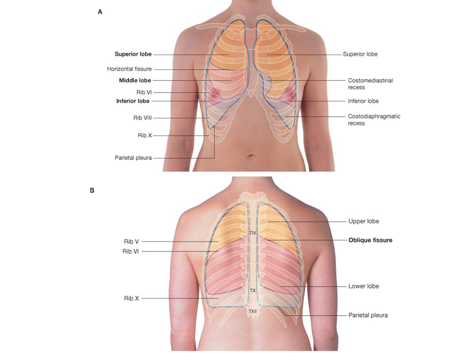
View?

44
Ribs & thoracic vertebrae
articulations

47
Summary…. Please go over joint movements!!
Think about a joint injury you’ve had (sprained ankle, knee or wrist). How did you do it? What happened to that joint after the injury?
. How did you do it What happened to that joint after the injury")
Similar presentations