Download presentation
Presentation is loading. Please wait.

1
Reading: Chapter 6 Pages 152 – 159 & 163-167 only! Omit Bone Development Bone Tissue

2
What are the functions of the skeletal system? list

3
long shortflatirregular Show bones

4
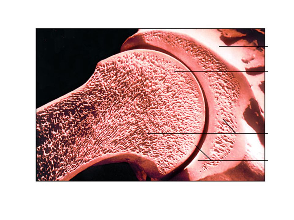
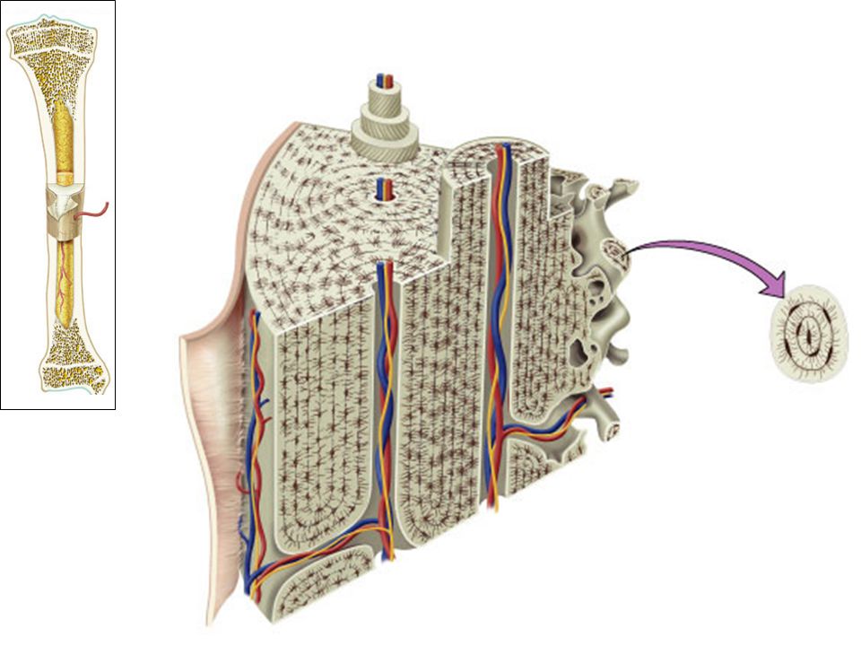
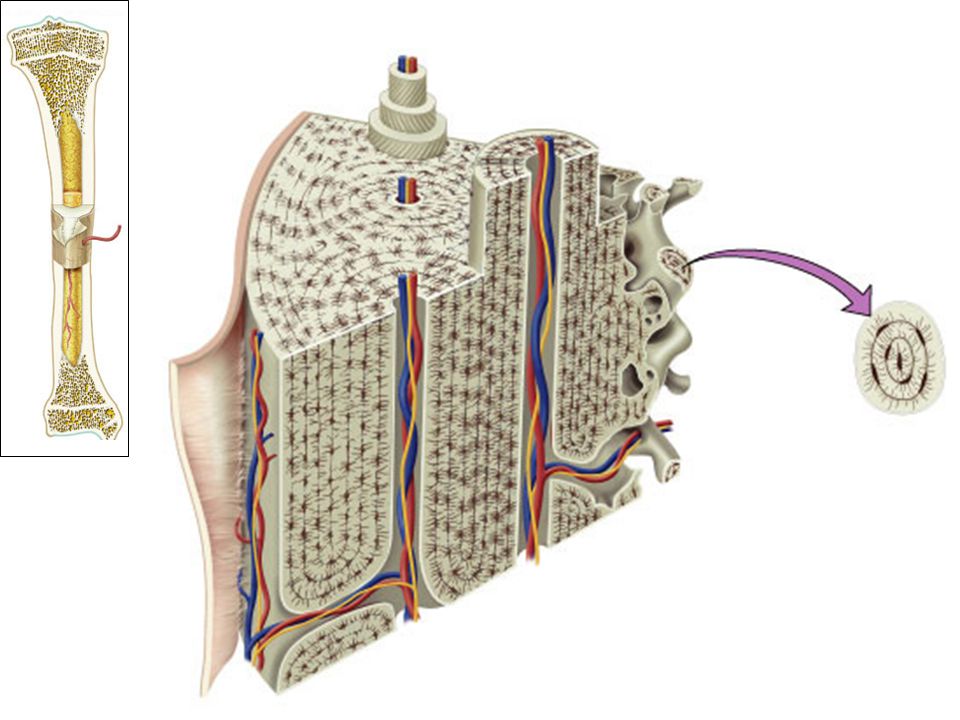
Fig. 6.2a(TE Art) Epiphysis Diaphysis Spongy bone Epiphyseal line Compact bone Medullary cavity Endosteum Articular cartilage Periosteum
 Epiphysis Diaphysis Spongy bone Epiphyseal line Compact bone Medullary cavity Endosteum Articular cartilage Periosteum.")
5
Fig. 6.2b(TE Art) Compact bone Spongy Bone (diploe) Suture
 Compact bone Spongy Bone (diploe) Suture")
8
Fig. 6.3a(TE Art) OsteoprogenitorOsteoblastsOsteocyte
 OsteoprogenitorOsteoblastsOsteocyte")
11
Fig. 6.3b(TE Art) Stem cellsOsteoclast Fusion Periosteum
 Stem cellsOsteoclast Fusion Periosteum")
13
Fig. 6.4d

14
Bone deposition = resorption

15
What would happen to bone if no calcium was available during development?

16
Shell-less chick

17
Drink milk?

18
“To name all of our bones!”

19
Fig. 7.1(TE Art) Skull Frontal bone Clavicle Maxilla Parietal bone Mandible Temporal bone Occipital bone Mandible Humerus Femur Tibia Calcaneus Fibula Ulna Radius Scapula Clavicle Scapula Vertebral column Pelvic girdle Patella Metacarpal bones Carpus Tarsus Thoracic cage Sternum Ribs Phalanges Metatarsal bones Phalanges Zygomatic bone axial appendicular
 Skull Frontal bone Clavicle Maxilla Parietal bone Mandible Temporal bone Occipital bone Mandible Humerus Femur Tibia Calcaneus Fibula Ulna Radius Scapula Clavicle Scapula Vertebral column Pelvic girdle Patella Metacarpal bones Carpus Tarsus Thoracic cage Sternum Ribs Phalanges Metatarsal bones Phalanges Zygomatic bone axial appendicular.")
20
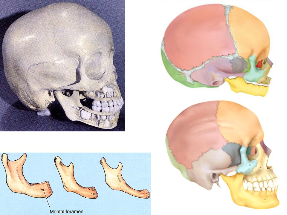
Crest Line Linea aspera Head Trochanters Epicondyles Condyles

22
Identify the humerus, radius and ulna (be able to label them). The humerus is ____________ to the radius. The radius is ____________ to the ulna (anatomical position). List the bones that articulate with the humerus. Anatomical position??
. List the bones that articulate with the humerus. Anatomical position .")
23
Fig. 8.4(TE Art) Olecranon AnteriorPosterior Head of radius Head of radius Neck of radius Styloid process Neck of radius Styloid process Styloid process Articular facets Head of ulna Interosseous membrane Ulna Radius Trochlear notch
 Olecranon AnteriorPosterior Head of radius Head of radius Neck of radius Styloid process Neck of radius Styloid process Styloid process Articular facets Head of ulna Interosseous membrane Ulna Radius Trochlear notch.")
24
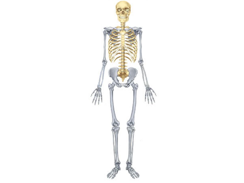
Axial skeleton

25
cranial facial

27
Fig. 7.4a(TE Art) Parietal bone Frontal bone Sphenoid bone Temporal bone Mandible Ethmoid bone Lacrimal bone Nasal bone Zygomatic bone Maxilla Occipital bone Mandibular condyle Styloid process Mastoid process
 Parietal bone Frontal bone Sphenoid bone Temporal bone Mandible Ethmoid bone Lacrimal bone Nasal bone Zygomatic bone Maxilla Occipital bone Mandibular condyle Styloid process Mastoid process.")
28
Fig. 7.3(TE Art) Parietal bone Frontal bone Lacrimal bone Vomer Mandible Sphenoid bone Ethmoid bone Nasal bone Nasal conchae Zygomatic bone Maxilla Temporal bone
 Parietal bone Frontal bone Lacrimal bone Vomer Mandible Sphenoid bone Ethmoid bone Nasal bone Nasal conchae Zygomatic bone Maxilla Temporal bone.")
29
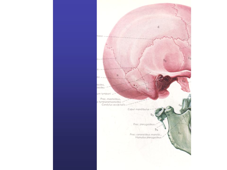
Fig. 7.5a(TE Art) Zygomatic bone External occipital protuberance Foramen magnum Carotid canal Palatine bone Temporal bone Occipital bone Parietal bone Occipital condyle Vomer Nasal choana
 Zygomatic bone External occipital protuberance Foramen magnum Carotid canal Palatine bone Temporal bone Occipital bone Parietal bone Occipital condyle Vomer Nasal choana.")
30
Animation - review

32
Fig. 7.8(TE Art) Frontal Ethmoid Maxillary Sphenoid Sinuses Mucous membrane: pseudostratified columnar epithelium, ciliated, goblet cells
 Frontal Ethmoid Maxillary Sphenoid Sinuses Mucous membrane: pseudostratified columnar epithelium, ciliated, goblet cells.")
34
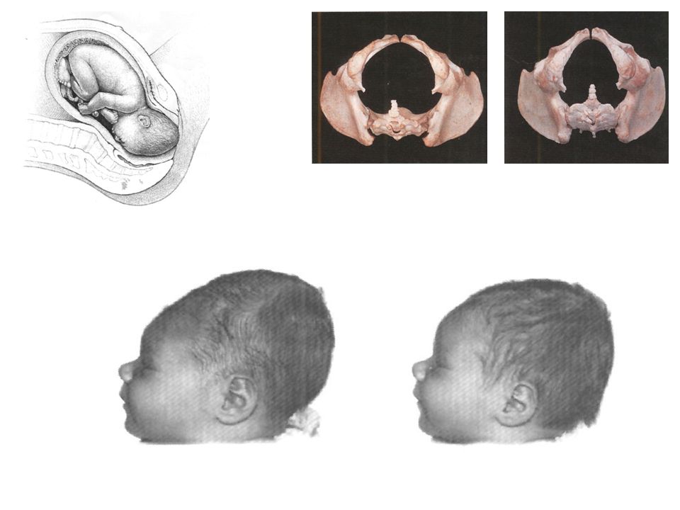
Fig. 7.31(TE Art) Coronal suture Lambdoid suture Squamous suture Anterior fontanel Sagittal suture Parietal bone Posterior fontanel Sphenoid bone Mandible Sphenoid fontanel
 Coronal suture Lambdoid suture Squamous suture Anterior fontanel Sagittal suture Parietal bone Posterior fontanel Sphenoid bone Mandible Sphenoid fontanel.")
38
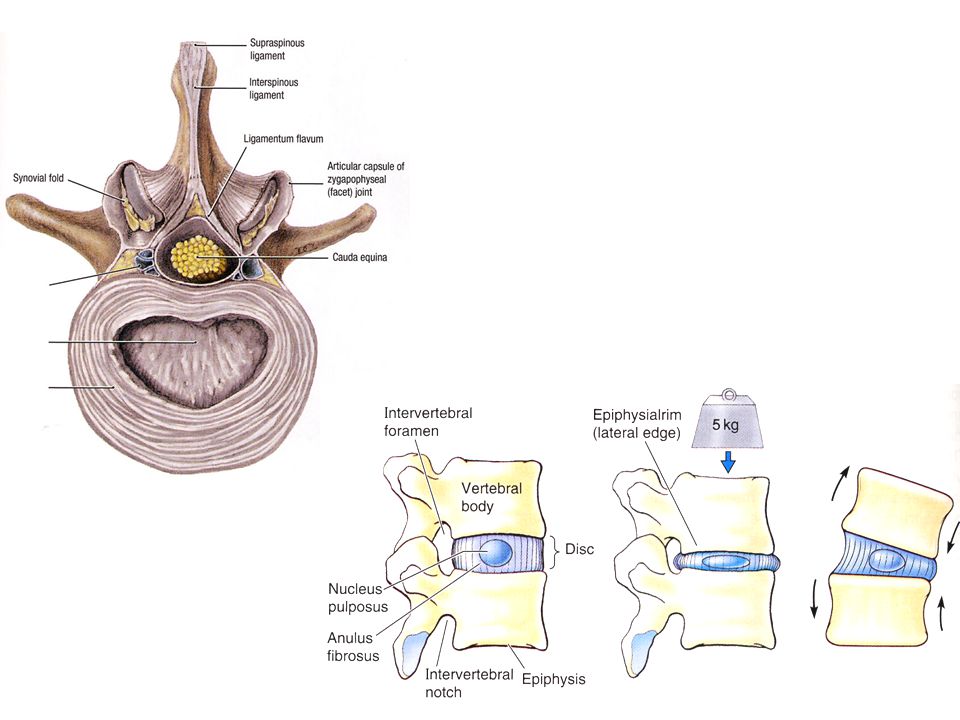
Spinous process Transverse process Body Nucleus pulposus Annulus fibrosus Intervertebral disc

42
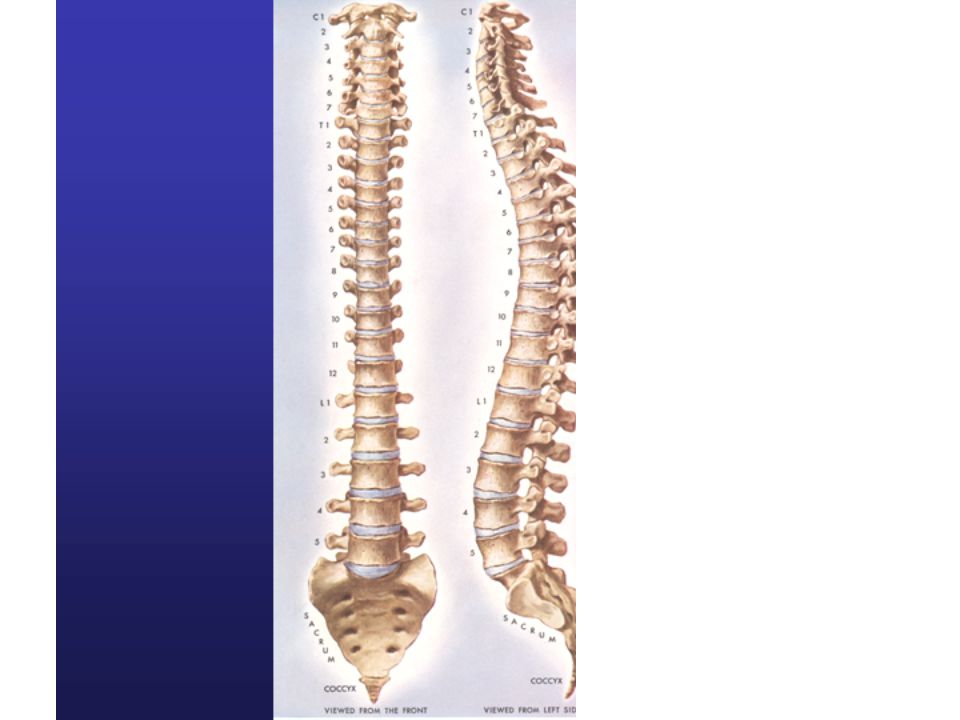
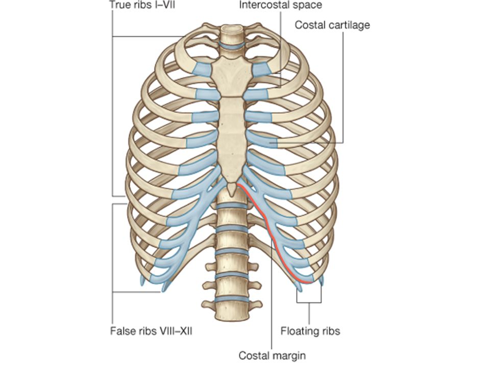
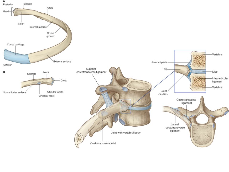
Cervical vertebrae Thoracic vertebrae Lumbar vertebrae Costal facet Costal facet

43
C1 or Atlas C2 or Axis Transverse foramen Superior articular facet Dens

44
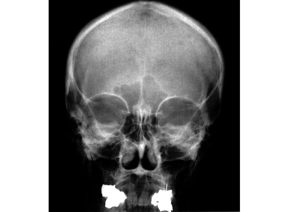
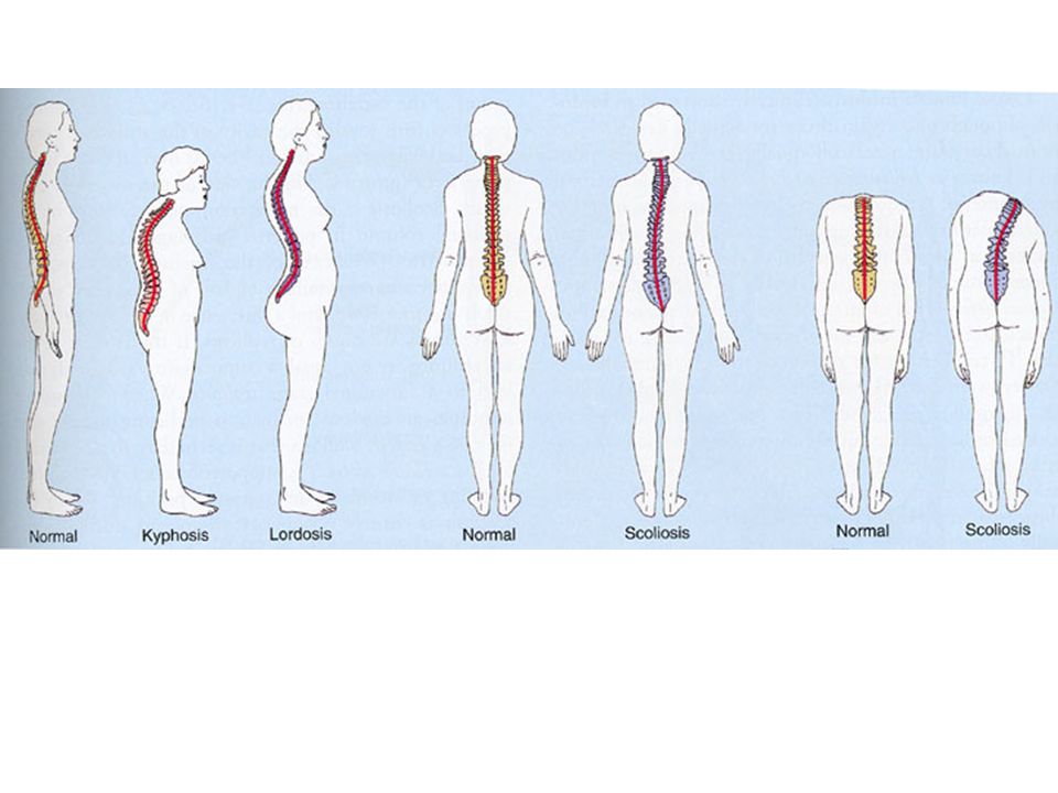
View?

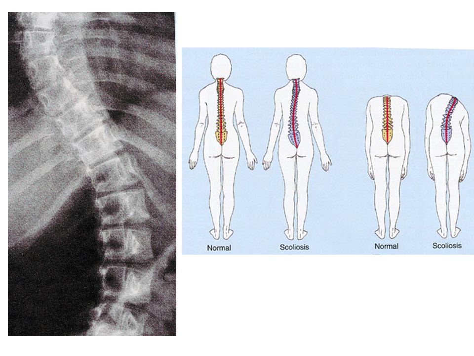
50
For tomorrow Please go over joint movements!! Think about a joint injury you’ve had (sprained ankle, knee or wrist). How did you do it? What happened to that joint after the injury?
. How did you do it. What happened to that joint after the injury .")
Similar presentations