Download presentation
Presentation is loading. Please wait.

1
Chapter 8 Joints

2
Joints (Articulations)
Articulation—site where two or more bones meet Functions of joints: Give skeleton mobility Hold skeleton together

3
Functional Classification of Joints
Based on amount of movement allowed by the joint Three functional classifications: Synarthroses—immovable Amphiarthroses—slightly movable Diarthroses—freely movable Synarthroses – sutures of the skull Amphiarthroses – vertebral bodies Diarthroses - glenohumeral

4
Structural Classification of Joints
Based on material binding bones together and whether or not a joint cavity is present Three structural classifications: Fibrous Cartilaginous Synovial Fibrous joints – sutures, syndemoses, and gomphoses joints – Bones joined by dense fibrous connective tissue No joint cavity Most are synarthrotic (immovable)
")
5
Fibrous Joints: Sutures
Rigid, interlocking joints containing short connective tissue fibers Allow for growth during youth In middle age, sutures ossify and are called synostoses

6
Fibrous Joints: Syndesmoses
Bones connected by ligaments (bands of fibrous tissue) Movement varies from immovable to slightly movable Examples: Synarthrotic distal tibiofibular joint Diarthrotic interosseous connection between radius and ulna
 Movement varies from immovable to slightly movable. Examples: Synarthrotic distal tibiofibular joint. Diarthrotic interosseous connection between radius and ulna.")
7
Fibrous Joints: Gomphoses
Peg-in-socket joints of teeth in alveolar sockets Fibrous connection is the periodontal ligament

8
Bones united by cartilage No joint cavity Two types:
Cartilaginous Joints Bones united by cartilage No joint cavity Two types: Synchondroses – typically epiphyseal plates Symphyses – pubic symphysis Synchondroses A bar or plate of hyaline cartilage unites the bones All are synarthrotic Symphyses Hyaline cartilage covers the articulating surfaces and is fused to an intervening pad of fibrocartilage Strong, flexible amphiarthroses

9
Synovial Joints Distinguishing features:
Articular cartilage: hyaline cartilage Joint (synovial) cavity: small potential space Articular (joint) capsule Outer fibrous capsule and an inner synovial membrane Synovial fluid: Viscous slippery filtrate of plasma + hyaluronic acid Lubricates and nourishes articular cartilage Three possible types of reinforcing ligaments: Capsular (intrinsic)—part of the fibrous capsule Extracapsular—outside the capsule Intracapsular—deep to capsule; covered by synovial membrane Rich nerve and blood vessel supply: Nerve fibers detect pain, monitor joint position and stretch Capillary beds produce filtrate for synovial fluid All are diarthrotic Include all limb joints; most joints of the body
 cavity: small potential space. Articular (joint) capsule. Outer fibrous capsule and an inner synovial membrane. Synovial fluid: Viscous slippery filtrate of plasma + hyaluronic acid. Lubricates and nourishes articular cartilage. Three possible types of reinforcing ligaments: Capsular (intrinsic)—part of the fibrous capsule. Extracapsular—outside the capsule. Intracapsular—deep to capsule; covered by synovial membrane. Rich nerve and blood vessel supply: Nerve fibers detect pain, monitor joint position and stretch. Capillary beds produce filtrate for synovial fluid. All are diarthrotic. Include all limb joints; most joints of the body.")
10
Ligament Joint cavity (contains synovial fluid) Articular (hyaline)
cartilage Fibrous capsule Articular capsule Synovial membrane Periosteum Figure 8.3

11
Synovial Joints: Friction-Reducing Structures
Bursae: Flattened, fibrous sacs lined with synovial membranes Contain synovial fluid Commonly act as “ball bearings” where ligaments, muscles, skin, tendons, or bones rub together

12
Synovial Joints: Friction-Reducing Structures
Tendon sheath: Elongated bursa that wraps completely around a tendon

13
Stabilizing Factors at Synovial Joints
Shapes of articular surfaces (minor role) Ligament number and location (limited role) Muscle tone, which keeps tendons that cross the joint taut Extremely important in reinforcing shoulder and knee joints and arches of the foot
 Ligament number and location (limited role) Muscle tone, which keeps tendons that cross the joint taut. Extremely important in reinforcing shoulder and knee joints and arches of the foot.")
14
Synovial Joints: Movement
Muscle attachments across a joint: Origin—attachment to the immovable bone Insertion—attachment to the movable bone Muscle contraction causes the insertion to move toward the origin Movements occur along transverse, frontal, or sagittal planes

15
Synovial Joints: Range of Motion
Nonaxial—slipping movements only Uniaxial—movement in one plane Biaxial—movement in two planes Multiaxial—movement in or around all three planes

16
Summary of Characteristics of Body Joints
Consult Table 8.2 for: Joint names Articulating bones Structural classification Functional classification Movements allowed

17
Movements at Synovial Joints
Gliding Angular movements: Flexion, extension, hyperextension Abduction, adduction Circumduction Rotation Medial and lateral rotation Gliding – One flat bone surface glides or slips over another similar surface Examples: Intercarpal joints Intertarsal joints Between articular processes of vertebrae

18
Movements at Synovial Joints
4. Special movements Supination, pronation Dorsiflexion, plantar flexion of the foot Inversion, eversion Protraction, retraction Elevation, depression Opposition

19
Angular Movements Movements that occur along the sagittal plane: Flexion—decreases the angle of the joint Extension— increases the angle of the joint Hyperextension—excessive extension beyond normal range of motion

20
Angular Movements Movements that occur along the frontal plane: Abduction—movement away from the midline Adduction—movement toward the midline Circumduction—flexion + abduction + extension + adduction of a limb so as to describe a cone in space

21
The turning of a bone around its own long axis Examples:
Rotation The turning of a bone around its own long axis Examples: Between C1 and C2 vertebrae Rotation of humerus and femur

22
Movements of radius around ulna:
Special Movements Movements of radius around ulna: Supination (turning hand backward) Pronation (turning hand forward)
 Pronation (turning hand forward)")
23
Special Movements Movements of the foot:
Dorsiflexion (upward movement) Plantar flexion (downward movement)
 Plantar flexion (downward movement)")
24
Special Movements Movements of the foot:
Inversion (turn sole medially) Eversion (turn sole laterally)
 Eversion (turn sole laterally)")
25
Movements in a transverse plane:
Special Movements Movements in a transverse plane: Protraction (anterior movement) Retraction (posterior movement)
 Retraction (posterior movement)")
26
Special Movements Elevation (lifting a body part superiorly) Depression (moving a body part inferiorly)
 Depression (moving a body part inferiorly)")
27
Opposition of the thumb
Special Movements Opposition of the thumb Movement in the saddle joint so that the thumb touches the tips of the other fingers

28
Classification of Synovial Joints
Six types, based on shape of articular surfaces: Plane Hinge Pivot Condyloid Saddle Ball and socket

29
Plane Joints Nonaxial joints Flat articular surfaces Short gliding movements Ex. Intercarpal Joint

30
Hinge Joints Uniaxial joints Motion along a single plane Flexion and extension only

31
Pivot Joints Rounded end of one bone conforms to a “sleeve,” or ring of another bone Uniaxial movement only

32
Condyloid (Ellipsoidal) Joints
Biaxial joints Both articular surfaces are oval Permit all angular movements

33
Allow greater freedom of movement than condyloid joints
Saddle Joints Biaxial Allow greater freedom of movement than condyloid joints Each articular surface has both concave and convex areas Ex. Thumbs (Carpo-metacarpal joints)
")
34
Ball-and-Socket Joints
Multiaxial joints The most freely moving synovial joints

35
Knee Joint Largest, most complex joint of body
Three joints surrounded by a single joint cavity: Femoropatellar joint: Plane joint Allows gliding motion during knee flexion Lateral and medial tibiofemoral joints between the femoral condyles and the C-shaped lateral and medial menisci (semilunar cartilages) of the tibia Allow flexion, extension, and some rotation when knee is partly flexed
 of the tibia. Allow flexion, extension, and some rotation when knee is partly flexed.")
36
Knee Joint At least 12 associated bursae
Capsule is reinforced by muscle tendons: E.g., quadriceps and semimembranosus tendons Joint capsule is thin and absent anteriorly Anteriorly, the quadriceps tendon gives rise to: Lateral and medial patellar retinacula Patellar ligament

37
Capsular and extracapsular ligaments
Knee Joint Capsular and extracapsular ligaments Help prevent hyperextension Intracapsular ligaments: Anterior and posterior cruciate ligaments Prevent anterior-posterior displacement Reside outside the synovial cavity

38
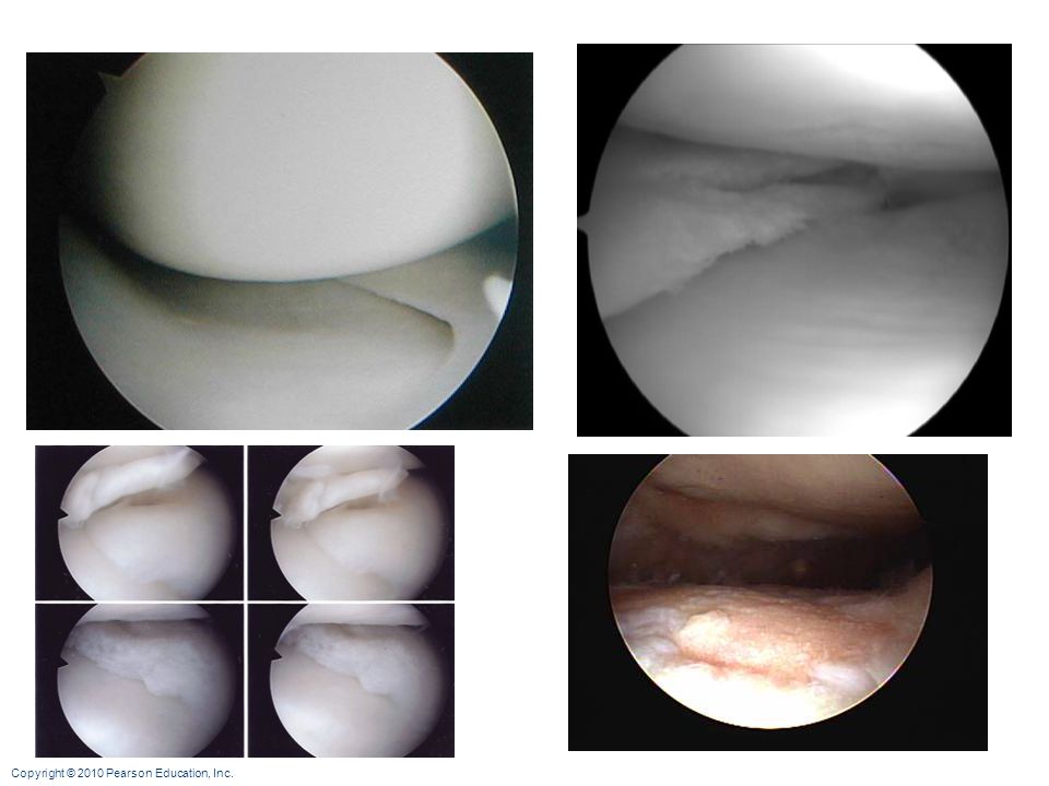
Lateral knee injury

39
Shoulder (Glenohumeral) Joint
Ball-and-socket joint: head of humerus and glenoid fossa of the scapula Stability is sacrificed for greater freedom of movement

40
Reinforcing ligaments:
Shoulder Joint Reinforcing ligaments: Coracohumeral ligament—helps support the weight of the upper limb Three glenohumeral ligaments—somewhat weak anterior reinforcements

41
Shoulder joint Reinforcing muscle tendons:
Tendon of the long head of biceps: Travels through the intertubercular groove Secures the humerus to the glenoid cavity Four rotator cuff tendons encircle the shoulder joint: Subscapularis Supraspinatus Infraspinatus Teres minor

42
Elbow Joint Radius and ulna articulate with the humerus Hinge joint formed mainly by trochlear notch of ulna and trochlea of humerus Flexion and extension only

43
Anular ligament—surrounds head of radius
Elbow Joint Anular ligament—surrounds head of radius Two capsular ligaments restrict side-to-side movement: Ulnar collateral ligament Radial collateral ligament

44
Hip (Coxal) Joint Ball-and-socket joint Head of the femur articulates with the acetabulum Good range of motion, but limited by the deep socket Acetabular labrum—enhances depth of socket

45
Hip Joint Reinforcing ligaments: Iliofemoral ligament
Pubofemoral ligament Ischiofemoral ligament Ligamentum teres

46
Common Joint Injuries Sprains Cartilage tears
The ligaments are stretched or torn Partial tears slowly repair themselves Complete ruptures require prompt surgical repair Cartilage tears Due to compression and shear stress Fragments may cause joint to lock or bind Cartilage rarely repairs itself Repaired with arthroscopic surgery

48
Common Joint Injuries Dislocations (luxations)
Occur when bones are forced out of alignment Accompanied by sprains, inflammation, and joint immobilization Caused by serious falls or playing sports Subluxation—partial dislocation of a joint

49
Inflammatory and Degenerative Conditions
Bursitis An inflammation of a bursa, usually caused by a blow or friction Treated with rest and ice and, if severe, anti-inflammatory drugs Tendonitis Inflammation of tendon sheaths typically caused by overuse Symptoms and treatment similar to bursitis

50
Arthritis >100 different types of inflammatory or degenerative diseases that damage joints Most widespread crippling disease in the U.S. Symptoms; pain, stiffness, and swelling of a joint Acute forms: caused by bacteria, treated with antibiotics Chronic forms: osteoarthritis, rheumatoid arthritis, and gouty arthritis

51
Osteoarthritis (OA) Common, irreversible, degenerative (“wear-and-tear”) arthritis 85% of all Americans develop OA, more women than men Probably related to the normal aging process

52
Osteoarthritis (OA) More cartilage is destroyed than replaced in badly aligned or overworked joints Exposed bone ends thicken, enlarge, form bone spurs, and restrict movement Treatment: moderate activity, mild pain relievers, capsaicin creams, glucosamine and chondroitin sulfate

53
Rheumatoid Arthritis (RA)
Chronic, inflammatory, autoimmune disease of unknown cause Usually arises between age 40 and 50, but may occur at any age; affects 3 times as many women as men Signs and symptoms include joint pain and swelling (usually bilateral), anemia, osteoporosis, muscle weakness, and cardiovascular problems
, anemia, osteoporosis, muscle weakness, and cardiovascular problems.")
54
Rheumatoid Arthritis RA begins with synovitis of the affected joint Inflammatory blood cells migrate to the joint, release inflammatory chemicals Inflamed synovial membrane thickens into a pannus Pannus erodes cartilage, scar tissue forms, articulating bone ends connect (ankylosis)
")
55
Figure 8.15

Similar presentations

>")


 and type of substance.>")
 Weakest parts of the skeleton Weakest parts of the skeleton Articulation – site where two or more bones meet Articulation – site.>")

