Download presentation
Presentation is loading. Please wait.

1
Skeleton Chapter 7

2
Skeletal System The bones of your skeleton are called OSSEUS TISSUE. Bone tissue is a type of connective tissue. Your skeleton performs several important functions.

3
Functions SUPPORT and PROTECTION BODY MOVEMENT – muscles “pull” on bones BLOOD CELL FORMATION – hemopoiesis occurs in red bone marrow STORAGE of inorganic salts – especially calcium phosphate, but also magnesium, sodium, potassium, carbonates and others

4
Organization normally 206 bones 2 Main Divisions: AXIAL APPENDICULAR

5
Axial AXIAL (purple): head, neck, trunk SKULL, HYOID BONE (upper neck, under jaw, mandible) VERTEBRAL COLUMN (spine/backbone) THORACIC CAGE (rib cage-12 pairs) STERNUM
: head, neck, trunk SKULL, HYOID BONE (upper neck, under jaw, mandible) VERTEBRAL COLUMN (spine/backbone) THORACIC CAGE (rib cage-12 pairs) STERNUM")
6
Appendicular APPENDICULAR (white): limbs and bones connecting the limbs to the: PECTORAL GIRDLE (scapula & clavicle), UPPER LIMBS (arms) PELVIC GIRDLE (coxal bones), LOWER LIMBS (legs)
: limbs and bones connecting the limbs to the: PECTORAL GIRDLE (scapula & clavicle), UPPER LIMBS (arms) PELVIC GIRDLE (coxal bones), LOWER LIMBS (legs)")
7
Bone Structure LONG BONE = typical bone Major parts: EPIPHYSIS – expanded ends of bone. ARTICULATES (forms a joint) with another bone. DIAPHYSIS – shaft of the bone ARTICULAR CARTILAGE – hyaline cartilage covering the ends of bones PERIOSTEUM – tough membrane-like covering over entire bone, except for articular cartilage. Connects with tendons and ligaments. Forms bone tissue.
 with another bone. DIAPHYSIS – shaft of the bone ARTICULAR CARTILAGE – hyaline cartilage covering the ends of bones PERIOSTEUM – tough membrane-like covering over entire bone, except for articular cartilage. Connects with tendons and ligaments. Forms bone tissue..")
8
Bone Structure MEDULLARY CAVITY – hollow chamber within the diaphysis connects to spaces in spongy bone. Filled with soft specialized tissue called bone marrow. Red Marrow – mainly in spongy bone in adults. Produces blood cells Yellow Marrow – fat storage. Replaces much of the red marrow in diaphysis through childhood ENDOSTEUM – lining of the medullary cavity

9
2 types of bone tissue 1.COMPACT (cortical) BONE – wall of the diaphysis, solid, strong 2. SPONGY (cancellous) BONE – epiphysis. Covered with a thin layer of compact bone. Many branching, bony “plates”
 BONE – epiphysis. Covered with a thin layer of compact bone. Many branching, bony plates .")
10
Microscopic Structure MATRIX composed of collagen and inorganic salts OSTEOCYTES (mature bone cells) are enclosed in tiny chambers called LACUNAE and form concentric “ring” (layers) around a passageway called the HAVERSION CANAL The osteocytes are connected by minute passages called CANALICULI (canaliculus) through which tiny “branches” or processes pass
 are enclosed in tiny chambers called LACUNAE and form concentric ring (layers) around a passageway called the HAVERSION CANAL The osteocytes are connected by minute passages called CANALICULI (canaliculus) through which tiny branches or processes pass")
11
Microscopic Structure The circular layers of matrix material and osteocytes, along with the haversian canal, forms a unit called a HAVERSIAN SYSTEM. Compact bone tissue is formed in this way The HAVERSIAN CANALS are interconnected by passages called VOLKMANN’S CANALS. All of these canals contain blood vessels and nerve fibers

13
End of day one

14
Bone Development and Growth There are two types of bone based on the way the bones form 1. INTRAMEMBRANOUS BONES = broad, flat bones of the skull. These bones form from membrane-like sheets of connective tissue 2. ENCHONDRAL BONES = all other bones.

15
How bones develop Bones first form as hyaline cartilage. The cartilage then gradually changes into bone tissue – a process called OSSIFICATION, which begins near the middle of the diaphysis in an area called the PRIMARY OSSIFICATION CENTER. Later the bone begins to ossify in the epiphysis – these areas are called the SECONDARY OSSIFICATION CENTERS.

16
How bones develop As long as growth is occurring, a “band” of cartilage (called an epiphyseal disk, growth plate) remains between the diaphysis and the epiphysis. Bones increase in length as these cartilage cells continue to reproduce and ossify Cartilage cells form cells called OSTEOBLASTS. These cells produce bone matrix and once the cell is enclosed in bone matrix it is considered to be a mature bone cell and called an OSTEOCYTE

17
How bones develop Growth in thickness of bone occurs as osteoblasts from beneath the periosteum produce bone matrix and built compact bone just underneath the periosteum Bone tissue is constantly being replaced as special cells called OSTEOCLASTS dissolve bone tissue from within the medullary cavity – a process called RESORPTION. At the same time new bone tissue is being added to the outside of the bone.

18
3 Basic Types of Joints (articulations): SYNARTHROTIC – immoveable joint, such as bones in the skull, these junctions are called SUTURES AMPHIARTHOTIC – slightly moveable joint, vertebrae DIARTHROTIC – freely moveable joint, such as shoulders, hips, knees, elbows, wrists, fingers… these joints are enclosed within a fibrous capsule which contains a lubricating fluid called SYNOVIAL fluid. These are called SYNOVIAL JOINTS.

19
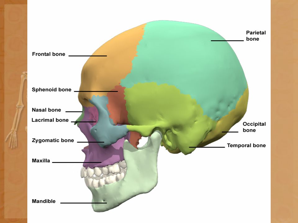
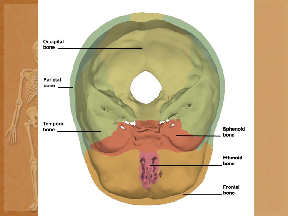
Bones of the skull Bones of the Skull Frontal - anterior portion above eyes Parietal – one on each side of the skull, just behind frontal bone Occipital – forms the back of the skull and base of the cranium Temporal – forms parts of the sides and base of cranium Sphenoid – wedged between several other bones in anterior portion of the cranium Maxilla – forms upper jaws Mandible – lower jaws, only moveable bone of the skull

22
Sutures Coronal – between frontal and parietal bones Lambdoidal – between occipital and parietal bones Squamosal – between temporal and parietal bones Sagittal - between parietal bones

23
Fontanels Fontanels - “soft spots” of an infant’s skull, see page 138 anterior fontanel, posterior fontanel, sphenoid fontanel, mastoid fontanel

24
Foramen Magnum Foramen Magnum – Large opening through the underside of the skull, spinal cord enters skull

25
Rest of Bones Ribs – Thoracic Cage - 12 pairs True Ribs – first 7 pairs, attach directly to STERNUM by costal cartilage False Ribs – last five pairs Floating ribs – last two pairs Pectoral Girdle: Shoulder 2 clavicles (collar bones) 2 scapula (shoulder blade)
 2 scapula (shoulder blade)")
26
Rest of Bones Arms: Upper arm – humerus. Lower arm – radius and ulna. Wrist – 8 small bones called carpels Fingers – Metacarpels, Phalanges

27
Rest of Bones Pelvic Girdle: Hips - Two large bones called COXAL BONES Legs: Upper leg (thigh) - FEMUR. Lower leg – tibia & fibula. Ankle and Upper foot – 7 bones called TARSALS Largest is the heel bone called the CALCANEOUS Toes Metatarsals, Phalanges

28
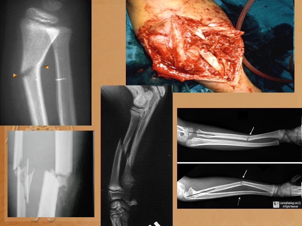
What about Broken Bones? A complete fracture is when the bone has broken into two pieces. A greenstick fracture is when the bone cracks on one side only, not all the way through. A single fracture is when the bone is broken in one place. A comminuted (say: kah-muh-noot-ed) fracture is when the bone is broken into more than two pieces or crushed. A bowing fracture, which only happens in kids, is when the bone bends but doesn't break An open fracture is when the bone is sticking through the skin.
 fracture is when the bone is broken into more than two pieces or crushed. A bowing fracture, which only happens in kids, is when the bone bends but doesn t break An open fracture is when the bone is sticking through the skin..")
30
Femur Fracture Surgery http://www.youtube.com/watch?v=ITwniRKtKwM

Similar presentations


>")


 Support- framework.>")

 Mineral Storage of Calcium and Phosphate Red Blood Cell Production (long.>")
 fibers along with water and mineral salts (calcium hydroxide & calcium.>")


