Download presentation
Presentation is loading. Please wait.

1
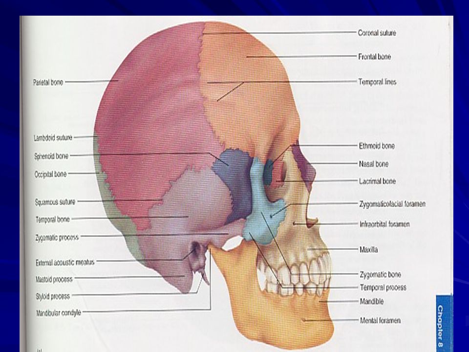
Bones of Skull and cranial cavity
DR.CHANDRALEKHA

4
Cranial bones There are 8 cranial bones 1 frontal bone.
1 occipital bone. 1 sphenoid bone 1 Ethmoid bone 2 parietal bones 2 Temporal bones

6
cranium The cranium consists of two major parts
The calvaria (skull cap) forms the roof and the walls The base of the cranial cavity is divided into three regions. 1. Anterior cranial fossa 2. Middle cranial fossa 3. Posterior cranial fossa
 forms the roof and the walls. The base of the cranial cavity is divided into three regions. 1. Anterior cranial fossa. 2. Middle cranial fossa. 3. Posterior cranial fossa.")
9
The skull Cranial cavity Orbital cavity Nasal cavity Buccal cavity
Middle ear cavity Inner ear cavity Paranasal air sinuses

13
Frontal bone Frontal bone forms the forehead.
It forms anterior wall and about one – third of the roof of the cranial cavity. Frontal tuber Superciliary arch Glabella Supra orbital margin Nasion Coronal suture Frontal air sinus

16
Frontal bone

17
Parietal bones There are 4 borders 4 angles. Sutures: sagittal suture
Coronal suture Lambdoid suture Squamous suture Lines: superior temporal and inferior temporal lines. Parietal foramen

20
Temporal bones This bone has 4 different parts
1. Squamous part which has two prominent features zygomatic process and mandibular fossa 2. Tympanic part is a small ring of bone that borders the external acoustic meatus The styloid process

21
Temporal bones 3. Mastoid part– has mastoid air sinus
infection and inflammation of this air sinus is known as Mastoiditis Mastoid notch -- medial to the mastoid process Stylomastoid foarmen Mastoid foarmen—at its posterior end 4. Pterous part: anterior surface forms part of the middle cranial fossa The posterior surface forms the part of posterior cranial fossa Posteromedial surface has Internal acoustic meatus The carotid canal--- a passage of internal carotid artery Jugular foramen: IJV,9,10,11 NERVES, SIGMOID SINUS

23
Occipital bone It has three parts 1. Squamous part 2. condylar part.
3. basilar part Foramen magnum---transmits spinal cord to the cranial cavity Hypoglossal canal---anterior to the occipital condyle Condylar canal--- posterior to the occipital condyle

24
Occipital bone External occipital protruberance Superior nuchal line
Inferior nuchal line

26
Sphenoid bone Body of sphenoid Pair of greater wings
Pair of lesser wings Optic foramen Anterior clinoid process Superior orbital fissure Sella turcica

28
Foramens in the sphenoid bone
Foramen rotundum Forman ovale Foramen spinosum Foramen lacerum--- occurs at the junction of sphenoid, temporal, and occipital bones

29
Sphenoid bone Sphenoidal air sinus occur within the body of the sphenoid bone. At the junction of the body and the greater wing there is a pair of pterygoid process which has medial pterygoid and lateral pterygoid plate . Between the two pterygoid plate is the Pterygoid fossa.

31
Ethmoid bone Crista galli Cribriform plate of ethmoid
Perpendicular plate of ethmoid bone Superior and middle conchae are the extension of ethmoid bone On each side of the perpendicular plate of ethmoid bone, anterior, middle and posterior ethmoidal air sinuses are seen

33
Facial bones There are 14 facial bones 2 maxillae 2 nasal bones
2 palatine bones 2 inferior nasal conchae 2 zygomatic bones 2 lacrimal bones 1 vomer 1mandible

35
Maxillae Has body, palatine process,
Frontal process, zygomatic process Alveolar process Palatine process forms the roof of the mouth cavity, and floor of the nasal cavity (hard palate) Near the anterior margin of palatine process, just behind the incisors , is an incisive foramen Body– infra orbital foramen transmits infra orbital vessels and nerves
 Near the anterior margin of palatine process, just behind the incisors , is an incisive foramen. Body– infra orbital foramen transmits infra orbital vessels and nerves.")
36
MAXILLA

37
Palatine bones horizontal plate of Palatine bones form the posterior part of the hard palate Orbital plate of palatine bones form part of the floor of the orbital cavity. Perpendicular plate of palatine bone forms the lateral wall of the nasal cavity Posterolateral corner of the hard palate--- two large greater palatine foramina.

39
NASAL BONE Two small rectangular bones form the bridge of the nose and support cartilages that give shape to the nose. NASAL BONE

40
Lacrimal bones This bone forms the medial wall of the orbit
Lacrimal fossa- depression in it , lodges the lacrimal sac.

41
INFERIOR NASAL CONCHA The inferior nasal conchae are the largest of the three conchae in the lateral wall of the nasal cavity.

42
VOMER The vomer forms the inferior half of the nasal septum.

43
Zygomatic bones Has maxillary process Frontal process Temporal proces
This bones from the prominence of cheek Zygomatico facial foramen and zygomatico temporal foramen

45
Mandible Mandible is the strongest bone of the skull.
It forms the lower jaw Provides attachments of muscles of facial expression and muscles of mastication It has body, ramus, and angle The point of chin is the mental protruberance. Mental foramen

48
Bones associated with the skull
There are 7 bones associated with the skull but not considered part of it. Three ear ossicles on each side namely malleus, incus and stapes (6bones) The hyoid bone between the chin and the larynx
 The hyoid bone between the chin and the larynx.")
49
Incudo malleolar joint Incudostapedial joint
Ear ossicles Malleus consists of head, neck, anterior process, lateral process & handle. Incus has a body & long process. Stapes smallest and medially placed ossicle consists of the head, neck two limbs or crura, footplate or base. Joints are incudo malleolar joint (a saddle joint) & incudostapedial joint ( a ball & socket joint). Incudo malleolar joint Incus Malleus Stapes Incudostapedial joint
 & incudostapedial joint ( a ball & socket joint). Incudo malleolar joint. Incus. Malleus. Stapes. Incudostapedial joint.")
50
Hyoid bone The hyoid bone is suspended from the styloid process of the skull by stylohyoid ligament and stylohyoid muscles. It has body pair of greater horn and pair of lesser horn

52
The skull in the infancy and childhood
The spaces between the unfused cranial bones are called fontanels The bones are joined at these points only by fibrous membranes. The anterior, posterior, sphenoid (2)and mastoid fontanels(2) In total there are 6 fontanels Anterior fontanels fuse between 18 to 24 months after birth. All the other fontanels ossify before the infant is a year old.
and mastoid fontanels(2) In total there are 6 fontanels. Anterior fontanels fuse between 18 to 24 months after birth. All the other fontanels ossify before the infant is a year old.")
54
The skull in the infancy and childhood
The frontal bone and the mandible are separate bones at birth but fuse medially in early childhood The frontal bones usually fuse by 6 year but in some children the metapic suture persists between them. Traces of these sutures are evident in some adult skull

55
The skull in the infancy and childhood
The face of the new born is flat and the cranium is large. To accommodate the growing brain, the skull grows more rapidly than the rest of the skeleton during childhood.

Similar presentations