Download presentation
Presentation is loading. Please wait.

1
The Skeletal System Chapter 36 Section 1 Notes

2
Keys Lecture Outline – The Skeletal System PowerPoint Notes textbook questions

3
Skeletal System Functions MMMMakes up body framework - gives body shape SSSSupports the body PPPProtects vital internal organs

4
MMMMakes up body framework - gives body shape SSSSupports the body PPPProtects vital internal organs

5
Functions continued… PPPProvides for movement SSSStores mineral reserves PPPProduces red blood cells

6
Bone Composition BBBBone is living tissue. IIIIt is a solid network of cells and protein fibers surrounded by deposits of minerals. CCCComponents: 332% Organic materials (collagen and bone cells) 443% Minerals (calcium and phosphorous) 225% Water
 443% Minerals (calcium and phosphorous) 225% Water.")
7
Bone Cells There are four main types of bone cells in bone tissue. Osteogenic cells respond to traumas, such as fractures, by giving rise to osteoblasts and osteoclasts. Osteoblasts (bone-forming cells) synthesize and secrete unmineralized ground substance and are found in areas of high metabolism within the bone. Osteocytes are mature bone cells made from osteoblasts that have made bone tissue around themselves. Osteoclasts are large cells that break down bone tissue. They are very important to bone growth, healing, and remodeling.
 synthesize and secrete unmineralized ground substance and are found in areas of high metabolism within the bone. Osteocytes are mature bone cells made from osteoblasts that have made bone tissue around themselves. Osteoclasts are large cells that break down bone tissue. They are very important to bone growth, healing, and remodeling..")
8
Skeletal Tissue 4 main types Compact bone Spongy bone Cartilage Fibroblasts spongy compactcartilagefibroblasts Ligament

9
Anatomy of a Typical Long Bone Femur Structure of Bone

10
Structure of bone Taking a closer look: A cross- section of the long bone. Periosteum covers bone, is a place for tendon and ligament attachment, and brings blood, lymph vessels and nerves into the bone. Compact bone is a dense layer of bone tissue composed of cylinders or tubes of mineral crystals and protein fibers, that give bone its strength. Spongy bone is the inside layer of compact bone that is actually quite strong but lacy in appearance and contains red marrow which produces blood cells. Bone marrow (primarily yellow marrow) stores fat that serves as an energy reserve and contains blood vessels and nerve cells. Copyright © 2003 Pearson Education, Inc., publishing as Benjamin Cummings
 stores fat that serves as an energy reserve and contains blood vessels and nerve cells. Copyright © 2003 Pearson Education, Inc., publishing as Benjamin Cummings.")
11
Structure of Bone Here is another diagram Just to help give you that visual to remember all of this! Haversian canal Haversian canal are interconnected networks of tubes that blood vessels and nerves run through. Blood vessels carry nourishment to the living bone tissue as well as removing wastes Osteocytes are responsible for bone growth and changes in the shape of bone and can either deposit or absorb calcium salts

12
Structure of Bone Notice… Hyalin cartilage covers the ends of bones where they articulate (join) with other bones. As adulthood is reached, the epiphyseal plate (growth plate) is replaced by bone and fuses, thus completing growth.
 is replaced by bone and fuses, thus completing growth..")
13
Structure of Bone What parts do you remember? Let’s Quiz Ourselves! BBBBlood vessels BBBBone marrow CCCCompact bone HHHHaversian canal OOOOsteocyte PPPPeriosteum SSSSpongy bone 4 6 7 1 3 2 5 1 { 2323 5 6 7 4

14
Bones of the skeleton contain a combination of spongy and compact bone. Do you recognize the bone at the left? What classification (type) of bone is it? What type of bone marrow is found within the spaces of the spongy bone? Skull Bone A flat bone Red Marrow
 of bone is it. What type of bone marrow is found within the spaces of the spongy bone. Skull Bone A flat bone Red Marrow.")
15
Bone Formation Called Ossification- Process of producing bone from cartilage ________ is replaced by _________ which secrete ________deposits and then mature into __________(bone cells). ___________ break down bone and remove _________bone tissue when a bone is broken. Cartilage osteoblasts mineral osteocytes Osteoclasts damaged x

16
Bone Formation The _______ plate (epiphyseal disc) is an area of _______ in the _____of long bones where bone _________ occurs. lengthening growth cartilage ends Growth in Length

17
10 week fetus Cartilage bone of the skull Intramembranous ossification produces the roofing bones of the skull Primary ossification centers of the diaphyses (skeleton of the lower limb) Future hip bone Bone Formation The basic shape of a long bone, such as an arm bone, is first formed as cartilage Bone growth begins long before birth.
 Future hip bone Bone Formation The basic shape of a long bone, such as an arm bone, is first formed as cartilage Bone growth begins long before birth.")
18
Bone Formation 12 week fetus 16 week fetus Ossification begins to take place up to seven months before birth

19
Bone Formation Babies are born with 350 bones, many are composed almost entirely of cartilage. Latter the cartilage cells will be replaced by cells that form the bones. (ossification) Long bones develop and grow through out childhood at the centers of ossification (growth plates) The SOFT SPOT of a babie’s skull will fuse around age 2, but growth of the skull continues until adulthood. The SOFT SPOT of a babie’s skull will fuse around age 2, but growth of the skull continues until adulthood.
 Long bones develop and grow through out childhood at the centers of ossification (growth plates) The SOFT SPOT of a babie’s skull will fuse around age 2, but growth of the skull continues until adulthood. The SOFT SPOT of a babie’s skull will fuse around age 2, but growth of the skull continues until adulthood..")
20
Bone Formation BBBBetween the ages of 16 and 25 years, all of the cartilage of the epiphyseal disc is replaced by bone. This is called closure of the epiphyseal disc, and the bone lengthening process stops. Stages of Ossification

21
Bone Formation GROWTH INW IDTH

22
Bones of the Skeleton The adult skeleton contains _____ bones The adult skeleton contains _____ bones 206

23
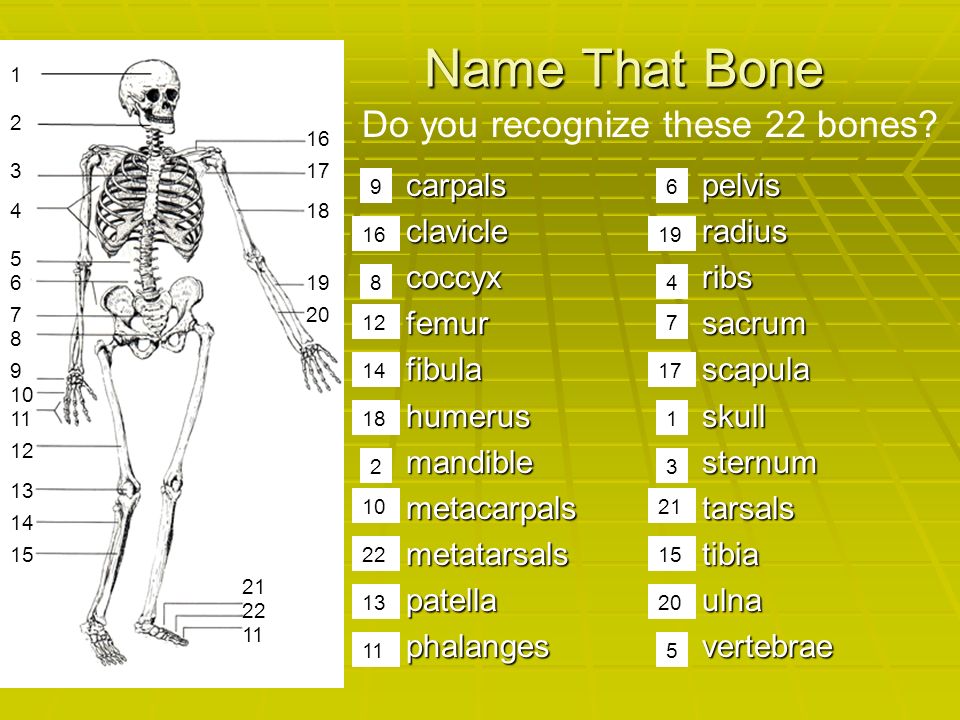
Name That Bone carpals clavicle coccyx femur fibula humerus mandible metacarpals metatarsals patella phalanges pelvis radius ribs sacrum scapula skull sternum tarsals tibia ulna vertebrae Do you recognize these 22 bones? 21 22 20 15 19 1417 18 13 1 4 32 7 6 5 9 8 10 16 11 21 22 20 19 12 18 15 16 17 13 14 10 9 8 6 5 4 3 2 1 11 7 12
24
Divisions of Skeleton Axial and Appenicular Skeletons

25
Axial Skeleton TTTTHE AXIAL SKELETON - CONSIST OF THE SKULL, VERTEBRAL COLUMN, AND THE RIB CAGE SSSSkull VVVVertebral column RRRRib cage (ribs + sternum) Skull
 Skull")
26
Skull Bones * The Skull consists of 8 CRANIAL BONES + 13 FACIAL BONES 13 FACIAL BONES * The Ears consists of 6 BONES and 6 BONES and * Floating in the throat is 1 HYOID BONE Inner Ear

27
Rib Cage AAAAlso called the Thoracic Cage 11112 pairs of RIBS 7777 true ribs 5555 false ribs 2222 floating ribs 1 1 1 1 STERNUM (breastbone)
")
28
Vertebral Column TTTThe Vertebral Column (Spinal Column or Backbone) 7777 CERVICAL (NECK) VERTEBRAE, 11112 THORACIC 5555 LUMBAR, 5555 FUSED VERTEBRAE INTO 1 SACRUM, 4444 SMALL FUSED VERTEBRAE INTO 1 COCCYX (YOUR TAIL BONE)
 7777 CERVICAL (NECK) VERTEBRAE, 11112 THORACIC 5555 LUMBAR, 5555 FUSED VERTEBRAE INTO 1 SACRUM, 4444 SMALL FUSED VERTEBRAE INTO 1 COCCYX (YOUR TAIL BONE)")
29
Appendicular Skeleton THE APPENDICULAR SKELETON – consists of bones of the: ARMS (upper limbs) LEGS (lower limbs) SHOULDER GIRDLE (pectoral girdle) HIP GIRDLE (pelvic girdle)
 LEGS (lower limbs) SHOULDER GIRDLE (pectoral girdle) HIP GIRDLE (pelvic girdle)")
30
Shoulder Girdles and Arms The Shoulder girdle is also called the pectoral girdle Consists of 4 bones Upper limbs consist of 60 bones (the hands and wrist contain 54 separate bones).
.")
31
Hip Girdles and Legs The hip girdle is also called the pelvic girdle Consists of 2 bones Lower limbs consist of 60 bones (the ankles and feet contain 52 separate bones)
")
32
Comparison of Skletons The Human Skeleton is homologous to skeletons of other animals. Once you learn the bones in a human, you can identify the bones in other animals. cat rat horse

33
Bone Classification by Shape 5555 Types LLLLong SSSShort FFFFlat IIIIrregular ssssesamoid

34
Shapes of Bones LLLLong bones are longer than they are wide and work as levers. The bones of the upper and lower extremities (ex. humerus, tibia, femur, ulna, metacarpals, etc.) are of this type. SSSShort bones are short, cube- shaped, and found in the wrists and ankles. FFFFlat bones have broad surfaces for protection of organs and attachment of muscles (ex. cranial bones, ribs, and bones of hip and shoulder girdles). IIIIrregular bones are all others that do not fall into the previous categories. They have varied shapes, sizes, and surface features and include the bones of the vertebrae and a few in the skull.
 are of this type. SSSShort bones are short, cube- shaped, and found in the wrists and ankles. FFFFlat bones have broad surfaces for protection of organs and attachment of muscles (ex. cranial bones, ribs, and bones of hip and shoulder girdles). IIIIrregular bones are all others that do not fall into the previous categories. They have varied shapes, sizes, and surface features and include the bones of the vertebrae and a few in the skull..")
35
Types of Bones 1. The humerus and femur are examples of _______ bone. 2. Tarsal and carpal bones are examples of _______ bone. 3. Sternum and many skull bones are examples of ________bone. 4. Vertebrae and the patella are examples of _______ bone. long short flat irregular

36
Joints JOINTS: WHERE TWO or MORE BONES MEET Joints are responsible for keeping bones far enough apart so they do not rub against each other as they move, preventing damage. At the same time, joints hold the bones in place. Different joints permit different amounts of movement. Joints are classified by the amount and type of movement they permit.

37
Classification of Joints Three Main Types Immovable- A fixed joint, one that allows no movement bones of skull, pelvis, and sacrum Slightly movable- joint that permits a small amount of restricted movement between vertebrae, two bones of lower leg Freely movable- Permit movement in one or more directions

38
Classification of Joints Immovable bones of skull, pelvis, and sacrum pelvis, and sacrum Slightly movable between vertebrae, two bones of lower leg two bones of lower leg Tibia and Fibula skullPelvis Vertebra Ribs

39
Freely Movable Joints TYPES OF FREELY MOVABLE JOINTS A. BALL AND SOCKET JOINT – Permits circular movement - the widest range of movement. SHOULDER Joint- which enables you to move your arm up, down, forward and backward, as well as to rotate it in a complete circle. HIP Joint- same range of motion.

40
Types of Freely Movable Joints Continued BBBB. HINGED JOINT - Permits a back- and-forth motion. TTTThe Knee- enables your leg to flex and extend. TTTThe Elbow -allows you to move your forearm forward and backward. TTTThe Phalanges CCCC. PIVOT JOINT - Permits rotation of one bone around another. TThe elbow enables your hand to turn over. (radius rotates around ulna) II t also allows you to turn your head from side to side. (atlas rotates around axis)
 II t also allows you to turn your head from side to side. (atlas rotates around axis).")
41
D. GLIDING JOINT - Permits a sliding motion of one bone over another. Found at ends of the collarbones, between wrist bones, and between anklebones. E. SADDLE JOINT- Permits movement in two planes. This type of joint is found at the base of the thumb Click Here

42
Anatomy of a Joint 2 or more bones Cartilage Joint capsule Synovial membrane Synovial fluid Fat Bursa Meniscus Ligaments Tendons Structures of a freely movable joint

43
Anatomy of a Joint Cartilage - at the joint, the bones are covered with cartilage, which is wear-resistant and helps reduce the friction of movement. Joint capsule- is a thick, tough layer that envelops the joint cavity forming a membrane or sac that adheres firmly to the periosteum of the articulating bones

44
Anatomy of a Joint Synovial membrane - a tissue that lines the joint and seals it into a joint capsule. The synovial membrane secretes synovial fluid. Synovial fluid - a clear, sticky fluid secreted by the synovial membrane to lubricate the joint.

45
Anatomy of a Joint Fat- Helps pad and cushion the joint. Bursa- fluid-filled sac between adjacent structures such as ligaments or bones which help reduce friction in a joint, cushion it, and absorb shock. Meniscus- wedge shaped cartilage, curved like the letter "C" at the inside and outside of each knee. A strong stabilizing tissue, helps the knee joint carry weight, and glide and turn in many directions. It also keeps your femur and tibia from grinding against each other.

46
Anatomy of a Joint lllligaments - tough, elastic bands of connective tissue ssssurround the joint to give support and limit the joint's movement. AAAAttach bone to bone ttttendons – another type of tough connective tissue o o o on each side of a joint attached to muscles that control movement of the joint. AAAAttach muscle to bone Knee Joint

47
Skeletal Disorders A broken bone is known as a fracture. This can simply be a crack or buckle in the structure of the bone, or a complete break, producing two or more fragments. Fractures Fractures

48
Skeletal Disorders The repair of bone fractures is similar to embryonic bone formation. Bone Fracture Repair

49
Consists of more than 100 different conditions The common denominator for all these conditions is joint pain Osteoarthritis- nick-named “wear and tear” arthritis Rheumatoid arthritis is one of the most crippling forms of arthritis. It is characterized by chronic inflammation of the lining of joints. Arthritis- inflammation of the joints Arthritis

50
Skeletal Disorders Osteoporosis- literally means "porous bones“ Occurs when a body's blood calcium level is low and calcium from bones is dissolved into the blood to maintain a proper balance. Over time, bone mass and bone strength decrease. As a result, bones become dotted with pits and pores, weak and fragile, and break easily. Other factors besides age can lead to osteoporosis, such as a diet low in calcium and protein, a lack of vitamin D, smoking, excessive alcohol drinking, and insufficient weight-bearing exercises to stress the bones. Osteoporosis

51
Skeletal Disorders Childhood disorder involving softening and weakening of the bones. It is primarily caused by lack of vitamin D, calcium, or phosphate Rickets

52
Skeletal Disorders Condition involving complex lateral and rotational curvature and deformity of the spine. Typically classified as: Idiopathic (unknown cause) Congenital (caused by vertebral abnormality present at birth) possibly inherited Secondary symptom of another condition, such as cerebral palsy or muscular dystrophy Scoliosis
 Congenital (caused by vertebral abnormality present at birth) possibly inherited Secondary symptom of another condition, such as cerebral palsy or muscular dystrophy Scoliosis.")
53
Skeletal Disorders Kyphosis can be thought of as an arching of the spine in which the top of the arch is seen in the back This condition is sometimes referred to as “humpback” or “hunchback” Caused by inflammation of vertebrae, poor posture, or congenital abnormality Kyphosis

54
Skeletal Disorders Lordosis is the increase of the spinal posterior concavity. In most cases the cause is unknown and the disorder appears from the onset of skeletal growth. This condition is also referred to as “swayback”. Lordosis

55
Skeletal Disorders IIIInfection of bone or bone marrow, usually caused by bacteria. TTTThe infective process encompasses all of the bone components, including the bone marrow PPPPus is produced within the bone, which may result in an abscess which then deprives the bone of its blood supply. BBBBecause of the particulars of their blood supply, the tibia, femur, humerus, and vertebral bodies are especially prone Osteomyelitis

56
The most common type of malignant bone cancer, accounting for 35% of primary bone malignancies. Usually occurs in the area where the body of cartilage (that separates the epiphyses and the diaphysis) of tubular long bones is located. 50% of cases occur around the knee. Osteosarcoma
 of tubular long bones is located. 50% of cases occur around the knee. Osteosarcoma.")
57
This is the end of the show. Hope you enjoyed it! Adios Amigos

58
Math problem You have 1 less ankle bone on each leg (7) compared to the number of wrist bones(8). The number of feet and hand bones is the same (19) each. So you have 54 total hand + wrist bones and 52 total foot and ankle bones So why is the total number of bones in the lower limbs (60) the same as the total number in the upper limbs (60)? answer- add the 2 knee caps So you have 54 total hand + wrist bones and 52 total foot and ankle bones So why is the total number of bones in the lower limbs (60) the same as the total number in the upper limbs (60)? answer- add the 2 knee caps link this to slide 29 link this to slide 29
 each. So you have 54 total hand + wrist bones and 52 total foot and ankle bones So why is the total number of bones in the lower limbs (60) the same as the total number in the upper limbs (60). answer- add the 2 knee caps So you have 54 total hand + wrist bones and 52 total foot and ankle bones So why is the total number of bones in the lower limbs (60) the same as the total number in the upper limbs (60). answer- add the 2 knee caps link this to slide 29 link this to slide 29.")
Similar presentations


