Download presentation
Presentation is loading. Please wait.

1
The Skeletal System

2
How many bones are in the body?
206

3
There are 5 main functions of bones
Framework- support muscles Protection- surrounds vital organs, etc. Levers- attach to muscles to provide movement Produce blood cells- red, white, and platelets Storage- calcium

4
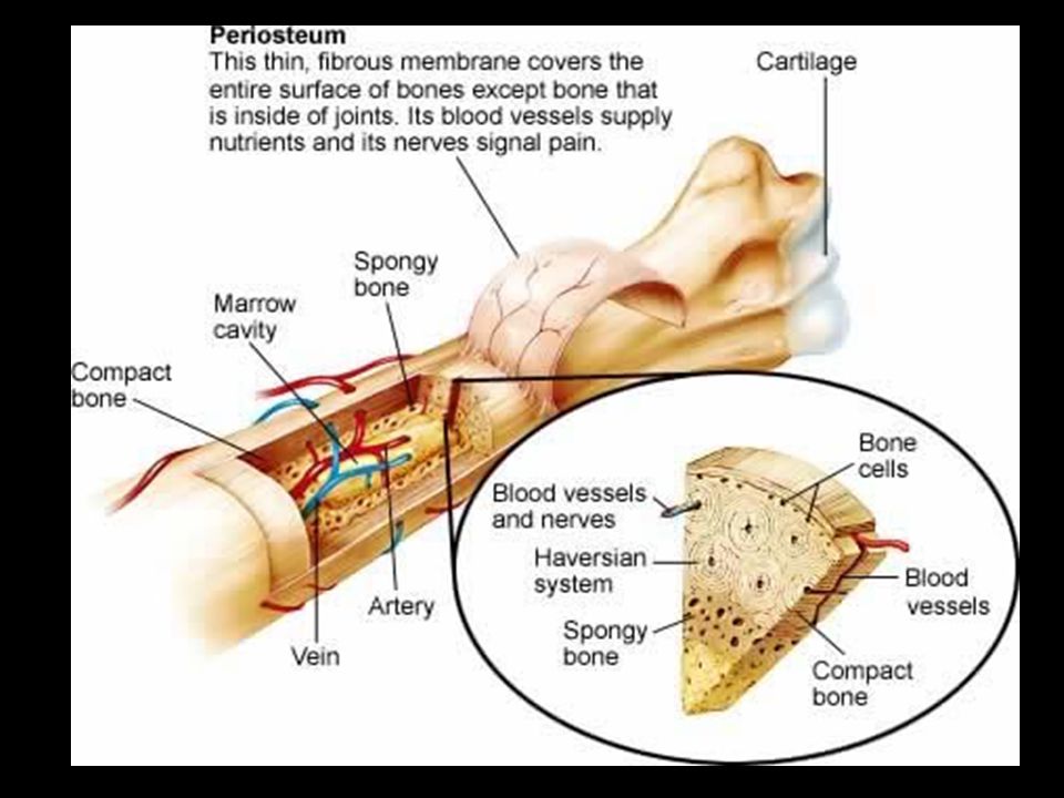
Parts of long bones Long bones are bones of extremities
Periosteum-tough membrane covering outside of bones Contains osteoblasts which are special cells that form new bone tissue

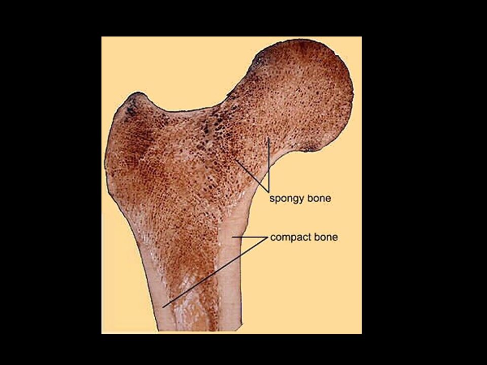
5
Spongy Bone Located at the ends of long bones
Has many small, open spaces that make bones lightweight Filled with a substance called marrow yellow composed of fat cells red produces red blood cells

7
Cartilage Smooth, slippery, thick layer of tissue
Covers the ends of bones Does not contain blood vessels or minerals Flexible and acts as a shock absorber Makes movement easier by reducing friction

9
Two sections of a skeleton
Axial Skeleton 2. Appendicular Skeleton

10
Axial Skeleton Forms main trunk of the body
Composed of the skull, spinal column, ribs, and sternum

11
Appendicular Skeleton
Forms extremities (arms and legs) Composed of shoulder girdle, arm bones, pelvic girdle, and leg bones
 Composed of shoulder girdle, arm bones, pelvic girdle, and leg bones.")
12
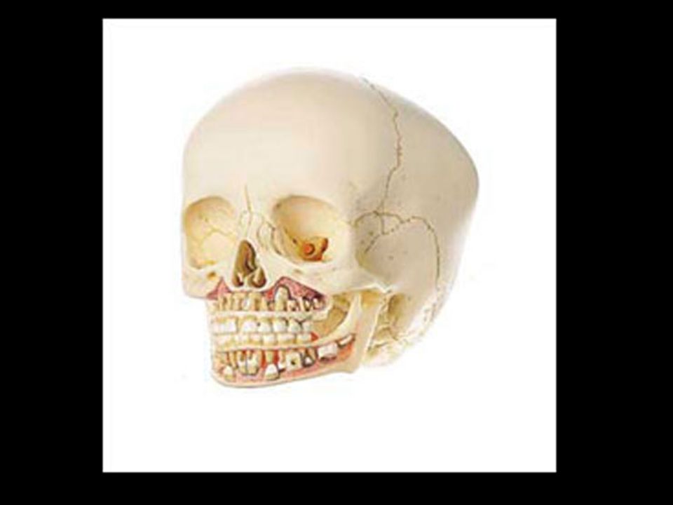
Skull Composed of cranial and facial bones

13
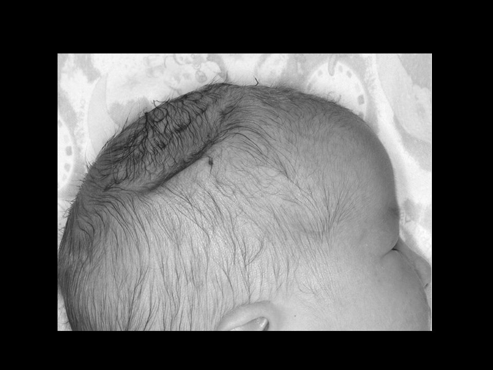
Cranium Spherical structure that surrounds and protects the brain
At birth, the cranium is not solid bone Spaces are called fontanels “soft spots” that allow for brain growth Turns into solid bone by 18 months Composed of 8 bones Frontal Two parietal Two temporal Occipital Ethmoid Sphenoid

14
Facial bones Fourteen facial bones Main bones of the face
Mandible: lower jaw Maxilla: two bones forming upper jaw Zygomatic: two cheek bones Nasal: five bones in upper part of nose Lacrimal: two bones at inner aspect of eyes Palatine: two bones of hard palate or roof of mouth

15
Areas where cranial bones have joined together
Sutures Areas where cranial bones have joined together

17
Sinuses Air spaces in the bones of the skull
Act as a resonating chamber for the voice Lined with mucous membranes

18
Foramina (Foramen) Openings in the bones Allow nerves and blood vessels to enter or leave bone
 Openings in the bones Allow nerves and blood vessels to enter or leave bone")
19
Vertebrae Spinal column is composed of 26 bones called vertebrae Protects the spinal cord Provides support for head and trunk

20
Main sections of vertebral column:
Cervical: 7 neck vertebrae Thoracic: 12 vertebrae in back of chest, attached to ribs Lumbar: 5 vertebrae by waist Sacrum: 1 large vertebra on back of pelvic girdle Coccyx: 1 fused vertebra called “tailbone”

21
Intervertebral disks Pads of cartilage tissue that separate vertebrae Act as shock absorbers Permit bending and twisting movements of vertebral column

23
12 pairs of long, slender bones
Ribs (costae) 12 pairs of long, slender bones Attach to thoracic vertebrae on dorsal surface of body
 12 pairs of long, slender bones. Attach to thoracic vertebrae on dorsal surface of body.")
24
True ribs False ribs Floating ribs First 7 pairs of ribs
Attach directly to sternum on front of body False ribs Next 5 pairs of ribs First three pairs attach to cartilage of rib above Floating ribs Last 2 pairs of false ribs No attachment on front of body

25
Sternum “Breastbone” Consists of three parts
Manubrium or upper region Gladiolus: body or center area Xiphoid process: small piece of cartilage at bottom (landmark for chest compressions) Two clavicles attach to the manubrium by ligaments Ribs attach to sternum with cartilage
 Two clavicles attach to the manubrium by ligaments. Ribs attach to sternum with cartilage.")
26
Shoulder or pectoral girdle
Two clavicles or “collarbones” Two scapulas or “shoulder bones” Scapula provides for attachment of upper arm bone

27
Bones of the arm Humerus: upper arm bone
Radius: lower arm bone on thumb side that rotates around ulna to allow the hand to turn freely Ulna: larger bone of lower arm that contains a projection called the olecranon process at upper end, forming “elbow”

28
Wrist and hands Carpals: 8 wrist bones on each hand
Metacarpals: 5 bones on each hand to form palm Phalanges: 14 bones on each hand to form a thumb and fingers 3 on each finger 2 on each thumb

29
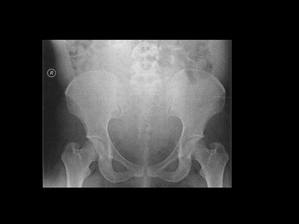
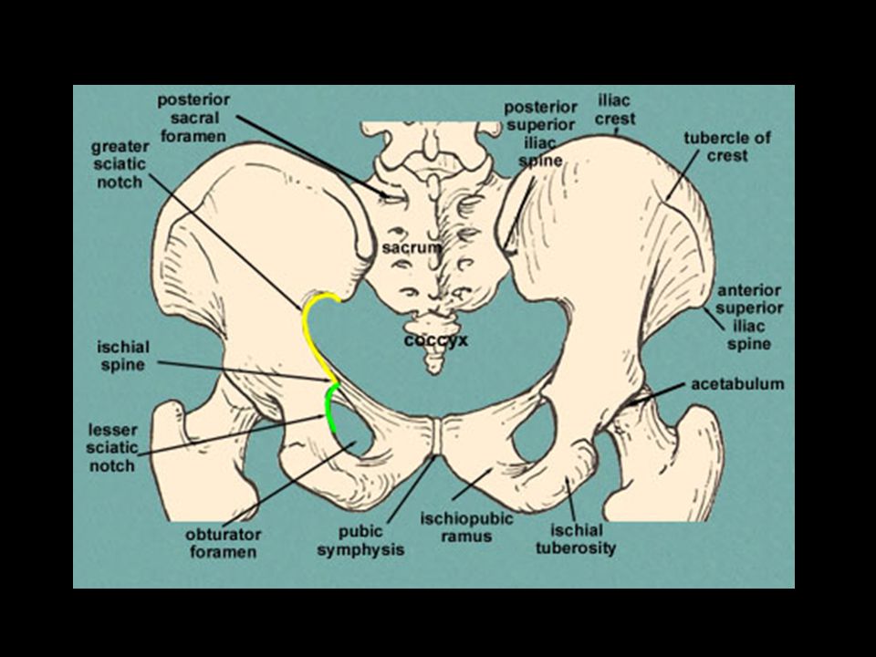
Pelvic Girdle Made of two os coxae or “hip bones”
Join with sacrum on dorsal part of body Join together at a joint called the symphysis pubis on ventral part of body

31
Each os coxae is made of three bones that are fused or joined
Ilium Ischium Pubis Contains 2 recessed areas or sockets called acetabulum that provide for attachment of smooth rounded head of the femur Obturator foramen Opening between the ischium and pubis Allows for passage of nerves and blood vessels to and from the legs

33
Bones of the legs Femur: Thigh bone Patella: Knee cap
Tibia: Larger weight bearing bone of lower leg. Commonly called the “shin bone” Fibula: slender smaller bone of the lower leg that attaches to the proximal end of the tibia

34
Ankles and Feet Tarsals: 7 bones of ankles Calcaneous- Heel bone
Metatarsals: 5 bones forming instep of foot Phalanges 14 bones on each foot Form the toes 2 on each great toe 3 on each of other toes

35
Joints

36
Joints Ligaments: areas where 2 or more bones join together
connective tissue bands that hold long bones together

37
3 Main Types of Joints Diarthrosis or synovial Freely movable
Ex: “Ball-and-socket joints” of the shoulder and hip Ex: Hinge joints of the elbow and knee

38
2. Amphiarthrosis 3. Synarthrosis Slightly movable
Ex: is the attachment of the ribs to the thoracic vertebrae Ex: symphysis pubis or joint between two pelvic bones 3. Synarthrosis Immovable Examples are the suture joints of the cranium

39
Diseases and Abnormal Conditions

40
Group of diseases involving an inflammation of the joints
Arthritis Group of diseases involving an inflammation of the joints

41
Two main types: Osteoarthritis Chronic disease that occurs with aging Symptoms: Joint pain, stiffness, aching, limited range of motion Treatment: rest, heat/cold applications, ASA, anti-inflammatory medications, steroid injections, special exercises

42
2. Rheumatoid Chronic inflammatory disease of joints Three times more common in women Often begins between age 35-45 Scar tissue forms and atrophy of bone and muscle occurs Permanent deformity and immobility Treatment: Rest and prescribed exercise Anti-inflammatory medications: ASA and steroids Surgery, or arthroplasty to replace damaged joints such as hips or knees

44
Inflammation of bursae, (small fluid-filled sacs surrounding joints)
Bursitis Inflammation of bursae, (small fluid-filled sacs surrounding joints)
")
45
Bursitis frequently affects shoulders, elbows, hips, or knees
Symptoms: Severe pain, limited movement, accumulation of fluid in joint Treatment: Pain medications and rest Injections of steroids and anesthetics into joint Aspiration of joint Physical therapy to preserve joint motion

48
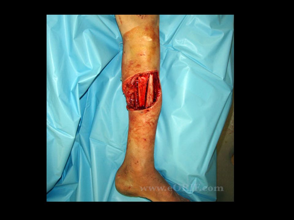
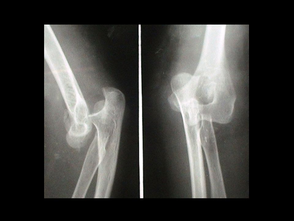
A crack or break in a bone
Fractures A crack or break in a bone

49
Types of fractures: Greenstick: bone is bent and splits, causing a crack or an incomplete break: common in children Simple: complete break with no damage to skin Compound: break in bone that ruptures through skin; increased chance of infection

50
Types of fractures, cont…
Impacted: broken fragments; or splinters into more than two pieces Spiral: severe twisting of a bone causing one or more breaks: common in skiing and skating accidents Depressed: broken piece of skull bone moves inward Colles: breaking and dislocation of the distal radius that causes a characteristic bulge at the wrist, caused by falling with outstretched hand.

51
Compound Fracture

55
Greenstick

56
Simple Fracture

58
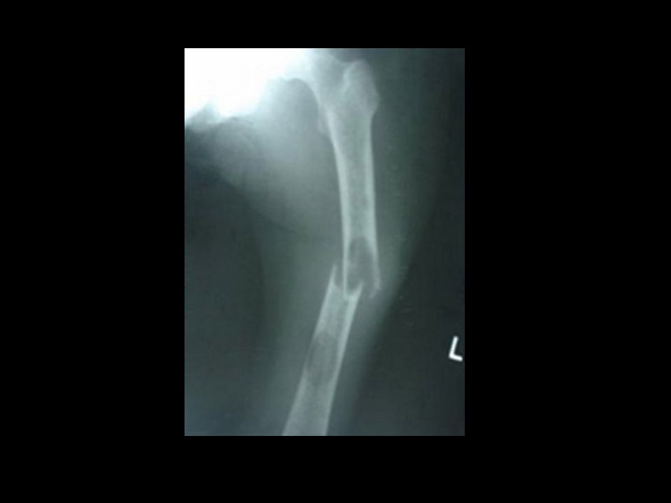
Spiral Fracture

59
Impacted Fracture

60
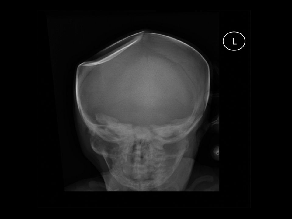
Depressed

63
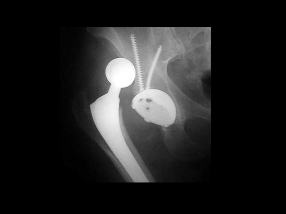
Process by which bone is put back into proper alignment
Reduction Process by which bone is put back into proper alignment Closed reduction: position bone in alignment, usually with traction, and apply cast or splint to maintain position Open reduction: surgical repair of bone and, at times, insertion of pins, plates, and other devices.

64
ORIF (Open Reduction Internal Fixation)
")
65
Bone is forcibly displaced from a joint
Disclocation Bone is forcibly displaced from a joint Frequently occurs in shoulders, fingers, knees, and hips Reduced and immobilized with splint, cast or traction

69
Twisting action tears ligaments at a joint
Sprain Twisting action tears ligaments at a joint Common sites are wrists and ankles Symptoms: pain, swelling, discoloration, limited movement Treatment: rest and elevations, immobilization with elastic bandage or splint, cold applications.

70
Inflammation of bone usually caused by pathogenic organism
Osteomyelitis Inflammation of bone usually caused by pathogenic organism Pathogen causes formation of abscess within bone and accumulation of pus in medullary canal Symptoms: pain at site, swelling, chills, fever Treatment: antibiotics for infection

71
Metabolic disorder with increased porosity or softening of bones
Osteoporosis Metabolic disorder with increased porosity or softening of bones Causes Deficiency of hormones, especially estrogen in females Prolonged lack of calcium in diet Sedentary lifestyle Tx: Increased intake of Ca and Vit D, exercise, medications such as Fosamax and Citracel, Estrogen replacement

73
Ruptured disk (herniated)
Interbertebral disk ruptures or protrudes out of place and causes pressure on the spinal nerve Most common site is lumbrosacral area: can occur anywhere on spinal column

74
Symptoms: severe pain, muscle spasm, impaired movement, and/or numbness
Treatment: Pain, anti-inflammatory, and muscle relaxant medications Rest and traction Physical and Massage therapy Chiropractic treatment Heat or cold applications Laminectomy: surgical removal of the protruding disk for severe cases Severe cases, spinal fusion can be performed to insert a screw/rod

75
Abnormal curvatures of spinal column
Kyphosis: “hunchback” bowing of thoracic area Scoliosis: side to side, or lateral curvature of spine Lordosis: swayback, inward curvature of lumbar spine

76
Kyphosis

77
Scoliosis

78
Lordosis

80
Causes of abnormal curvatures of spine:
Poor posture Congenital defects Structural defects Malnutrition Treatment: Therapeutic exercises, firm mattresses, and braces Surgical repair for severe deformities

Similar presentations