Download presentation
Presentation is loading. Please wait.

1
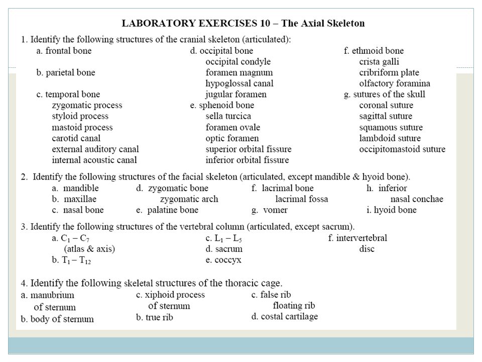
The Axial Skeleton & Fetal Skull
Exercise 9 The Axial Skeleton & Fetal Skull

3
Two Skeletal Divisions
Axial skeleton Bones around the body’s “axis” or center of gravity Appendicular skeleton Bones of the limbs or “appendages”

4
VERY IMPORTANT TABLE

5
Fig. 7-1b THE AXIAL SKELETON

6
Fig. 7-1a

7
SKULL Cranial skeleton Facial Skeleton Cranium, braincase face
Fig. 7-2

8
Fig. 7-3

9
TEMPORAL BONE Fig. 7-3

10
Fig. 7-3 TEMPORAL BONE

11
Fig. 7-3 TEMPORAL BONE

12
Fig. 7-3 TEMPORAL BONE

13
HYPOGLOSSAL CANAL IS UNDERNEATH THE CONDYLE
Fig. 7-3 OCCIPITAL BONE HYPOGLOSSAL CANAL IS UNDERNEATH THE CONDYLE

14
SPHENOID BONE Fig. 7-3

15
SPHENOID BONE Fig. 7-3

16
SPHENOID BONE Fig. 7-3

17
OLFACTORY FORAMINA ARE THE HOLES IN THE CRIBIFORM PLATE
ETHMOID BONE Fig. 7-3 OLFACTORY FORAMINA ARE THE HOLES IN THE CRIBIFORM PLATE

18
ETHMOID BONE Fig. 7-3

19
Figure 9.1a External anatomy of the right lateral aspect of the skull.
Coronal suture Frontal bone Sphenoid bone (greater wing) Parietal bone Temporal bone Ethmoid bone Lacrimal bone Squamous suture Lacrimal fossa Lambdoid suture Occipital bone Nasal bone Zygomatic process Zygomatic bone Occipitomastoid suture Maxilla External acoustic meatus Mastoid process Styloid process Mandible
 Parietal bone. Temporal bone. Ethmoid bone. Lacrimal bone. Squamous suture. Lacrimal fossa. Lambdoid suture. Occipital bone. Nasal bone. Zygomatic process. Zygomatic bone. Occipitomastoid suture. Maxilla. External acoustic meatus. Mastoid process. Styloid process. Mandible.")
20
Figure 9.1b External anatomy of the right lateral aspect of the skull.
Frontal bone Coronal suture Sphenoid bone (greater wing) Parietal bone Ethmoid bone Squamous suture Lacrimal bone Temporal bone Nasal bone Zygomatic process Lambdoid suture Lacrimal fossa Occipital bone Zygomatic bone Occipitomastoid suture Maxilla External acoustic meatus Mastoid process Styloid process Mandible 20
 Parietal bone. Ethmoid bone. Squamous suture. Lacrimal bone. Temporal bone. Nasal bone. Zygomatic process. Lambdoid suture. Lacrimal fossa. Occipital bone. Zygomatic bone. Occipitomastoid suture. Maxilla. External acoustic meatus. Mastoid process. Styloid process. Mandible. 20.")
21
Figure 9.2a Inferior view of the skull, mandible removed.
Maxilla (palatine process) Palatine bone (horizontal plate) Maxilla Zygomatic bone Sphenoid bone (greater wing) Temporal bone (zygomatic process) Foramen ovale Vomer Foramen spinosum Carotid canal External acoustic meatus Styloid process Mastoid process Jugular foramen Temporal bone (petrous part) Occipital condyle Parietal bone Occipital bone Foramen magnum 21
 Palatine bone (horizontal plate) Maxilla. Zygomatic bone. Sphenoid bone (greater wing) Temporal bone (zygomatic process) Foramen ovale. Vomer. Foramen spinosum. Carotid canal. External acoustic meatus. Styloid process. Mastoid process. Jugular foramen. Temporal bone (petrous part) Occipital condyle. Parietal bone. Occipital bone. Foramen magnum. 21.")
22
Figure 9.2b Inferior view of the skull, mandible removed.
Zygomatic arch Foramen ovale Carotid canal Styloid process Mastoid process Jugular foramen Occipital condyle Foramen magnum 22

23
Figure 9.3a-b Internal anatomy of the inferior portion of the skull.
Cribriform plate Ethmoid bone Crista galli Frontal bone Cribriform foramina Optic canal Sphenoid Foramen ovale sella turcica Hypoglossal canal Temporal bone (petrous part) Internal acoustic meatus Jugular foramen Parietal bone Occipital bone Foramen magnum 23
 Internal acoustic meatus. Jugular foramen. Parietal bone. Occipital bone. Foramen magnum. 23.")
24
Figure 9.3c Internal anatomy of the inferior portion of the skull.
Frontal bone Crista galli Ethmoid bone Cribriform plate Cribriform foramina Optic canal Sphenoid sella turcica Foramen ovale Temporal bone (petrous part) Jugular foramen Parietal bone Occipital bone Foramen magnum 24
 Jugular foramen. Parietal bone. Occipital bone. Foramen magnum. 24.")
25
Fig. 7-3 SKULL SUTURES

26
SKULL SUTURES Fig. 7-3

27
SKULL SUTURES Fig. 7-3

28
SKULL SUTURES Fig. 7-3

29
SKULL SUTURES Fig. 7-3

30
Figure 9.6b Anatomy of the anterior and posterior aspects of the skull.
Sagittal suture Parietal bone Lambdoid suture Occipital bone Temporal bone (mastoid process) Occipitomastoid suture Occipital condyle 30
 Occipitomastoid suture. Occipital condyle. 30.")
31
FACIAL BONES OF THE SKULL
Fig. 7-2

32
FACIAL BONES OF THE SKULL Shallow depression in this bone
Fig. 7-2 LACRIMAL FOSSA = Shallow depression in this bone

33
FACIAL BONES OF THE SKULL
Fig. 7-3 Zygomatic bone = blue Temporal bone = pink Zygomatic ARCH is a segment of each of these bones, your “cheekbone” is actually partially temporal bone and partially zygomatic bone…your zygomatic arch.

34
FACIAL BONES OF THE SKULL
Inferior nasal conchae (2) are FACIAL bones…. You already learned that the middle nasal conchae are part of the ethmoid bone, a CRANIAL bone. Fig. 7-3
 are FACIAL bones…. You already learned that the middle nasal conchae are part of the ethmoid bone, a CRANIAL bone. Fig")
35
FACIAL BONES OF THE SKULL
Fig. 7-3, 4 The VOMER is the inferior portion of your nasal septum.

36
FACIAL BONES OF THE SKULL
PALATINE BONE: posterior 1/3 of “roof of mouth” Fig. 7-3

37
FACIAL BONES OF THE SKULL
Fig. 7-2 2 MAXILLARY BONES: upper jaw

38
FACIAL BONES OF THE SKULL
Fig. 7-2 MANDIBLE (1): lower jaw
: lower jaw.")
39
Figure 9.6c Anatomy of the anterior and posterior aspects of the skull.
Parietal bone Frontal bone Sphenoid bone Ethmoid bone Nasal bones Temporal bone Zygomatic bone Maxilla Mandible 39

40
Figure 9.7 Detailed anatomy of the mandible and maxilla.
Maxilla, right lateral view Mandible, right lateral view 40

41
Figure 9.8 Bones that form the orbit.
Superior orbital fissure Roof of orbit Optic canal • sphenoid bone • frontal bone Medial wall • ethmoid bone Lateral wall of orbit • maxilla sphenoid bone • Lacrimal bone • zygomatic bone Nasal bone Floor of orbit Inferior orbital fissure • palatine bone Zygomatic bone • maxillary bone • Zygomatic bone 41

42
Doesn’t articulate with any other bone—unique!
Hyoid bone Not really a skull bone Doesn’t articulate with any other bone—unique! Fig. 7-12

43
Vertebral Column Cervical: C1-C7 Atlas = C1 Axis = C2 Fig. 7-16

44
All cervicals have holes in the sides
Cervical Vertebrae Atlas = C1 “no body” Axis = C2 All cervicals have holes in the sides Fig. 7-19

45
All thoracics have facets on the sides where ribs attach
Thoracic Vertebrae T1-T12 All thoracics have facets on the sides where ribs attach Fig. 7-20 Fig. 7-20

46
All lumbars have large “bodies”
Lumbar Vertebrae L1-L5 All lumbars have large “bodies” Fig. 7-21

47
Know posterior/anterior
Sacrum & Coccyx 5 fused vertebrae Know posterior/anterior 4 fused vertebrae Fig. 7-22

48
Intervertebral Discs Inter = in between What type of cartilage?
Fig. 7-18

49
Table 9.1 Regional Characteristics of Cervical, Thoracic, and Lumbar Vertebrae

50
Figure 9.11 The vertebral column.
2 3 Cervical curvature (concave) 7 vertebrae, C1 – C7 4 5 C7 (vertebra prominens) 6 7 T1 Spinous process 2 3 Transverse processes 4 5 Thoracic curvature (convex) 12 vertebrae, T1 – T12 6 7 8 9 Intervertebral discs 10 11 Intervertebral foramen 12 L1 2 Lumbar curvature (concave) 5 vertebrae, L1 – L5 3 4 5 Sacral curvature (convex) Sacrum 5 fused vertebrae Coccyx 4 fused vertebrae Anterior view Right lateral view 50
 7 vertebrae, C1 – C C7 (vertebra prominens) T1. Spinous process Transverse processes Thoracic curvature (convex) 12 vertebrae, T1 – T Intervertebral discs Intervertebral foramen. 12. L1. 2. Lumbar curvature (concave) 5 vertebrae, L1 – L Sacral curvature (convex) Sacrum 5 fused vertebrae. Coccyx 4 fused vertebrae. Anterior view. Right lateral view. 50.")
51
Figure 9.12 Abnormal spinal curvatures
Scoliosis Kyphosis Lordosis 51

52
Figure 9.13 A typical vertebra, superior view.
52

53
Figure 9.14 The first and second cervical vertebrae.
Superior view of atlas (C1) Inferior view of atlas (C1) Posterior Superior view of axis (C2) 53
 Inferior view of atlas (C1) Posterior. Superior view of axis (C2) 53.")
54
Figure 9.15 Superior and right lateral views of typical vertebrae.
Superior View Right Lateral View C2 Cervical Thoracic Lumbar 54

55
Figure 9.16 Sacrum and coccyx.
Anterior view Posterior view 55

56
Bony Thorax Fig. 7-23 STERNUM

57
FALSE RIBS : FLOATING RIBS (11-12)
Bony Thorax Fig. 7-23 TRUE RIBS (1-7) FALSE RIBS (8-12) FALSE RIBS : FLOATING RIBS (11-12)
 FALSE RIBS (8-12) FALSE RIBS : FLOATING RIBS (11-12)")
58
Bony Thorax Fig. 7-23 COSTAL CARTILAGE NOT “COASTAL”

59
Figure 9.17 The thoracic cage.
Manubrium Body Sternum True ribs (1–7) Xiphoid process T2 T3 T4 T5 Heart False ribs (8–12) T9 L1 Vertebra Costal cartilage Floating ribs (11, 12) 59
 Xiphoid process. T2. T3. T4. T5. Heart. False ribs (8–12) T9. L1 Vertebra. Costal cartilage. Floating ribs (11, 12) 59.")
60
Figure 9.18 Structure of a typical true rib and its articulations.
Transverse costal facet (for tubercle of rib) 60
 60.")
61
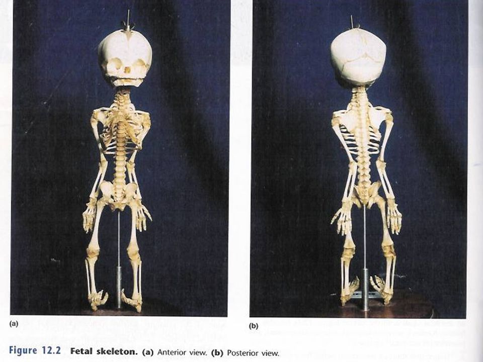
The Fetal Skeleton

63
Fontanels: indentations between bones of fetal skull (fibrous membranes), which ossify as the child ages (20-22 months)
, which ossify as the child ages (20-22 months)")
65
Face: cranium Head:body Ossification centers Frontal bone Vertebrae
Sternum Patellae Coxal Rib cage Carpals tarsals

66
Figure 9.19 Skull of a newborn.
Occipital bone Frontal bone Posterior fontanelle Parietal bone Sphenoidal fontanelle Ossification center Ossification center Posterior fontanelle Parietal bone Anterior fontanelle Mastoid fontanelle Frontal bone Frontal suture Occipital bone Anterior Temporal bone (squamous part) Superior view Left lateral view Anterior fontanelle Anterior fontanelle Parietal bone Frontal suture Parietal bone Frontal bone Frontal bone Sphenoidal fontanelle Occipital bone Maxilla Sphenoidal fontanelle Temporal bone (squamous part) Mastoid fontanelle Mandible Anterior view Left lateral view 66
 Superior view. Left lateral view. Anterior fontanelle. Anterior fontanelle. Parietal bone. Frontal suture. Parietal bone. Frontal bone. Frontal bone. Sphenoidal fontanelle. Occipital bone. Maxilla. Sphenoidal fontanelle. Temporal bone (squamous part) Mastoid fontanelle. Mandible. Anterior view. Left lateral view. 66.")
67
Figure 9.19a Skull of a newborn.
Occipital bone Posterior fontanelle Ossification center Parietal bone Anterior fontanelle Frontal bone Frontal suture Anterior Superior view 67

68
Figure 9.19b Skull of a newborn.
Frontal bone Parietal bone Sphenoidal fontanelle Ossification center Posterior fontanelle Mastoid fontanelle Occipital bone Temporal bone (squamous part) Left lateral view 68
 Left lateral view. 68.")
69
Figure 9.19c Skull of a newborn.
Anterior fontanelle Frontal suture Parietal bone Frontal bone Sphenoidal fontanelle Maxilla Mandible Anterior view 69

70
Figure 9.19d Skull of a newborn.
Anterior fontanelle Parietal bone Frontal bone Occipital bone Sphenoidal fontanelle Temporal bone (squamous part) Mastoid fontanelle Left lateral view 70
 Mastoid fontanelle. Left lateral view. 70.")
71
Review Figure 9.1 71

72
Review Figure 9.2 72

73
Review Figure 9.3 (curvature) (curvature) (curvature) (curvature) 73
 (curvature) (curvature) (curvature) 73")
74
Review Figure 9.4 L1 vertebra 74

75
Review Figure 9.5 75

Similar presentations