Download presentation
Presentation is loading. Please wait.

1
Systemic Veins 1. Characteristics of Veins
2. Composition of Systemic Veins 3. Veins of Head,Neck And Upper Limb As Well As Thorax, Abdomen And Lower Limb Guo Ling,MD,PhD Department of Anatomy

2
a. Veins convey the blood
Characteristics of Vein a. Veins convey the blood from peripheral organs to the heart. b. Tributaries, venules drain to big veins. (It is not called a branch but a tributary,from a small D to a large D.) c. No pulse of vein able to be felt in living body.
 c. No pulse of vein able. to be felt in living body.")
3
2.The systemic veins are divided into two sets: Superficial veins
Deep veins ① Located beneath the skin ② No correspondingly accompanied arteries 1) Superficial Veins (subcutaneous vein)
 Superficial Veins. (subcutaneous vein)")
4
2) Deep Veins (venae comitantes) ①accompanied with the
corresponding arteries ②having the same names as those described in the corresponding arteries

5
3. There are valves in veins of extremities The valves open toward
the heart to prevent the backflow of blood into the peripheral organs.

6
Composition of Systemic Veins 1. Superior vena cava
and its tributaries 2. Inferior vena cava 3. Veins of the heart

7
Composition and Tributaries
of Superior Vena Caval System Subclavian Vein Internal jugular V Venous angle Left / right brachiocephalic Vs Superior vena cava Right atrium

8
Collectiing Scope of Superior Vena Cava
It receives the blood from head , neck, upper limbs & thorax.

9
⑤Internal jugular V Veins of Head & Neck ①Superior temporal V
②Facial V ③Maxillary V ④External jugular V ⑤Internal jugular V

10
Veins of Neck (anterior view)
")
11
The facial V communicates
with the cavernous sinus Sup. Ophthalmic V Cavernous sinus Facial V

12
Veins of Upper Limbs 1.Superficial V 1) cephalic(lat.)
2) basilic(med.) 3 )median viens Clinical Applications A) intravenous injection B) blood sampling C) blood/ liquid transfusion D) introduction of catheters to the heart or the coronary arteries
 basilic(med.) 3 )median viens. Clinical Applications. A) intravenous injection. B) blood sampling. C) blood/ liquid. transfusion. D) introduction of. catheters to the heart or. the coronary arteries.")
13
2. Deep Veins Subclavian Vein Axillary Vein Brachial Vein Radial V ein
Ulnar Vein Subclavian Vein

14
② ① Collections? ③ Thoracic Veins ③hemiazygos Vein Endings?
② Accessory hemiazygos Vein ② ① ③hemiazygos Vein Collections? Endings? ③

15
Veins in Abdominal Cavity
----Inferior Vena Caval System 1. Composition Left / Right common iliac vein 2.Tributaries 1) parietal 2) visceral 3) paired, 4) unpaired 3. Drainage lower half of body 4. End right atrium
 parietal. 2) visceral. 3) paired, 4) unpaired. 3. Drainage. lower half of body. 4. End. right atrium.")
16
Tributaries of Inferior Vena Cava 1) Parietal tributaries
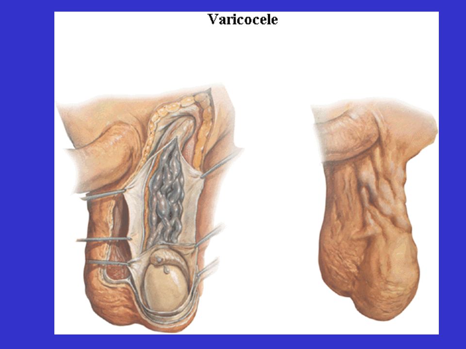
2) Visceral tributaries ① Renal V ②Testicular V (ovarian V) ③Hepatic V--3 tributaries (submerged in the liver)
 Visceral tributaries. ① Renal V. ②Testicular V. (ovarian V) ③Hepatic V--3 tributaries. (submerged in the liver)")
18
Veins in Pelvis Internal iliac vein The venous plexuses
in the pelvis are: 1)Vesical venous plexus 2)Uterine venous plexus 3)Rectal venous plexus All the plexuses lack valves to enable the blood to freely flow forward or backward (bi-directional flows).
Vesical venous plexus. 2)Uterine venous plexus. 3)Rectal venous plexus. All the plexuses. lack valves to enable. the blood to freely flow. forward or backward. (bi-directional flows).")
19
Veins of Lower Limb 1) Superficial Veions ① Great saphenous V
tributaries: a) Superficial epigastric V b) Superficial iliac circumflex V c) External pudendal V d) Superficial medial femoral V e) Superficial lateral femoral V ② Small saphenous V (origin,route,ending)
 Superficial epigastric V. b) Superficial iliac circumflex V. c) External pudendal V. d) Superficial medial femoral V. e) Superficial lateral femoral V. ② Small saphenous V. (origin,route,ending)")
20
2) Deep Veins Common iliac V External iliac V Femoral V Popliteal V
Anterior tibial V Posterior tibial V Common iliac V

21
Splenic V Superior mesenteric V ④ Hepatic Portal Vein
1. Composition: splenic V, superior mesenteric V (both union behind the head of pancreas) Superior mesenteric V Splenic V
 Superior mesenteric V. Splenic V.")
22
2.Origin: capillaries of unpaired organs of abdomen
3.End: hepatic sinuses liver sinus Hepatic portal V

23
Hepatic V Hepatic portal V

24
3. Collection Coverage Unpaired organs in abdominal cavity include:
gallbladder, pancreas, spleen and digestive canal from stomach downward, except for the liver.

25
4.Tributaries of Hepatic Portal Vein (1) Splenic V (2) Superior mesenteric V (3) Inferior (4) Left gastric V (5) Right gastric V
 Right gastric V.")
26
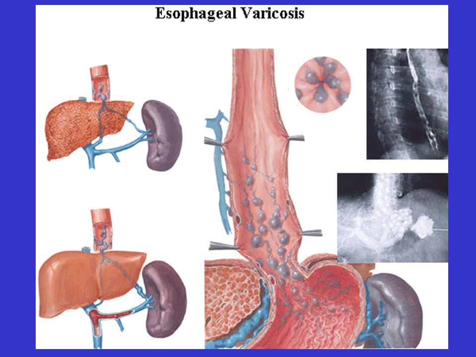
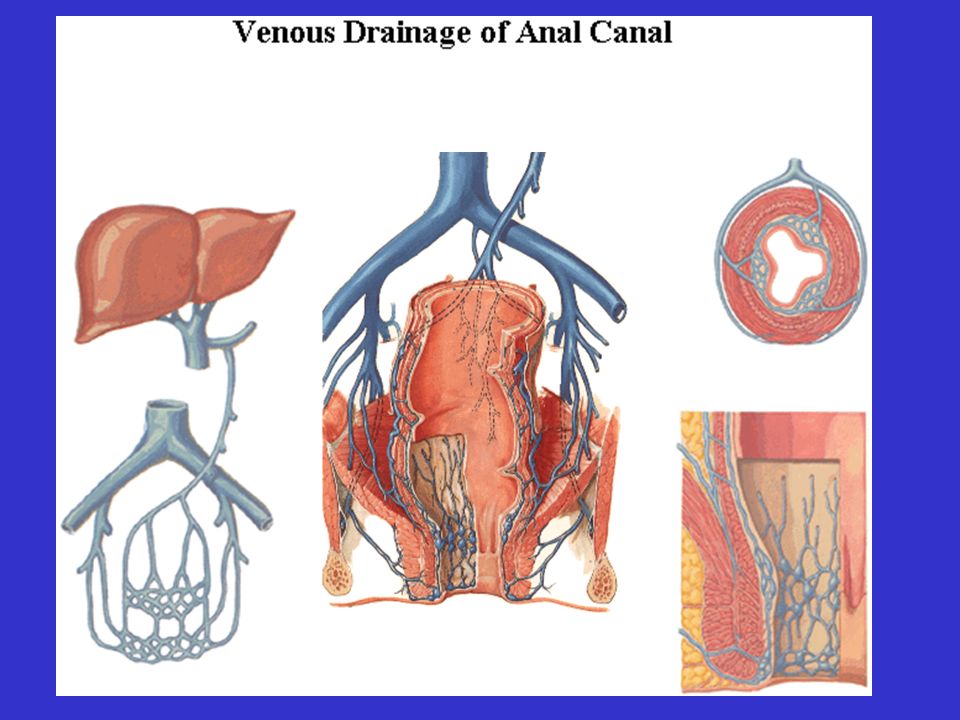
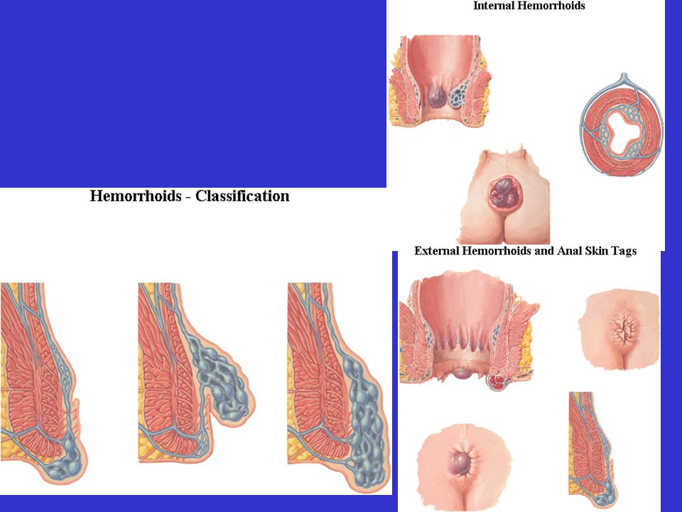
2) Rectal venous plexus 1) Esophageal venous plexus
5. Anastomoses a. position: low part b. sigificance : broken,vomiting blood. a. position: rectum b. sigificance : blocked,hemorrhoids. 1) Esophageal venous plexus 2) Rectal venous plexus 3)Periumbilical venous plexus a. position: around umbilicus b. siginificance: blocked, visibly swellen.
 Esophageal venous plexus. 2) Rectal venous plexus. 3)Periumbilical. venous plexus. a. position: around umbilicus. b. siginificance: blocked, visibly swellen.")
30
bile reabsorbed (Nutrients) Food
6. Characteristics of Hepatic Portal V Transporting thesubstances absorbed by stomach & intestines, include nutrients, toxins, drugs & wastes, etc. bile reabsorbed (Nutrients) Food
 Food.")
31
(2) Diversion of Blood Stream ▲The blood from superior mesenteric V
flows into the right lobe. ▲ The blood from splenic V flows into the left lobe.

32
(3) The both ends of hepaticportal vein are capillaries.
 The both ends of hepaticportal vein are capillaries.")
33
The substances absorbed by the small intestine
are carried out of body with blood and urine though the following pathway: Small intestine→sup.mesenteric V → hepatic portal V → Liver sinusoids →hepatic V → inf.vena cava → R.atrium → R.ventricle → pulmonary trunk,and L. R pulmonary arteries → lungs→ pulmonary Vs → L. atrium → L.ventricle → aorta → renal As → kidneys→ ureters→Urinary bladder → urethra

34
Glucose is injected into median cubital V ,
Trace the sugar until it run out of the body: Cephalic V →Subclavian V → Branchiocephalic V → Sup. Vena cava → R.atrium……(continue… please think about the subsequent course !!! )
")
35
Clinical Consideration What may happen after the hepatic
portal vein is obstructed ? a) building collateral circulations b) disfunctions of digestive tract c) toxin to brain d) poor nutrition supply
 building collateral circulations. b) disfunctions of digestive tract. c) toxin to brain. d) poor nutrition supply.")
Similar presentations