Download presentation
Presentation is loading. Please wait.

1
بسم الله الرحمن الرحيم

2
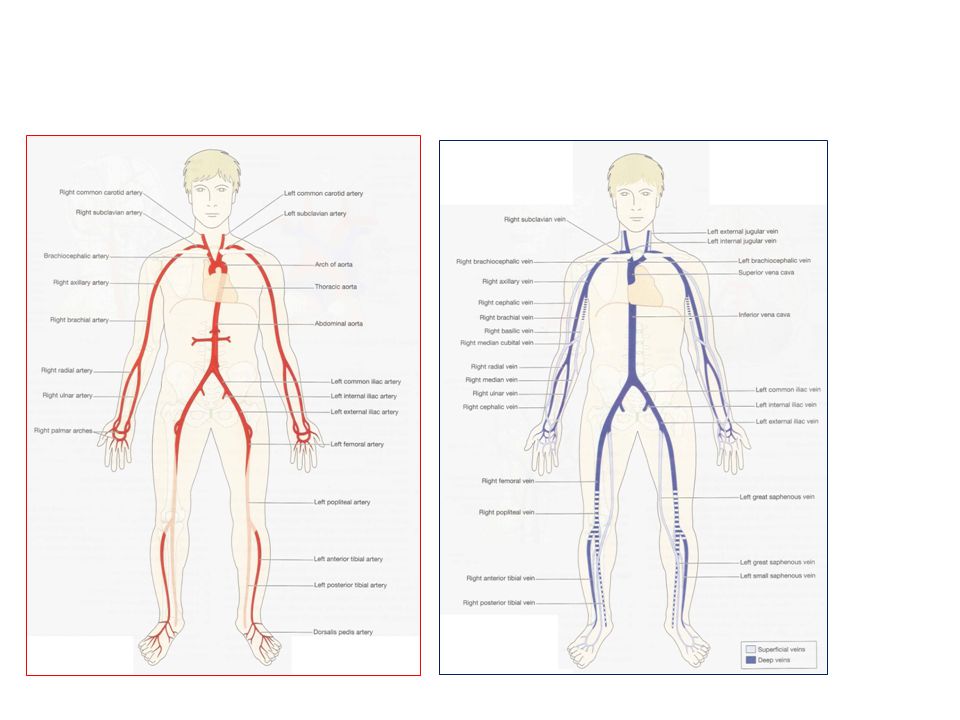
Systemic or general circulation
The blood pumped out from the left ventricle is carried by the branches of the aorta around the body and is returned to the right atrium of the heart by the superior and inferior venae cavae.

4
Aorta It begins at the upper part of the left ventricle and, after passing upwards for a short way, it arches backwards and to the left. It then descends behind the heart through the thoracic cavity a little to the left of the thoracic vertebrae. At the level of the D12 it passes behind the diaphragm then downwards in the abdominal cavity to the level of the L4, where it divides into the right and left common iliac arteries.

5
Throughout its length the aorta gives off numerous branches
Throughout its length the aorta gives off numerous branches. Some of the branches are paired, e.g. the right and left renal arteries, and some are single e.g. the coeliac artery. Thoracic aorta This part of the aorta is above the diaphragm and is described in three parts: ascending aorta arch of the aorta descending aorta in the thorax.

6
Arch of the aorta Three branches: brachiocephalic artery or trunk.
left common carotid artery. left subclavian artery. The brachiocephalic artery is divides into the right common carotid artery and the right subclavian artery.

7
Circulation of blood to the head and neck
Arterial supply The paired arteries supplying the head and neck are the common carotid arteries and the vertebral arteries. Carotid arteries. The right common carotid artery is a branch of the ?. The left common carotid artery arises from ?. They pass upwards on either side of the neck and have the same distribution on each side. The common carotid arteries are devided into: Extennal carotid artery internal carotid artery

8
External carotid artery supplies the superficial soft tissue structures of the head and neck.
The internal carotid artery is a major contributor to the circulus arteriosus (circle of Willis) which supplies the greater part of the brain. It ascends to the base of the skull and passes through the carotid foramen in the temporal bone.
 which supplies the greater part of the brain. It ascends to the base of the skull and passes through the carotid foramen in the temporal bone.")
9
(circle of Willis): two internal carotid arteries and two vertebral arteries. The vertebral arteries arise from the subclavian arteries, pass upwards through the foramina in the transverse processes of the cervical vertebrae, enter the skull through the foramen magnum, then join to form the basilar artery.

10
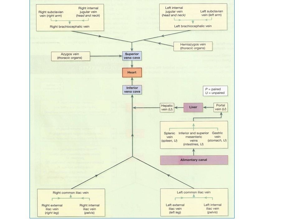
VENOUS DRAINAGE OF THE HEAD AND NECK
Superficial structures Superficial veins external jugular vein

11
Deep structures Dural (brain)venous sinuses
Superior and inferior sagittal sinuses Internal jugular vein

13
Circulation of blood to the upper limb
Arterial supply The subclavian arteries. The right subclavian artery arises from ?. the left branches from ? axillary arteries brachial artery radial and ulnar arteries.

14
Venous return from the upper limb
The veins of the upper limb are divided into two groups: deep and superficial veins. The deep veins follows the course of the arteries and have the same names. Both systems end in the brachiocephalic veins on either side which join together forming SVC.

15
Descending aorta in the thorax
Starts as….? Then It extends downwards on the anterior surface of the bodies of the thoracic vertebrae to the level of the D12, where it continuous as abdominal aorta. The descending aorta in the thorax gives off many branches which supply the walls of the thoracic cavity and the organs within the cavity e.g. Bronchi, the lungs, oesophagus, intercostal muscles ….

16
Venous return from the thoracic cavity
Most of the venous blood from the organs in the thoracic cavity is drained into the azygos vein and the hemiazygos vein. both veins end in a way or another in the SVC.

17
Abdominal aorta The abdominal aorta is a continuation of the thoracic aorta. It descends in front of the bodies of the vertebrae to the level of L4, where it divides into the 2 CIA. Many branches arise from the abdominal aorta, some of which are paired and some unpaired.

18
Paired branches: e.g. Renal , the suprarenal arteries. Testicular arteries supply the testes in the male. Ovarian arteries supply the ovaries in the female. Unpaired branches: e.g. The coeliac artery.gives 3 branches (splenic, hepatic, lt gastric) The superior mesenteric artery. Gives branches to small intestin and rt side of colon. The inferior mesenteric artery.gives branches to lt colon and rectum.
 The superior mesenteric artery. Gives branches to small intestin and rt side of colon. The inferior mesenteric artery.gives branches to lt colon and rectum.")
19
Venous return from the abdominal organs
The IVC is formed when right and left common iliac veins join at the level of L5. This is the largest vein in the body and it conveys blood from all parts of the body below the diaphragm to the right atrium of the heart. It passes through the central tendon of the diaphragm at the level of the 8th thoracic vertebra. Paired testicular, ovarian, renal and adrenal veins join the inferior vena cava. Blood from the remaining organs in the abdominal cavity passes through the liver via the portal circulation before entering the inferior vena cava.

20
IVC Portal circulation PV hepatic veins
1st Capillary bed of digestive system, spleen, pancreas PV liver Hepatic sinusoids 2nd capillary bed hepatic veins IVC

21
Portal circulation In this way blood with a high concentration of nutrients absorbed from the stomach and intestines, goes to the liver first. In the liver certain modifications take place, including the regulation of nutrient supply to other parts of the body.

22
Portal vein This is formed by the union of these veins each of which drains blood from the area supplied by the corresponding artery.

23
Circulation of blood to the pelvis and lower limb
Arterial supply Common iliac arteries The right and left common iliac arteries are formed …? Then each one is divided into: internal iliac artery. external iliac artery. The internal iliac artery supply the organs within the pelvic cavity. largest branches is the uterine artery. The external iliac artery runs obliquely downwards into the thigh where it becomes the femoral artery.

24
Femoral artery → popliteal artery →
1-The anterior tibial artery → dorsalis pedis artery. 2-The posterior tibial artery → peroneal artery.

25
Venous return There are both deep and superficial veins in the lower limb. Blood entering the superficial veins passes to the deep veins through communicating veins. Movement of blood towards the heart is partly dependent on contraction of skeletal muscles. Backward flow is prevented by a large number of valves.

26
Deep veins. The deep veins accompany the arteries and their branches and have the same names.
The femoral vein → the external iliac vein + internal iliac vein →common iliac vein→ inferior vena cava Superficial veins: small saphenous vein joins the popliteal vein. great saphenous vein joins the femoral vein. Many communicating veins join the superficial veins and deep veins of the lower limb.

Similar presentations


 The first artery to branch off the aortic arch that will then divide into the right common carotid.>")


