Download presentation
Presentation is loading. Please wait.

1
The Skeletal System Health Sciences Diane A. Young

2
Structure of the Bone A form of connective tissue. Bones have their own system of blood vessels and nerves allowing circulation to occur within the bone.

3
Functions of the Bone Serve as a framework for the body, giving the body structure and support. Protect internal structures – Brain – Spinal cord Act as a storage area for calcium. – Calcium is used in the blood if the diet does not provide enough calcium

4
Functions of the Bones Cont’d Produce blood cells (Hemopoeisis) – Red bone marrow produces blood cells Movement- muscles contract and pull bones to produce movement
 – Red bone marrow produces blood cells Movement- muscles contract and pull bones to produce movement")
5
hemo = blood poiesis = to make Hemopoiesis Red bone marrow produces: red blood cells white blood cells platelets Red blood cells purple: platelets green/gold: WBC

6
Red Marrow Red Blood Cell production In adults: ribs vertebrae ends of humerus, pelvis femur

8
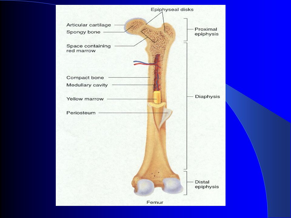
Tissues of Bone Red bone marrow- located in spongy bone and manufactures red blood cells. Cartilage – smooth, rubbery blue-white connective tissue. Acts as a shock absorber between bones. (outer ear and tip of nose) Articular cartilage – covers the surfaces of bones that form joints to make smooth joint movement possible and to protect the bones from rubbing against each other. Meniscus – curved fibrous cartilage found in some joints, such as the knee and the temporomandibular joint of the jaw.
 Articular cartilage – covers the surfaces of bones that form joints to make smooth joint movement possible and to protect the bones from rubbing against each other. Meniscus – curved fibrous cartilage found in some joints, such as the knee and the temporomandibular joint of the jaw..")
9
Anatomic Landmarks of a Bone Diaphysis – the shaft of a long bone Epiphysis – wide end of a long bone Proximal epiphysis – end nearest the midline of the body Distal epiphysis – end located farthest from the midline Foramen – opening through which blood vessels, nerves and ligaments pass. Process – normal projection on the surface that serves as attachments for muscles and tendons.

11
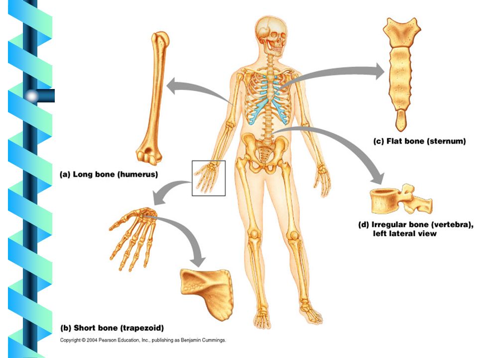
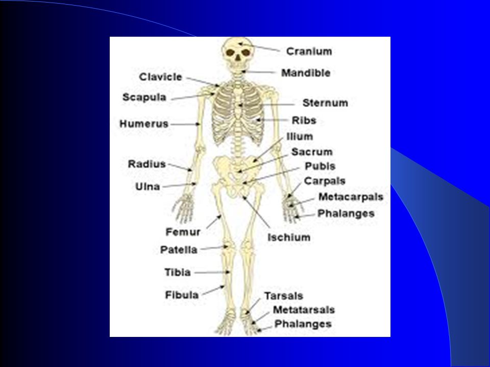
Types of Bone Long bones (longer than their width) – Humerus – Radius – Ulna – Femur – Tibia – Fibula
 – Humerus – Radius – Ulna – Femur – Tibia – Fibula")
12
Types of Bone Cont’d Short bones (length and width are nearly equal) – Wrist and hand – Ankle and feet Flat bones (two layers of bone divided by a narrow span) – Skull – Sternum – Ribs – Shoulder blade
 – Wrist and hand – Ankle and feet Flat bones (two layers of bone divided by a narrow span) – Skull – Sternum – Ribs – Shoulder blade")
13
Types of Bone Cont’d Irregular bones (do not fit into other three groups) – Face – Spine – Hip
 – Face – Spine – Hip")
15
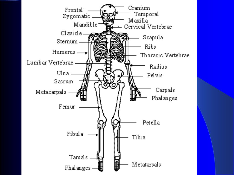
Groups of Bones Skeleton is divided into two groups Axial – included 80 bones found in the skull, vertebrae, ribs and sternum Appendicular – 126 bones found in the arms, hands, legs, feet, and pelvis.

16
Axial Skeleton

17
Vertebral Column Cervical Vertebrae (7) Thoracic Vertebrae (12) Lumbar Vertebrae (5) Sacrum Coccyx
 Thoracic Vertebrae (12) Lumbar Vertebrae (5) Sacrum Coccyx")
18
Thoracic Cage Sternum True Ribs (7) False Ribs (3) Floating Ribs (2)
 False Ribs (3) Floating Ribs (2)")
19
Appendicular Skeleton

20
Humerus

21
Ulna & Radius

22
Hand Bones

24
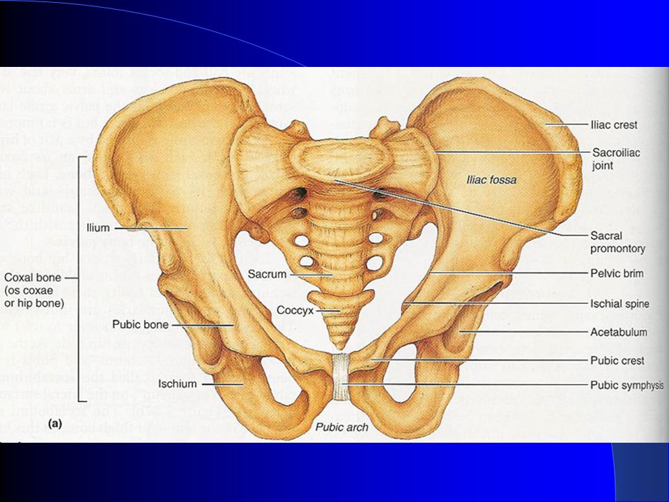
FEMALE MALE Pelvic Girdle

25
The female pelvis …. · Bones are thinner and lighter · Pelvis is more shallow · Hips are wider · Sacrum is shorter and wider

26
The Lower Limb (Legs) Femur Patella Tibia Fibula Tarsals Metatarsals Phalanges
 Femur Patella Tibia Fibula Tarsals Metatarsals Phalanges")
27
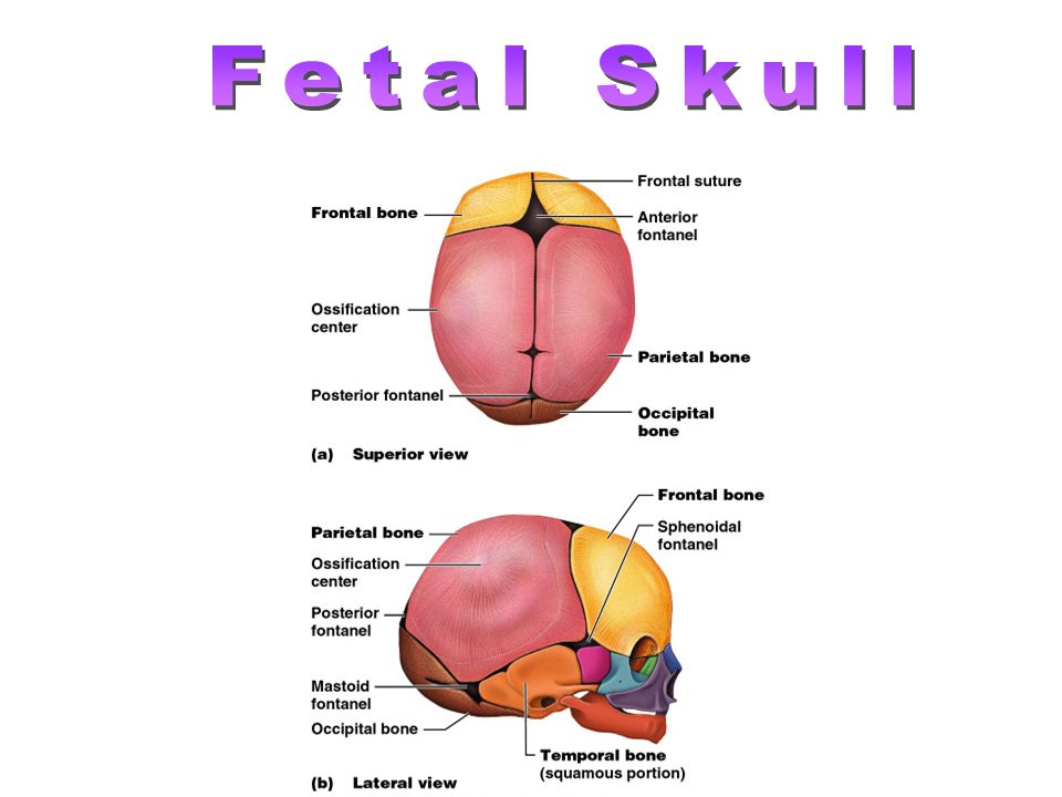
Joints The point where two bones meet Three main groups of joints Immovable joints – cranium (suture joints) Slightly movable joints – vertebral discs, symphisis pubis, sacroiliac joints Freely movable joints – shoulder joint, elbow, wrist, finger joints, knee and ankle joints
 Slightly movable joints – vertebral discs, symphisis pubis, sacroiliac joints Freely movable joints – shoulder joint, elbow, wrist, finger joints, knee and ankle joints")
28
(Synarthrosis) immovable joints Skull suture pubis symphisis
 immovable joints Skull suture pubis symphisis")
29
(Amphiarthrosis) Slightly Movable Joint
 Slightly Movable Joint")
30
(Diarthroses) Freely movable joints pelvis ligaments femur
 Freely movable joints pelvis ligaments femur")
31
Ligaments Ligaments connect to bones and hold bones together. Joints are formed where the bones meet.

33
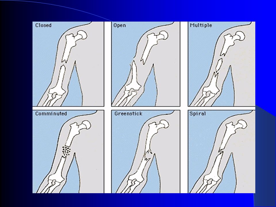
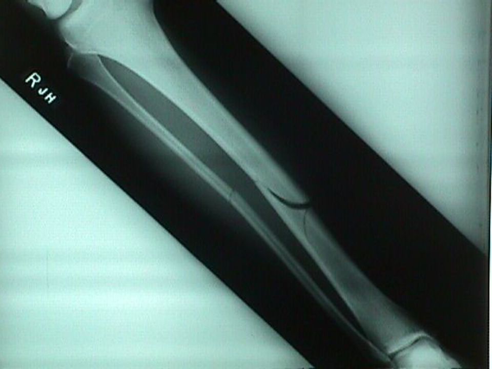
Disorders of the Skeleton Arthritis – inflammation of the joints. Degenerative joint diseases – cause changes in the structure of a joint. Fractures – a break in a bone

34
Types of Fractures Simple fracture – occurs when the bone is broken but the skin is not broken. Compound fracture – occurs when the bone is broken and penetrates the skin. Comminuted fracture – occurs when the bone breaks and there are bone fragments in the tissue. Greenstick fracture – occurs when the bone is bent and splits causing an incomplete break. Most common in children.

37
Repair of Fractures hematoma callus bony callus bone remodeling

38
Bone cells that aid in remodeling Osteoblast – builds new bone Osteocyte – mature bone cell Osteoclast – eats bone

39
Rickets- vitamin D deficiency deficiency Osteomalacia- soft bones, inadequate mineralization in bones, lack of vitamin D Pagets Disease- spotty weakening in the bones, excessive and abnormal bone remodelin Rheumatoid arthritis- autoimmune reaction Diseases of the Skeletal System

40
Osteoporosis- bone reabsorption outpaces bone deposit; bones become lighter and fracture easier Risk Factors: age, gender (more in women) diet poor in Ca ++ and protein abnormal vitamin D receptors smoking
 diet poor in Ca ++ and protein abnormal vitamin D receptors smoking")
41
Rheumatoid Arthritis

42
Rickets

43
Osteomalacia

44
Osteoporosis 29 40 84 92

46
cartilage calcified cartilage bone epiphyseal plate epiphyseal line Endochondral Ossification 2 o ossification center Fetus: 1 st 2 months AdultChildhood Just before birth

47
275 bones 12 weeks (6-9 inches long)
")
Similar presentations


 Triglyceride storage.>")


 2.Protection: skull, vertebrae,>")

