Download presentation
Presentation is loading. Please wait.

1
The skeletal system Structure and function of bone Organization of the skeleton Joints

2
Functions of bone (skeleton) Support and protection Blood cell formation Mineral storage (calcium especially) Site for muscle attachment body movement
 Support and protection Blood cell formation Mineral storage (calcium especially) Site for muscle attachment body movement")
3
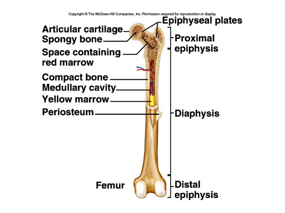
Bones classified by shape: long, short, flat, irregular, round Bone enclosed in periosteum, which is continuous with tendons and ligaments blood vessels in periosteum Epiphysis- ends spongy bone contains red marrow compact bone, articular cartilage Diaphysis- middle compact bone medullary cavity- contains yellow marrow (fat) lined with endosteum (squamous epithelium)
 lined with endosteum (squamous epithelium)")
5
Compact bone osteocytes within lacunae arranged in concentric circles called lamellae This surround a central canal; complex is called Haversian system Canaliculi connect osteocytes to central canal and to each other

7
Prenatal development skeleton is mostly cartilaginous Cartilage cells and then osteoblasts start to deposit minerals Cartilaginous disk (epiphyseal disk) remains in epiphysis Cells eventually stop dividing
 remains in epiphysis Cells eventually stop dividing")
9
Adults continually break down and build up bone Osteoclasts remove damaged cells and release calcium into blood Osteoblasts remove calcium from blood and build new matrix. They become trapped osteoclasts

10
Types of bone breaks Simple- skin is not pierced Compound- skin is pierced Complete- bone is broken in half Partial- broken lengthwise but not into two parts Greenstick- incomplete break on outer arc Comminuted- broken into several pieces Spiral- twisted

11
Fracture repair Hematoma- blood clot in space between edges of break Fibrocartilage callus- begins tissue repair Bony callus- osteoblasts produce trabeculae (structural support) of spongy bone and replace fibrocartilage Remodeling- osteoblasts build new compact bone, osteoclasts build new medullary cavity
 of spongy bone and replace fibrocartilage Remodeling- osteoblasts build new compact bone, osteoclasts build new medullary cavity")
13
Axial skeleton skull (cranium and facial bones) hyoid bone (anchors tongue and muscles associated with swallowing) vertebral column (vertebrae and disks) thoracic cage (ribs and sternum) Appendicular skeleton pectoral girdle (clavicles and scapulae) upper limbs (arms) pelvic girdle (coxal bones, sacrum, coccyx) lower limbs (legs)
 hyoid bone (anchors tongue and muscles associated with swallowing) vertebral column (vertebrae and disks) thoracic cage (ribs and sternum) Appendicular skeleton pectoral girdle (clavicles and scapulae) upper limbs (arms) pelvic girdle (coxal bones, sacrum, coccyx) lower limbs (legs)")
14
posterior view p. 135

15
Axial skeleton supports and protects organs of head, neck and trunk Appendicular skeleton- bones of limbs and bones that anchor them to the axial skeleton Articulation- where joints are formed

16
22 bones in skull 6 in middle ears 1 hyoid bone 26 in vertebral column 25 in thoracic cage 4 in pectoral girdle 60 in upper limbs 60 in lower limbs 2 in pelvic girdle 206 bones in all

17
The skull 8 sutured bones in cranium Facial bones: 13 sutured bones, 1 mandible Cranium encases brain attachments for muscles sinuses

19
Allows for growth

20
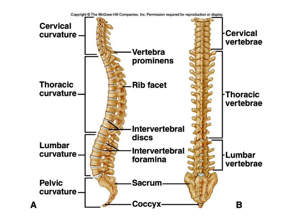
Vertebral column 7 cervial vertebrae 12 thoracic 5 lumbar 1 sacrum (5 fused 1 coccyx (4 fused) Vertebrae vary in size and morphology
 Vertebrae vary in size and morphology")
22
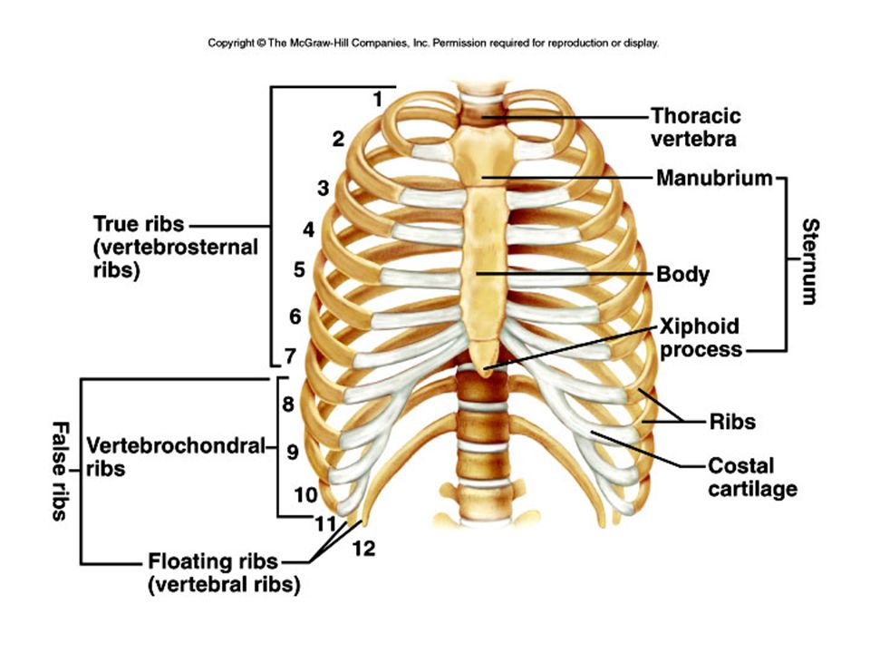
Thoracic cage ribs thoracic vertebrae sternum costal cartilages True ribs are directly attached to the sternum (first seven pairs) Three false ribs are joined to the 7 th rib Two pairs of floating ribs
 Three false ribs are joined to the 7 th rib Two pairs of floating ribs")
24
Clavicles and scapulae Help brace shoulders Attachment sites for muscles

26
Bones of upper limb Humerus (upper arm) Radius; ulna Carpals, metacarpals, phalanges Bones of lower limb Femur Patella Tibia, fibula Tarsals, metatarslas, phalanges
 Radius; ulna Carpals, metacarpals, phalanges Bones of lower limb Femur Patella Tibia, fibula Tarsals, metatarslas, phalanges")
Similar presentations


identifying the four bone types. 6) Identify bones that compose the skeletal system. 6.2) identifying.>")


 Mineral Storage of Calcium and Phosphate Red Blood Cell Production (long.>")
 fibers along with water and mineral salts (calcium hydroxide & calcium.>")


 hyoid bone (anchors tongue and muscles associated with swallowing) vertebral column (vertebrae and disks)>")


