Download presentation
Presentation is loading. Please wait.

1
By Greyson Curry and Ryan Wardin THE SKELETAL SYSTEM

2
Introduction Presentation Questions Work Sheet LESSON PLAN

3
The framework of the body made up of bones and other connective tissues, which protects and supports the body tissues and other organs. INTRODUCTION

4
Bones are made of calcium, phosphorus, sodium, and other minerals. Bones are made of a hard outer layer, that is very strong Inside is a spongy bone layer which is like a honeycomb In the middle is a jelly like bone marrow Bone marrow cells are constantly being made Bones are made of living cells, when one breaks many more a produced to rebuild it Calcium is key in keeping bones strong There are 206 bones in the adult human body, this varies for every organism. BONES

5
Two types, red and yellow The yellow marrow fills the bone cavity with fatty connective tissues. The red produces the red blood cells and platelets, white blood cells. If the body is starving it uses the fat in the yellow marrow as energy. Bone marrow transplants have been done successfully. BONE MARROW

6
Ossification is the growth of bones. This begins in the fetus. Bones are all fibrous or cartilaginous in the fetus, these are templates for bone to grow. Actual bone develops through intramembranous ossification. The osteoblasts and the osteoclasts are the two primarily responsible cells in the reconstructive process. Osteoblast form new bone tissue when the body is active(tendons flexing activates them). This thickens the bone. Osteoclasts dissolve bone tissue by secreting enzymes. OSSIFICATION AND RECONSTRUCTION
. This thickens the bone. Osteoclasts dissolve bone tissue by secreting enzymes. OSSIFICATION AND RECONSTRUCTION.")
7
Haversian Canals are a system of canals within compact bone. These canals contain blood, lymph, and nerves. The osteon is a tube like structure that contains a central Haversian canal. Surrounding the central canal is bone matrix that form osteocytes. Osteocytes are found within lacunae. Osteocytes form lamellae, lamellae is concentric layers of hard bone matrix. OSSIFICATION AND RECONSTRUCTION

8
OSSIFICATION

9
This is made up of the vertebral column( coccyx, sacrum, and spine), the hyoid bone, the Skull, the sternum, and the ribs. These components are aligned along the long axis of the body The cranial bones are in the skull, they are rounded, flat and fused together to protect the brain. There are eight cranial bones. The facial bones are also in the skull, they are the blueprint for facial muscles, they form the jaw and the eye sockets. There are fourteen facial bones in the skull. AXIAL SKELETON

10
The vertebral column has two sets of fused bones and three vertebrae. These are the vertebrae, cervical, thoracic, and lumbar. The vertebrae form a column of bone that protects the spinal cord. The twelve pairs of long flattened bones that protect the heart lungs and other internal organs are the ribs. The first seven ribs are called vertebrosternal or true ribs because they connect to the sternum. Ribs eight through ten connect indirectly to the sternum and are called false ribs. Ribs eleven and twelve are called floating ribs, because they aren’t attached to anything. AXIAL SKELETON

11
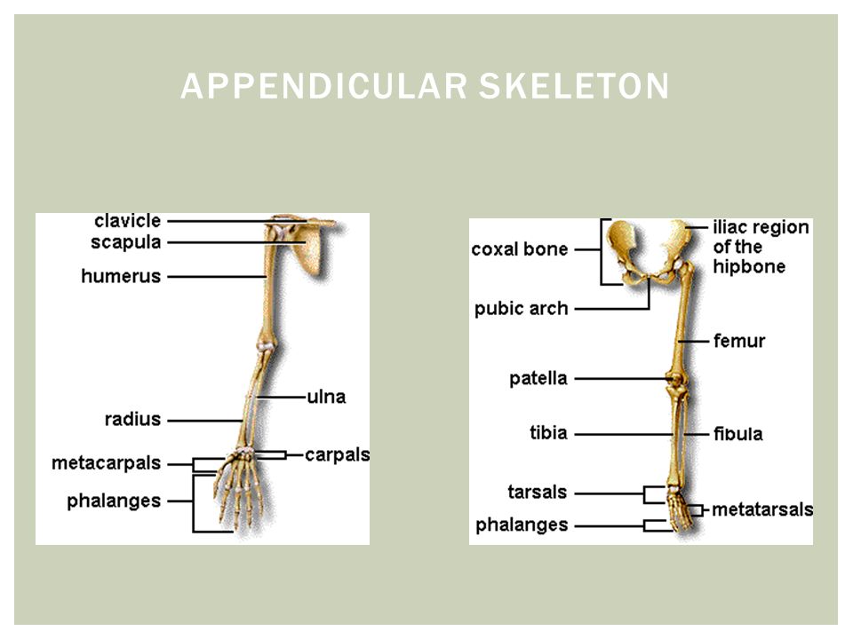
The appendicular skeleton includes the bones of the lower extremities(thigh, knee, leg, foot), the pectoral (shoulder) girdle, the bones of the upper extremities (arms, forearms, and hands), the pelvic (hip) girdle. These components are outside the main body axis. Two scapulae and two clavicles make up the pectoral girdle bones. The humorous, ulna, radius, carpals, meta carpals, ulna, and phalanges form the upper extremities bones. The carpals make the wrist, the metacarpals form the hand, and the phalanges form the fingers. The radius and ulna are the lower arm and the humorous is the upper arm. APPENDICULAR SKELETON

12
The pelvic girdle is the two hip bones. Fused bones form the coxa (hip bones). The pelvis is a bowl shaped cavity that protect abdominal organs. The pelvis is formed by the two coxae, the sacrum, and the coccyx. The femur is the heaviest bone in the body, it connects with the pelvic girdle at the hip joint. The tibia and femur meet at the knee, suspended in front of it is the patella. The fibula connect to the tibia and provides support to the ankle. In the foot are the tarsals, metatarsal, and phalanges, which are all specialized for weight bearing. These bones form a system of arches that allow for large amount of weight to be held. APPENDICULAR SKELETON

14
Joints are the place where two bones meet or connect. Ball and Socket: allow movement in any direction like twisting turning and swinging Hinge: the convex surface of one bone fits into the concave surface of a second bone. Gliding: are held in place by ligaments. The joint operates when the surfaces of two flat plates glide over one another, such as the bones in your wrists, feet and ankles Pivot: these joints allow rotation around an axis, one bone rotating around the end of another bone. You can turn your head from side to side due to the pivot joint in your neck. JOINTS

15
The skeletal system is very complex system of cells, tissues, and organs working together to provide stable framework and support for the body. CONCLUSION

16
WORKS CITED Http://hes.ucfsd.org/gclaypo/skelweb/skel01.html www.bidmc.org http://www.pennmedicine.org/health_info/body_guide/reftext/html/ske l_sys_fin.html

Similar presentations


 Mineral Storage of Calcium and Phosphate Red Blood Cell Production (long.>")
 Floating Ribs.>")


