Download presentation
Presentation is loading. Please wait.

1
Dr Hussein Farghaly, MD PSMMC Radionuclide Brain Imaging Master Watermark Image: http://williamcalvin.com/BrainForAllSeasons/img/bonoboLH-humanLH-viaTWD.gifhttp://williamcalvin.com/BrainForAllSeasons/img/bonoboLH-humanLH-viaTWD.gif 1

2
Syllabus Contents Cerebral Anatomy Cerebral Perfusion Imaging Radiopharmaceuticals Methodology Dosimetry Clinical Applications Cisternography Radiopharmaceuticals Methods Pharmacokinetics Dosimetry Clinical Applications

3
Lobes, the Cerebral Cortex, and Cortical Regions of the Brain

4
Cerebrum - The largest division of the brain. It is divided into two hemispheres, each of which is divided into four lobes. Cerebrum Cerebellum http://williamcalvin.com/BrainForAllSeasons/img/bonoboLH-humanLH-viaTWD.gif

5
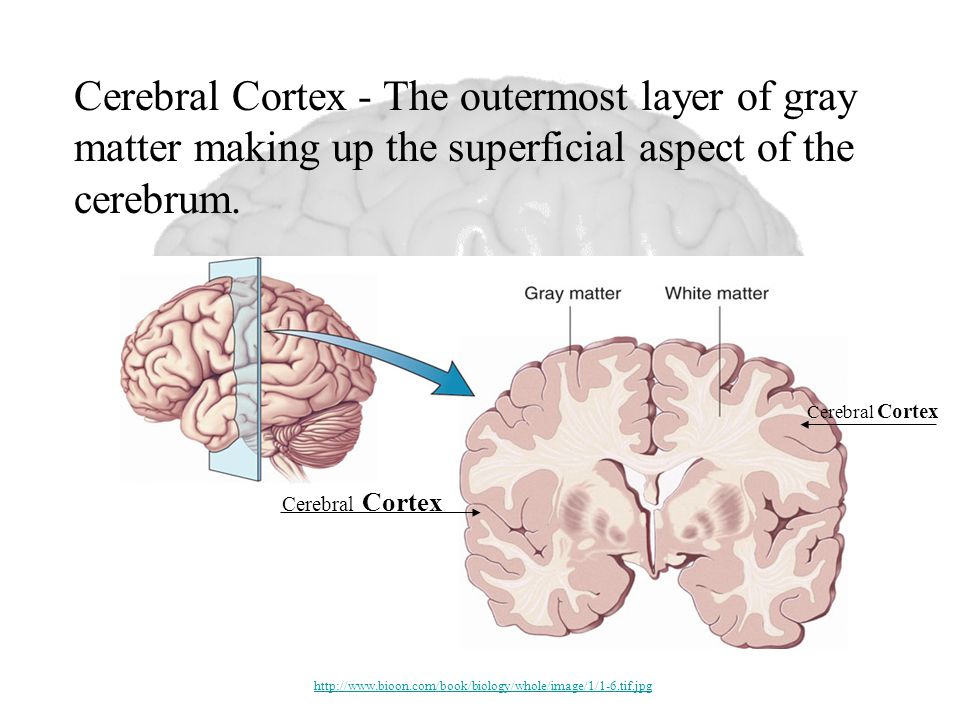
Cerebral Cortex Cerebral Cortex - The outermost layer of gray matter making up the superficial aspect of the cerebrum. http://www.bioon.com/book/biology/whole/image/1/1-6.tif.jpg
6
Cerebral Features: Sulci – Small grooves dividing the gyri – Central Sulcus – Divides the Frontal Lobe from the Parietal Lobe Fissures – Deep grooves, generally dividing large regions/lobes of the brain – Longitudinal Fissure – Divides the two Cerebral Hemispheres – Transverse Fissure – Separates the Cerebrum from the Cerebellum – Sylvian/Lateral Fissure – Divides the Temporal Lobe from the Frontal and Parietal Lobes Gyri – Elevated ridges “winding” around the brain.

7
Gyri (ridge) Fissure (deep groove) Sulci (groove) http://williamcalvin.com/BrainForAllSeasons/img/bonoboLH-humanLH-viaTWD.gif
 Fissure (deep groove) Sulci (groove)")
8
Longitudinal Fissure Transverse Fissure Sylvian/Lateral Fissure Central Sulcus http://www.bioon.com/book/biology/whole/image/1/1-8.tif.jpghttp://www.dalbsoutss.eq.edu.au/Sheepbrains_Me/human_brain.gif Specific Sulci/Fissures:

9
Lobes of the Brain (4) Frontal Parietal Occipital Temporal * Note: Occasionally, the Insula is considered the fifth lobe. It is located deep to the Temporal Lobe. http://www.bioon.com/book/biology/whole/image/1/1-8.tif.jpg

10
Lobes of the Brain - Frontal The Frontal Lobe of the brain is located deep to the Frontal Bone of the skull. (Investigation: Phineas Gage) It plays an integral role in the following functions/actions: - Memory Formation - Emotions - Decision Making/Reasoning - Personality Investigation (Phineas Gage) Modified from: http://www.bioon.com/book/biology/whole/image/1/1-8.tif.jpghttp://www.bioon.com/book/biology/whole/image/1/1-8.tif.jpg
 It plays an integral role in the following functions/actions: - Memory Formation - Emotions - Decision Making/Reasoning - Personality Investigation (Phineas Gage) Modified from:")
11
Frontal Lobe - Cortical Regions Primary Motor Cortex (Precentral Gyrus) – Cortical site involved with controlling movements of the body. Broca’s Area – Controls facial neurons, speech, and language comprehension. Located on Left Frontal Lobe. – Broca’s Aphasia – Results in the ability to comprehend speech, but the decreased motor ability (or inability) to speak and form words. Olfactory Bulb - Cranial Nerve I, Responsible for sensation of Smell
 to speak and form words. Olfactory Bulb - Cranial Nerve I, Responsible for sensation of Smell.")
12
Primary Motor Cortex/ Precentral Gyrus Broca’s Area Orbitofrontal Cortex Olfactory Bulb Modified from: http://www.bioon.com/book/biology/whole/image/1/1-8.tif.jpghttp://www.bioon.com/book/biology/whole/image/1/1-8.tif.jpg Regions

13
Lobes of the Brain - Parietal Lobe The Parietal Lobe of the brain is located deep to the Parietal Bone of the skull. It plays a major role in the following functions/actions: - Senses and integrates sensation(s) - Spatial awareness and perception (Proprioception - Awareness of body/ body parts in space and in relation to each other) Modified from: http://www.bioon.com/book/biology/whole/image/1/1-8.tif.jpghttp://www.bioon.com/book/biology/whole/image/1/1-8.tif.jpg
 - Spatial awareness and perception (Proprioception - Awareness of body/ body parts in space and in relation to each other) Modified from:")
14
Parietal Lobe - Cortical Regions Primary Somatosensory Cortex (Postcentral Gyrus) – Site involved with processing of tactile and proprioceptive information. Somatosensory Association Cortex - Assists with the integration and interpretation of sensations relative to body position and orientation in space. May assist with visuo-motor coordination. Primary Gustatory Cortex – Primary site involved with the interpretation of the sensation of Taste.

15
Primary Somatosensory Cortex/ Postcentral Gyrus Primary Gustatory Cortex Somatosensory Association Cortex Regions Modified from: http://www.bioon.com/book/biology/whole/image/1/1-8.tif.jpghttp://www.bioon.com/book/biology/whole/image/1/1-8.tif.jpg

16
Lobes of the Brain – Occipital Lobe The Occipital Lobe of the Brain is located deep to the Occipital Bone of the Skull. Its primary function is the processing, integration, interpretation, etc. of VISION and visual stimuli. Modified from: http://www.bioon.com/book/biology/whole/image/1/1-8.tif.jpghttp://www.bioon.com/book/biology/whole/image/1/1-8.tif.jpg

17
Occipital Lobe – Cortical Regions Primary Visual Cortex – This is the primary area of the brain responsible for sight - recognition of size, color, light, motion, dimensions, etc. Visual Association Area – Interprets information acquired through the primary visual cortex.

18
Primary Visual Cortex Visual Association Area Regions Modified from: http://www.bioon.com/book/biology/whole/image/1/1-8.tif.jpghttp://www.bioon.com/book/biology/whole/image/1/1-8.tif.jpg

19
Lobes of the Brain – Temporal Lobe The Temporal Lobes are located on the sides of the brain, deep to the Temporal Bones of the skull. They play an integral role in the following functions: - Hearing - Information Retrieval (Memory and Memory Formation) Modified from: http://www.bioon.com/book/biology/whole/image/1/1-8.tif.jpghttp://www.bioon.com/book/biology/whole/image/1/1-8.tif.jpg
 Modified from:")
20
Temporal Lobe – Cortical Regions Primary Auditory Cortex – Responsible for hearing Primary Olfactory Cortex – Interprets the sense of smell once it reaches the cortex via the olfactory bulbs. (Not visible on the superficial cortex) Wernicke’s Area – Language comprehension. Located on the Left Temporal Lobe.
 Wernicke’s Area – Language comprehension. Located on the Left Temporal Lobe..")
21
Primary Auditory Cortex Wernike’s Area Primary Olfactory Cortex (Deep) Conducted from Olfactory Bulb Regions Modified from: http://www.bioon.com/book/biology/whole/image/1/1-8.tif.jpghttp://www.bioon.com/book/biology/whole/image/1/1-8.tif.jpg
 Conducted from Olfactory Bulb Regions Modified from:")
22
Click the Region to see its Name Korbinian Broadmann - Learn about the man who divided the Cerebral Cortex into 52 distinct regions: http://en.wikipedia.org/wiki/Korbinian_Brodmann http://en.wikipedia.org/wiki/Korbinian_Brodmann Modified from: http://www.bioon.com/book/biology/whole/image/1/1-8.tif.jpghttp://www.bioon.com/book/biology/whole/image/1/1-8.tif.jpg

23
A: Primary Motor Cortex * This graphic representation of the regions of the Primary Motor Cortex and Primary Sensory Cortex is one example of a HOMUNCULUS: Homunculus

24
The five major senses

25
Diencephalon Thalamus- filters sensory information, controls mood states and body movement associated with emotive states Hypothalamus- ‘Central control’ for pituitary gland. Regulates autonomic, emotional, endocrine and somatic function. Has a direct involvement in stress and mood states. (http://training.seer.cancer.gov /module_anatomy/unit5_3_ne rve_org1_cns.html)
.")
26
Hindbrain Cerebellum- regulates equilibrium, muscle tone, postural control, fine movement and coordination of voluntary muscle movement. Pons- Relay station between cerebrum and cerebellum

27
Medulla oblongata- Conscious control of skeletal muscles, balance, co-ordination regulating sound impulses in the inner ear, regulation of automatic responses such as heart rate, swallowing, vomiting, coughing and sneezing

28
Basal Ganglia- Control of muscle tone, activity, posture, large muscle movements and inhibit unwanted muscle movements. Substatia Nigra- Produces dopamine is connected to the basal ganglia. – EPSE’s (Barlow and Durand, 2005)
.")
29
The Limbic system Amygdala- mediates and controls major affective mood states such as friendship, love, affection, fear, rage and aggression. Hippocampus- Memory, particularly the ability to turn short term memory into long term memory. Alzheimer's disease. (Barlow and Durand, 2005)
.")
30
Blood supply to the brain 19/09/2011, EM II.

31
Circle of Willis

32
anterior cerebral middle cerebral posterior cerebral

33
Veins drainig the brain

34
Protection and Blood Supply Meninges- Dura mater and Pia mater CSF- 2 main functions ; shock absorption and mediation of blood vessels and brain tissue in exchange of nutrients. Circle of Willis –carotid arteries and baliser arteries. Blood brain Barrier- Protect the brain from chemicals in the blood. Made up of tightly packed Endothelial cells/capillaries making it difficult to penetrate. (http://training.se er.cancer.gov/mo dule_anatomy/uni t5_3_nerve_org1 _cns.html)
.")
35
Structure of a Neuron ( This image has been released into the public domain by its author, LadyofHats. This applies worldwide.)public domainLadyofHats
public domainLadyofHats.")
36
Acetylcholine (ACh) Norepinephrine (NE) Dopamine (DA) Serotonin (5HT) Amino Acid (Glutamate, GABA) Neurotransmitters
 Norepinephrine (NE) Dopamine (DA) Serotonin (5HT) Amino Acid (Glutamate, GABA) Neurotransmitters")
37
Synaptic transmission Calcium ion channels Synapse Lock and key effect reuptake (This image has been released into the public domain by its author)public domain
public domain")
38
Brain SPECT and PET Radiopharmaceuticals

39
Resources Images: http://www.dalbsoutss.eq.edu.au/Sheepbrains_Me/human_brain.gif http://www.bioon.com/book/biology/whole/image/1/1-8.tif.jpg http://www.bioon.com/book/biology/whole/image/1/1-6.tif.jpg http://williamcalvin.com/BrainForAllSeasons/img/bonoboLH-humanLH-viaTWD.gif http://www.math.tu-dresden.de/~belov/brain/motorcor2.gif Larson, Gary. The Far Side. Phineas Gage: http://www.sruweb.com/~walsh/gage5.jpg http://soma.npa.uiuc.edu/courses/bio303/Image7.jpg http://en.wikipedia.org/wiki/Phineas_Gage http://science- education.nih.gov/nihHTML/ose/snapshots/multimedia/ritn/Gage/Broken_brain1.htmlhttp://science- education.nih.gov/nihHTML/ose/snapshots/multimedia/ritn/Gage/Broken_brain1.html

Similar presentations

 Neuropeptides 1) ACh 2) Amino Acids 3) Biogenic Amines.>")
 CNS = Brain + spinal cord Surface anatomy includes.>")


 Pia mater -inner membrane, contains.>")

 Lobes of the brain (Forebrain) Midbrain/Hindbrain Midbrain/Hindbrain Protection.>")
 CNS –brain –spinal cord.>")

 Pia mater -inner membrane, contains.>")


