Download presentation
Presentation is loading. Please wait.

1
The Human Brain Master Watermark Image: http://williamcalvin.com/BrainForAllSeasons/img/bonoboLH-humanLH-viaTWD.gifhttp://williamcalvin.com/BrainForAllSeasons/img/bonoboLH-humanLH-viaTWD.gif

2
Lobes, the Cerebral Cortex, and Cortical Regions of the Brain

3
Cerebrum - The largest division of the brain. It is divided into two hemispheres, each of which is divided into four lobes. Cerebrum Cerebellum http://williamcalvin.com/BrainForAllSeasons/img/bonoboLH-humanLH-viaTWD.gif

4
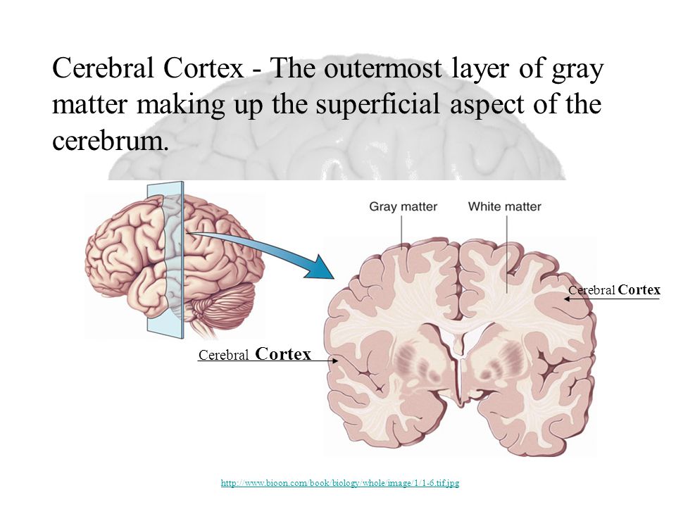
Cerebral Cortex Cerebral Cortex - The outermost layer of gray matter making up the superficial aspect of the cerebrum. http://www.bioon.com/book/biology/whole/image/1/1-6.tif.jpg
5
Architectural Features of the Cortex: Sulci – Small grooves dividing the gyri Fissures – Deep grooves, generally dividing large regions/lobes of the brain Gyri – Elevated ridges “winding” around the brain.

6
Gyri (ridge) Fissure (deep groove) Sulci (groove) http://williamcalvin.com/BrainForAllSeasons/img/bonoboLH-humanLH-viaTWD.gif
 Fissure (deep groove) Sulci (groove)")
7
Longitudinal Fissure Transverse Fissure Sylvian/Lateral Fissure Central Sulcus http://www.bioon.com/book/biology/whole/image/1/1-8.tif.jpghttp://www.dalbsoutss.eq.edu.au/Sheepbrains_Me/human_brain.gif Specific Sulci/Fissures:

8
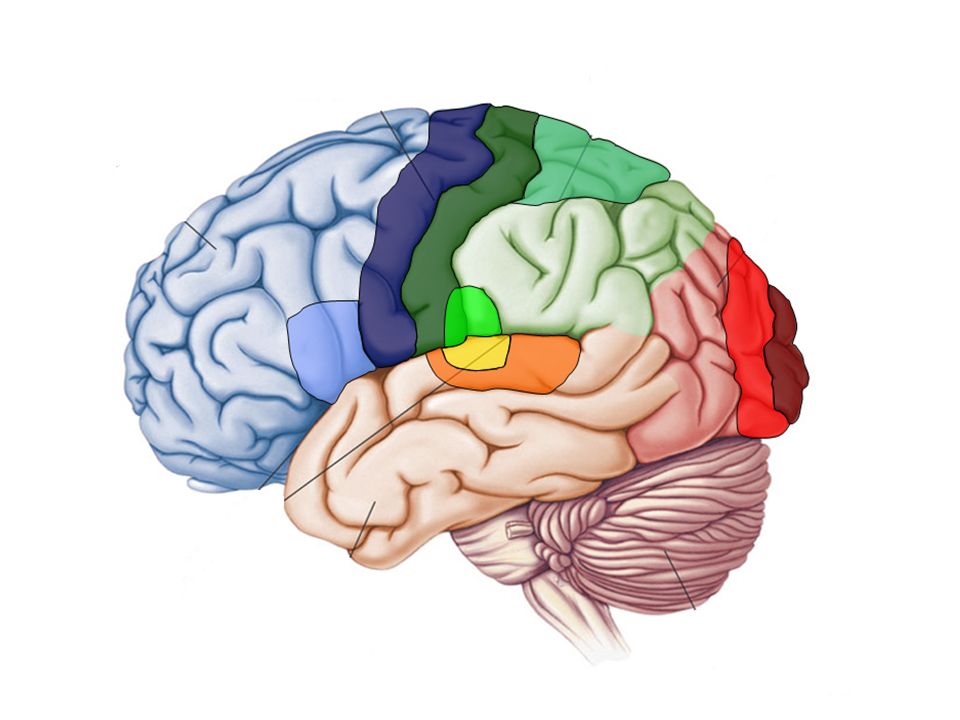
Lobes of the Brain (4) Frontal Parietal Occipital Temporal http://www.bioon.com/book/biology/whole/image/1/1-8.tif.jpg
 Frontal Parietal Occipital Temporal")
9
Lobes of the Brain - Frontal The Frontal Lobe of the brain is located deep to the Frontal Bone of the skull. It plays an integral role in the following functions/actions: - Memory Formation - Emotions - Decision Making/Reasoning - Personality Modified from: http://www.bioon.com/book/biology/whole/image/1/1-8.tif.jpghttp://www.bioon.com/book/biology/whole/image/1/1-8.tif.jpg

10
Frontal Lobe - Cortical Regions Orbitofrontal Cortex – Site of Frontal Lobotomies Primary Motor Cortex (Precentral Gyrus) – Cortical site involved with controlling movements of the body. Broca’s Area – Controls facial neurons, speech, and language comprehension. Located on Left Frontal Lobe. – Broca’s Aphasia – Results in the ability to comprehend speech, but the decreased motor ability (or inability) to speak and form words. Olfactory Bulb - Cranial Nerve I, Responsible for sensation of Smell * Desired Effects: - Diminished Rage - Decreased Aggression - Poor Emotional Responses * Possible Side Effects: - Epilepsy - Poor Emotional Responses - Perseveration (Uncontrolled, repetitive actions, gestures, or words)
 to speak and form words. Olfactory Bulb - Cranial Nerve I, Responsible for sensation of Smell * Desired Effects: - Diminished Rage - Decreased Aggression - Poor Emotional Responses * Possible Side Effects: - Epilepsy - Poor Emotional Responses - Perseveration (Uncontrolled, repetitive actions, gestures, or words).")
11
Primary Motor Cortex/ Precentral Gyrus Broca’s Area Orbitofrontal Cortex Olfactory Bulb Modified from: http://www.bioon.com/book/biology/whole/image/1/1-8.tif.jpghttp://www.bioon.com/book/biology/whole/image/1/1-8.tif.jpg Regions

12
Lobes of the Brain - Parietal Lobe The Parietal Lobe of the brain is located deep to the Parietal Bone of the skull. It plays a major role in the following functions/actions: - Senses and integrates sensation(s) - Spatial awareness and perception (Proprioception - Awareness of body/ body parts in space and in relation to each other) Modified from: http://www.bioon.com/book/biology/whole/image/1/1-8.tif.jpghttp://www.bioon.com/book/biology/whole/image/1/1-8.tif.jpg
 - Spatial awareness and perception (Proprioception - Awareness of body/ body parts in space and in relation to each other) Modified from:")
13
Parietal Lobe - Cortical Regions Primary Somatosensory Cortex (Postcentral Gyrus) – Site involved with processing of tactile and proprioceptive information. Somatosensory Association Cortex - Assists with the integration and interpretation of sensations relative to body position and orientation in space. Primary Gustatory Cortex – Primary site involved with the interpretation of the sensation of Taste.

14
Primary Somatosensory Cortex/ Postcentral Gyrus Primary Gustatory Cortex Somatosensory Association Cortex Regions Modified from: http://www.bioon.com/book/biology/whole/image/1/1-8.tif.jpghttp://www.bioon.com/book/biology/whole/image/1/1-8.tif.jpg

15
Lobes of the Brain – Occipital Lobe The Occipital Lobe of the Brain is located deep to the Occipital Bone of the Skull. Its primary function is the processing, integration, and interpretation of VISION and visual stimuli. Modified from: http://www.bioon.com/book/biology/whole/image/1/1-8.tif.jpghttp://www.bioon.com/book/biology/whole/image/1/1-8.tif.jpg

16
Occipital Lobe – Cortical Regions Primary Visual Cortex – This is the primary area of the brain responsible for sight - recognition of size, color, light, motion, dimensions, etc. Visual Association Area – Interprets information acquired through the primary visual cortex.

17
Primary Visual Cortex Visual Association Area Regions Modified from: http://www.bioon.com/book/biology/whole/image/1/1-8.tif.jpghttp://www.bioon.com/book/biology/whole/image/1/1-8.tif.jpg

18
Lobes of the Brain – Temporal Lobe The Temporal Lobes are located on the sides of the brain, deep to the Temporal Bones of the skull. They play an integral role in the following functions: - Hearing - Organization/Comprehension of language - Information retrieval (Memory and Memory Formation) Modified from: http://www.bioon.com/book/biology/whole/image/1/1-8.tif.jpghttp://www.bioon.com/book/biology/whole/image/1/1-8.tif.jpg
 Modified from:")
19
Temporal Lobe – Cortical Regions Primary Auditory Cortex – Responsible for hearing Wernicke’s Area – Language comprehension. Located on the Left Temporal Lobe. - Wernicke’s Aphasia – Language comprehension is inhibited. Words and sentences are not clearly understood. Sentence formation may be inhibited or non-sensical.

20
Primary Auditory Cortex Wernike’s Area Modified from: http://www.bioon.com/book/biology/whole/image/1/1-8.tif.jpghttp://www.bioon.com/book/biology/whole/image/1/1-8.tif.jpg

21
Arcuate Fasciculus - A white matter tract that connects Broca’s Area and Wernicke’s Area through the Temporal, Parietal and Frontal Lobes. Allows for coordinated, comprehensible speech. Damage may result in: - Conduction Aphasia - Where auditory comprehension and speech articulation are preserved, but people find it difficult to repeat heard speech. Modified from: http://www.bioon.com/book/biology/whole/image/1/1-8.tif.jpghttp://www.bioon.com/book/biology/whole/image/1/1-8.tif.jpg

23
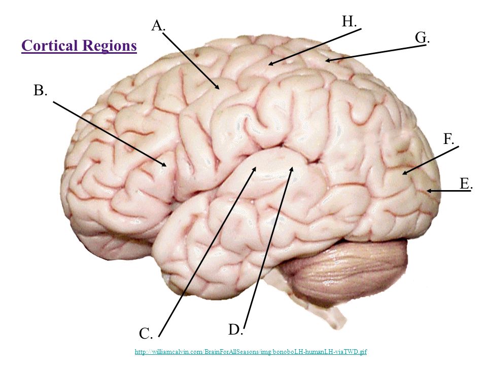
Lobes and Structures of the Brain B. A. C. D. E. F. G. http://williamcalvin.com/BrainForAllSeasons/img/bonoboLH-humanLH-viaTWD.gif

24
Lobes and Structures of the Brain B. A. (groove) C. (groove) D. E. F. G. B. Frontal Lobe G. Parietal Lobe F. Occipital Lobe D. Temporal Lobe A. Central Sulcus (groove) E. Transverse Fissure C. Sylvian/Lateral Fissure http://williamcalvin.com/BrainForAllSeasons/img/bonoboLH-humanLH-viaTWD.gif
 D. E. F. G. B. Frontal Lobe G. Parietal Lobe F. Occipital Lobe D. Temporal Lobe A. Central Sulcus (groove) E. Transverse Fissure C. Sylvian/Lateral Fissure")
25
Cortical Regions A. E. B. C. D. F. G. H. http://williamcalvin.com/BrainForAllSeasons/img/bonoboLH-humanLH-viaTWD.gif
26
A. Primary Motor Cortex/ Precentral Gyrus B. Broca’s Area H. Primary Somatosensory Cortex/ Postcentral Gyrus G. Somatosensory Association Cortex E. Primary Visual Cortex F. Visual Association Area C. Primary Auditory Cortex D. Wernike’s Area

27
Q: Assuming this comical situation was factually accurate, what Cortical Region of the brain would these doctors be stimulating? Copyright: Gary Larson

28
Homunculus: Maps of the Primary Motor Cortex and Primary Sensory Cortex Motor Map

29
Q: What do you notice about the proportions depicted in the aforementioned homunculus? Q: What is meant by depicting these body parts in such outrageous proportions? A: They are not depicted in the same scale representative of the human body. A: These outrageous proportions depict the cortical area devoted to each structure. - Ex: Your hands require many intricate movements and sensations to function properly. This requires a great deal of cortical surface area to control these detailed actions. Your back is quite the opposite, requiring limited cortical area to carry out its actions and functions, or detect sensation.

Similar presentations


 CNS = Brain + spinal cord Surface anatomy includes.>")


 CNS –brain –spinal cord.>")


