Download presentation
Presentation is loading. Please wait.

1
NSCI4700 Veterinary Anatomy and Physiology Andrea Dickie B.V.Sc.
Respiratory system NSCI4700 Veterinary Anatomy and Physiology Andrea Dickie B.V.Sc.

2
Learning outcomes By the end of this session you will be able to
Describe the components of the upper and lower respiratory tract State the function of each component Explain how air is inhaled and exhaled Explain how oxygen passes into the bloodstream from the lungs Describe the differences between respiration in mammals, birds & reptiles

3
Word Jumble A word jumble introduces some of the new terminology we will learn in this lecture. A number of words will appear on the next slide. You will have 60 seconds to try to remember as many word as possible without the aid of pen and paper. You will then have 60 seconds to write down as many as you can remember. Spelling doesn’t matter, just try to get the words down.

4
Word Jumble – 24 words terminal bronchioles lungs larynx alveolar sac respiration cellular metabolism paranasal sinuses diaphragm parietal pleura inspiration nasal turbinates epiglottis arytenoid cartilages alveoli duct hard palate nasopharynx cricoid cartilage expiration medisastinum nares primary bronchi thorax syrinx respiratory membrane

5
Cellular metabolism Cells requires oxygen
Cells produces carbon dioxide as a waste product Respiratory system’s prime responsibility is to bring in oxygen and to remove carbon dioxide

6
Primary Function of the Respiratory Tract
Respiratory system’s prime responsibility is to provide oxygen to and remove carbon dioxide from the cells of the body. This occurs in the alveoli.

7
Three types of Respiration
External respiration – action of bringing air (containing oxygen) into and out of the lungs and into the blood Internal respiration – the molecular exchange of oxygen and carbon dioxide between the blood and the body tissues Cellular respiration Aerobic respiration is the use of oxygen and glucose by the cells to produce energy.
 into and out of the lungs and into the blood. Internal respiration – the molecular exchange of oxygen and carbon dioxide between the blood and the body tissues. Cellular respiration. Aerobic respiration is the use of oxygen and glucose by the cells to produce energy.")
8
Secondary Functions Voice production via vocal chords within the larynx Controlling acid-base balance (pH level) – CO2 is carried around the body chemically bound to water, forming carbonic acid Temperature control – dogs pant to lose heat Smell – olfactory sense
 – CO2 is carried around the body chemically bound to water, forming carbonic acid. Temperature control – dogs pant to lose heat. Smell – olfactory sense.")
9
What Makes Up the Respiratory Tract
Upper Respiratory Tract - nose and nasal passages, para-nasal sinuses, and the pharynx Lower Respiratory Tract - larynx, trachea, bronchi and bronchioles, lungs

10
Which is the upper and lower respiratory system?

11
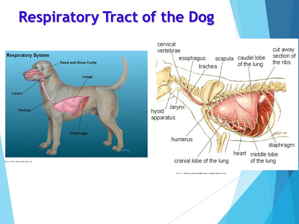
Respiratory Tract of the Dog
12
Upper Respiratory Tract
Pharynx Upper Respiratory Nostrils (nares) Nasal Passages Para-nasal sinuses Pharynx
 Nasal Passages. Para-nasal sinuses. Pharynx.")
13
Upper Respiratory Tract of the Dog

14
Lower Respiratory Tract
What can you identify on this diagram Lower Respiratory Larynx Trachea Lungs Bronchii Bronchioles Alveolar ducts Alveoli

15
Lower Respiratory Tract – Bronchi and Bronchioles
Bronchi – first division of the trachea Bronchioles – further division of the bronchi

16
Video Overview of Upper Respiratory System (Nose to Bronchi) 3 D Poor audio but good images – 3 min 30 sec Overview of the lower respiratory system (graphics not 100% clear play 15 sec to 1.58 min) List three things you have learned List at least one question you have from this video Further videos you may find useful – 3 min. – 7 minutes
 3 D Poor audio but good images – 3 min 30 sec v=OIU7Mdx4DTg Overview of the lower respiratory system v=o2OcGgJbiUk (graphics not 100% clear play 15 sec to 1.58 min) List three things you have learned List at least one question you have from this video Further videos you may find useful v=hc1YtXc_84A – 3 min. v=TQ24-WCsYN4 – 7 minutes")
17
UPPER RESPIRATORY SYSTEM ANATOMY

18
External Nares (Nose) http://gooddogsrule.com/?p=1099

19
Nasal Cavity Has receptors for smell Lined with mucous membrane that:
Moistens air Traps incoming foreign particles Has a ciliated epithelium that produces mucus

20
http://www. merckmanuals

21
Nasal Cavity Nasal Turbinate's Spiral shaped nasal turbinate's or conchae are found within the nasal cavity Theses are spiral bones covered by mucous membrane. Make air flow turbulent & gives it time to warm up. Nasal Turbinate's Wolf and a Dog nasal turbinate's

22
Sinuses Cavities within bones surrounding the nasal cavity
Most animals have four sinuses within the skull Bones surrounding the nasal cavity Lighten the skull Produce mucus that drains into the nasal cavity

24
Hard and Soft palates Hard palate – thin plate of bone, in the roof of the mouth that is part of the skull and is between the upper teeth. Soft Palate – The soft muscular tissue forming the back of the roof of the mouth. Does not contain bone. Feel inside your mouth to find the junction between the hard and soft palates.

25
Where do you think each section is on the diagram?
Pharynx Where do you think each section is on the diagram? The section of the digestive tract that extends from the mouth and nasal cavities to the larynx. Three regions of the pharynx Nasopharynx -behind nasal cavity. Oropharynx - middle region behind mouth. Laryngopharynx- caudal region attached to larynx nasopharynx laryngopharynx oropharynx

26
Pharynx What do you notice about the pathway of air and the pathway of food? Air moves into the larynx Food moves into the oesophagus. What structure prevents food entering the trachea? The epiglottis.

27
LOWER RESPIRATORY SYSTEM ANATOMY

28
What were the structures of the lower respiratory system?
Larynx Trachea Bronchi Bronchioles Lungs Alveoli On a piece of paper list three structures that are in the lower respiratory system

30
Can you identify the trachea, bronchi and the lungs Trachea
Why do the bronchi and bronchioles appear more white? Bronchi and Bronchioles Lungs Due to the cartilage in their walls Heart

31
Structures of the Larynx of the Dog
Formed from three cartilages Arytenoid, Cricoid Thyroid Epiglottis A flap that routes food to the oesophagus and air toward the trachea.

32
Structures of the Larynx
Vocal cords (vocal folds) Vibrate with expelled air to create sound In animals the larynx is suspended by bones called the hyoid apparatus (made up of 5 bones). In humans it is a single hyoid bone.
 Vibrate with expelled air to create sound. In animals the larynx is suspended by bones called the hyoid apparatus (made up of 5 bones). In humans it is a single hyoid bone.")
33
Larynx of the Dog epiglottis Arytenoid cartilages

34
The Larynx Functions Voice production
Prevention of foreign material being inhaled Control of airflow into and out of the lungs

35
The Larynx and Swallowing
Normally the larynx remains open to allow breathing. Swallowing is a complex action where the larynx is pushed up against the epiglottis, breathing stops and food is swallowed. Swallowing 2 minutes 29 sec

36
Trachea Takes air from the larynx to the lungs
Lined with ciliated epithelium that secretes mucous Cilia beat continuously in the opposite direction of incoming air Carries mucus loaded with dust and other debris away from lungs Walls are reinforced with C-shaped hyaline cartilage – keeps the trachea open. Open on the dorsal surface.

38
Lungs Occupies most of the thoracic cavity
Each lung is divided into lobes with species differences. Left lung 2 lobes – dog, pig, human, cow, rabbit 3 lobes – sheep, cat Right lung 3 lobes – human, cow 4 lobes – dog, pig, sheep, cat, rabbit Lungs of the dog

39
Why are they a different colour to the bone?
Where are the lungs on this x-ray? Why are they a different colour to the bone?

40
Pleura Pleura are the membranes covering the lung surface. two layers
Outer (Parietal) pleura lines the walls of the thoracic cavity - attaches to the ribs Inner Visceral pleura is attached to the surface of the lungs. Pleural fluid in the narrow space between layers of pleura to allow gliding
 pleura lines the walls of the thoracic cavity - attaches to the ribs. Inner Visceral pleura is attached to the surface of the lungs. Pleural fluid in the narrow space between layers of pleura to allow gliding.")
41
Lungs Figure 13.4b Slide 13.12b Copyright © 2003 Pearson Education, Inc. publishing as Benjamin Cummings

42
Bronchii, Bronchioles Formed by division of the trachea into primary bronchi, which enter the lungs Within the lungs, these divide into hundreds of bronchioles. Formed from cartilage (connective tissue) and smooth muscle (muscle tissue) Bronchioles can dilate or constrict depending on need for oxygen
 and smooth muscle (muscle tissue) Bronchioles can dilate or constrict depending on need for oxygen.")
43
Respiratory Tree Divisions
Trachea Primary bronchi Secondary bronchi Tertiary bronchi Bronchioles Terminal bronchioles

45
Bronchioles Smallest branches of the bronchi
All but the smallest branches have reinforcing cartilage Terminal bronchioles end in alveoli Figure 13.5a Copyright © 2003 Pearson Education, Inc. publishing as Benjamin Cummings

46
CROSS SECTIONAL VIEW OF THE THORACIC CAVITY
Mediastinum Area between the two lungs, where the pleural membranes meet What organ would we find in this space? CROSS SECTIONAL VIEW OF THE THORACIC CAVITY

47
Hilus The only point in the chest where each lung is attached
The lobes of the lung are mobile and slide around slightly within the thoracic cavity during inspiration and expiration Bronchus/blood vessels and lymph vessels enter at the hilus

48
END OF LECTURE ONE

49
Revision What structures are in the upper and lower respiratory systems? (list on board) What are the branches of the trachea?
 What are the branches of the trachea.")
50
Which is the upper and lower respiratory system?

51
The branching of the trachea

52
So where does respiration take place?
In the Respiratory Zone Structures Respiratory bronchioli Alveolar duct Alveoli

53
Alveoli An alveolus is a tiny air sac formed at the tip of the lungs' smallest airways, the bronchioles – millions in each lung. They are surrounded by capillaries. Fluid inside the alveoli, called surfactant, prevents them from collapsing and sticking together Gas exchange takes place within the alveoli Air inspired is: Rich in oxygen Low in carbon dioxide

55
Alveoli Structure of alveoli Alveolar duct Alveolar sacs
Gas exchange takes place within the alveoli in the respiratory membrane

56
Respiratory Membrane (Air-Blood Barrier)
Thin squamous epithelial layer lining alveolar walls Pulmonary capillaries cover external surfaces of alveoli and allow gas exchange

57
Respiratory Membrane (Air-Blood Barrier)
Figure 13.6 Slide 13.18b Copyright © 2003 Pearson Education, Inc. publishing as Benjamin Cummings

58
Gas Exchange Gas crosses the respiratory membrane by diffusion
Oxygen enters the blood Carbon dioxide enters the alveoli Macrophages add protection Surfactant coats gas-exposed alveolar surfaces. ur6XUiq4 – alveolar gas exchange 1 min 50 sec

59
Oxygen Movement in the Blood
The alveoli always have more oxygen than the blood Oxygen moves by diffusion towards the area of lower concentration from the alveoli to the blood Capillary blood gains oxygen

60
Gas Transport in the Blood
Oxygen transport in the blood Inside red blood cells attached to haemoglobin A small amount is carried dissolved in the plasma Carbon dioxide transport in the blood Most is transported in the plasma in the blood A small amount is carried inside red blood cells but at different sites to those of oxygen Alveoli and gas exchange

61
Alveolar gas exchange Air inspired is: 78% nitrogen 21% oxygen
0.1% argon 0.04% carbon dioxide Expired air for humans is: similar for other terrestrial mammals 16% oxygen 4.5% carbon dioxide

62
Exchange between the blood and the cells
Exchange of gases between blood and body cells An opposite reaction to what occurs in the lungs Carbon dioxide diffuses out of tissue to blood Oxygen diffuses from blood into tissue

63
Breathing, Gas Transport, and Exchange with cells Summary
Figure 13.10 Slide 13.35 Copyright © 2003 Pearson Education, Inc. publishing as Benjamin Cummings

64
Mechanics of Breathing
Two phases Inspiration – flow of air into lung Expiration – air leaving lung Breathing Video

65
Inspiration Diaphragm and intercostal muscles contract
The size of the thoracic cavity increases External air is sucked into the lungs

66
Breathing In (Inhalation, Inspiration)
Parietal pleura are attached to the rib cage and visceral pleura to the lung tissue Partial vacuum in pleural cavity around the lungs sucks these 2 layers together. Breathing In – expansion of the lungs. The intercostal muscles contract and pull the ribs up and out The parietal pleura moves with them & because the visceral pleura is ‘sucked’ onto it by the partial vacuum this also pulls the lungs up and out When the diaphragm contracts and flattens downward this also pulls the lung tissue down Pleural fluid provides lubrication and also helps hold the pleura together. Flexibility of the lungs allows them to be pulled wider & mould to the shape of the increased thoracic cavity

67
Inspiration Caudally Slide 13.22b Figure 13.7a
Copyright © 2003 Pearson Education, Inc. publishing as Benjamin Cummings

68
Exhalation Largely a passive process which depends on natural lung elasticity As muscles relax, air is pushed out of the lungs Forced expiration can occur mostly by contracting internal intercostal muscles to depress the rib cage

69
Exhalation Slide 13.23b caudally Figure 13.7b
Copyright © 2003 Pearson Education, Inc. publishing as Benjamin Cummings

70
The space inside the pleural cavity is tiny and consists of a negative pressure – this “sucks” the lungs against the thoracic lining of the chest.

71
Non Respiratory Air Movements
Examples Cough and sneeze – clears lungs of debris Yawn Hiccup

72
Resting Respiration Rates (for Reference Only)
Species Breaths per Minute Cat 16-40 Cow 26-50 Dog 18-34 Horse 10-14 Pig 32-58 Sheep 16-34

73
Respiratory Capacities
Figure 13.9 Slide 13.30 Copyright © 2003 Pearson Education, Inc. publishing as Benjamin Cummings

74
Control of Respiration
Breathing control centres in the medulla and pons in the brain Nervous system Chemical receptors and stretch receptors in the body tissues and aorta. Sense changes in oxygen and carbon dioxide Increased respiratory rate is often due to extra oxygen needs

75
Control of breathing - 2 Medulla Oblongata – In the brain stem
Controls rhythm Chemical receptors and Stretch receptors – In the body tissue Control depth and rate YdchQtdAU – 3 min 30 seconds – mor detail than needed.

76
Control of breathing Take a few short breathes – then pause for a few seconds. Then take another few breathes and pause for a few seconds. Now hold your breath What happens? Breathing is an involuntary process, controlled automatically by the brain Automatic breathing can be overridden, meaning you can choose when to breath, but involuntary control will always take over when necessary

77
Differences in birds and mammals
Larynx does not produce sound in birds Enlargement of trachea near bifurcation The syrinx – voice box – very complex in some birds – voice box Mesobronchii, ventrobronchii and parabronchii instead of bronchioles Air capillaries instead of alveoli Lungs fixed – do not inflate or deflate Syrinx of the Bird

78
Avian Respiration Oxygen is extracted on inspiration and expiration Air is not mixed as in mammals – air is always fresh and moves one direction The sternum (breastbone) moves significantly downwards, as well as ribs rotating outwards to draw air into the sacs. Vital that birds are gently restrained. Holding a bird "too tight" can easily cause the bird to suffocate. No diaphragm, air is moved in and out of the respiratory system through pressure changes in the air sacs. – 3 min 10 sec – good. Use this.
 moves significantly downwards, as well as ribs rotating outwards to draw air into the sacs. Vital that birds are gently restrained. Holding a bird too tight can easily cause the bird to suffocate. No diaphragm, air is moved in and out of the respiratory system through pressure changes in the air sacs. v=kWMmyVu1ueY – 3 min 10 sec – good. Use this.")
79
Air Sacs Air sacs thin walled and minor involvement in respiration. Most birds have 9 of these and they extend into the cavities of the bones. This is why birds bones are so light.

80
Birds

81
Avian respiration – Respiration in birds requires two respiratory cycles (inspiration, expiration, inspiration, expiration) to move the air through the entire respiratory system. In mammals, only one respiratory cycle is necessary. In the avian lung, the gas exchange occurs in the walls of microscopic tubules, called 'air capillaries.' First inspiration – some air moves through lungs and some air passes into posterior airsacs - warmed and humidified First expiration – passed to lungs where gas exchange occurs Second inspiration – moves out of lungs into anterior air sacs Second expiration – air passes back out up trachea
 to move the air through the entire respiratory system. In mammals, only one respiratory cycle is necessary. In the avian lung, the gas exchange occurs in the walls of microscopic tubules, called air capillaries. First inspiration – some air moves through lungs and some air passes into posterior airsacs - warmed and humidified. First expiration – passed to lungs where gas exchange occurs. Second inspiration – moves out of lungs into anterior air sacs. Second expiration – air passes back out up trachea. c= &aid=2721.")
82
Air Capillaries

83
Avian Respiratory System

84
Fish Muscles raise the mouth floor
Water is pushed toward the gills where gaseous exchange takes place Operculum opens as water is pushed over gills 1 min

85
Reptiles Tend to have very delicate lungs, so care needed in handling
No diaphragm, so the ribs and abdominal muscles responsible for all breathing movements – hence care with handling

86
Terminology of the Respiratory System
Alveoli - The sacs in the lungs where oxygen is transferred into the blood and carbon dioxide is transferred out of the blood. Bronchi - The largest branches from the trachea into the lungs Bronchioli - The small branches coming off of the bronchi Epiglottis – The flap on top of the larynx; keeps food out of the larynx and trachea while the animal is swallowing Larynx - Voice box; located at the top of the windpipe (trachea)
")
87
Terminology of the Respiratory System
Nasal - Pertaining to the nose Nasopharynx - The upper part of the pharynx, at the back of the nasal passages Oral - Pertaining to the mouth Oropharynx - The middle part of the pharynx, at the far back of the mouth Paranasal sinuses - Often just called “sinuses”; the tissue- lined cavities in the skull just behind and above the eyes and along the sides of the nose

88
Terminology of the Respiratory System
Pharynx - The passage between the back of the nose and the throat; it is separated (top to bottom) into the nasopharynx, oropharynx, and hypopharynx (throat) Pleura - The sac that encloses the lung Pulmonary - Pertaining to the lung Rhino - Pertaining to the nose Trachea – Windpipe, airway
 into the nasopharynx, oropharynx, and hypopharynx (throat) Pleura - The sac that encloses the lung. Pulmonary - Pertaining to the lung. Rhino - Pertaining to the nose. Trachea – Windpipe, airway.")
Similar presentations


. 2.Production of sound (vocal cords). 3.Pulmonary ventilation. 4. Inspiration (intercostals muscles lift.>")


