Download presentation
Presentation is loading. Please wait.

1
Respiratory System

2
objectives You will find out about: The structure and functions of the respiratory system How we breathe Gas exchange The effect of exercise on breathing rate

3
Function of the Respiratory System Slide 13.2 Copyright © 2003 Pearson Education, Inc. publishing as Benjamin Cummings Passageways to the lungs purify, warm, and humidify the incoming air To produce oxygenated blood To remove carbon dioxide

4
Organs of the Respiratory system Slide 13.1 Copyright © 2003 Pearson Education, Inc. publishing as Benjamin Cummings Nose Pharynx Larynx Trachea Bronchi Lungs – alveoli Figure 13.1

5
Slide 13.3b Copyright © 2003 Pearson Education, Inc. publishing as Benjamin Cummings Figure 13.2 Upper Respiratory Tract

6
The Nasal Cavity Slide 13.4a Copyright © 2003 Pearson Education, Inc. publishing as Benjamin Cummings Hairs and mucous trap particles within the air to prevent it entering the lungs and causing damage or infection. The air is warmed to within 1’C of body temperature as well as humidifying it to prevent drying and irritation. Site of olfactory epithelium giving sense of smell.

7
The throat has a dual function, one for the respiratory system and one for the digestive system. The epiglottis separates the two systems at the top of the trachea and the oesophagus.

8
The Larynx Slide 13.9a Copyright © 2003 Pearson Education, Inc. publishing as Benjamin Cummings This is made of cartilage. Its function is to generate sound which can then be turned into speech by the mouth, tongue and oral cavity. Vocal cords vibrate with expelled air. The entrance is protected by the epiglottis to prevent food going down the wrong way.

9
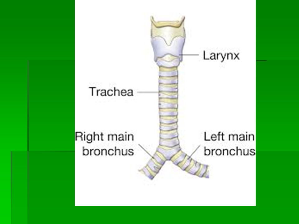
Trachea It is a tube like structure which has incomplete rings of cartilage around it to provide protection against crushing but also to allow food to pass through the oesophagus It is approx 25mm in diameter and 10- 16cm in length. It opens out into the bronchi at the top of the lungs

10
Trachea (Windpipe) Slide 13.10 Copyright © 2003 Pearson Education, Inc. publishing as Benjamin Cummings Connects larynx with bronchi Lined with ciliated mucosa Cilia beat continuously in the opposite direction of incoming air They expel mucus loaded with dust and other debris away from lungs Walls are reinforced with C-shaped hyaline cartilage

12
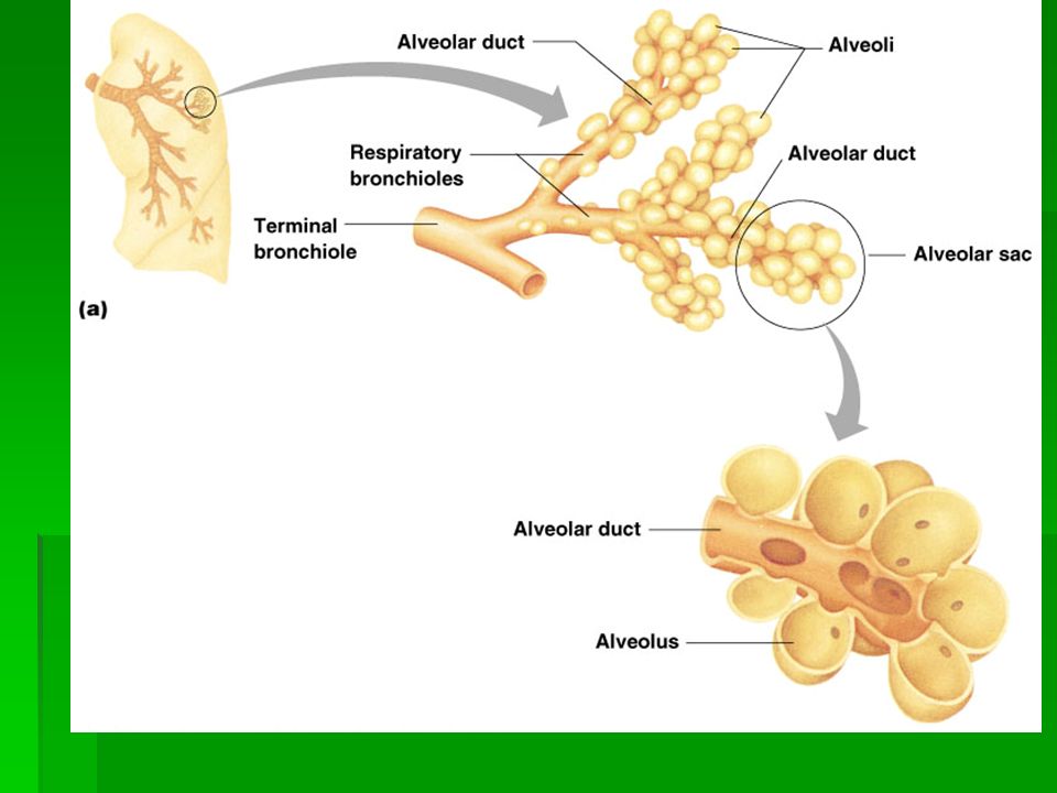
Primary Bronchi Slide 13.11 Copyright © 2003 Pearson Education, Inc. publishing as Benjamin Cummings Formed by division of the trachea These are large tubes that allow air into the lungs Bronchi subdivide into smaller and smaller branches called bronchioles. These end as tiny air sacs called alveoli.

14
Alveoli Lastly, the alveoli are air filled sacs where gas exchange takes place. There are approx. 700 million in a pair of human lungs

15
Lungs Slide 13.12a Copyright © 2003 Pearson Education, Inc. publishing as Benjamin Cummings Occupy most of the thoracic cavity Apex is near the clavicle (superior portion) Base rests on the diaphragm (inferior portion) Each lung is divided into lobes by fissures Left lung – two lobes Right lung – three lobes
 Base rests on the diaphragm (inferior portion) Each lung is divided into lobes by fissures Left lung – two lobes Right lung – three lobes.")
16
Coverings of the Lungs Slide 13.13 Copyright © 2003 Pearson Education, Inc. publishing as Benjamin Cummings Pleural membranes cover the lung surfaces Pleura lines the walls of the thoracic cavity Pleural fluid fills the area between layers of pleura to allow gliding The thorax is an air-tight cavity

17
https://www.youtube.com/watch?v=B1w3s9m3hIg

18
Breathing Breathing movements draw air in and out of the lungs. Due to pressure changes in the lungs brought about by movement of ribs and diaphragm

19
The diaphragm is a sheet of skeletal muscle that encloses the bottom of the rib cage. It is involved in the process of breathing

20
Inspiration Slide 13.22b Copyright © 2003 Pearson Education, Inc. publishing as Benjamin Cummings Figure 13.7a

21
Inspiration Slide 13.22a Copyright © 2003 Pearson Education, Inc. publishing as Benjamin Cummings Diaphragm contracts, flattening diaphragm external intercostal muscles contract moving ribs up and out. Volume of thorax increases External air is pulled into the lungs

22
Exhalation Slide 13.23b Copyright © 2003 Pearson Education, Inc. publishing as Benjamin Cummings Figure 13.7b

23
Exhalation Slide 13.23a Copyright © 2003 Pearson Education, Inc. publishing as Benjamin Cummings Largely a passive process which depends on natural lung elasticity Volume of thorax decreases Pressure increases Air moves out.

24
The diaphragm relaxes and returns to it original position. Elastic recoil of the lung and thoracic cavity tissue cause the volume to decrease, expelling air due to the pressure differential. This process is passive If the internal intercostal and abdominal muscles are used the process becomes active and is known as forced expiration. This allows a greater volume of air to pass from the lungs.

25
Respiratory Capacities Slide 13.30 Copyright © 2003 Pearson Education, Inc. publishing as Benjamin Cummings Figure 13.9

26
We can use a Spirometer to measure pulmonary ventilation The spirometer measures the tidal volume (TV) as you breath in and out or vital capacity
 as you breath in and out or vital capacity")
27
Tidal volume Normally only 0.5 litres of air move in and out with each breath.

28
Vital capacity Measured using a spirometer The maximum volume of air which can be breathed out following a forced inspiration. Varies with age, sex and size

29
Residual volume There is always some air left in the lungs following forced expiration. This is Residual volume and is normally around 1.2 litres

30
Inspiratory Reserve Volume is the volume of the lungs which the person fills when they take a deep breath. It is normally in the region of 2.5 to 3.0 litres above the TV Expiratory Reserve Volume is the volume exhaled by a person when they exhale as much air as possible from the lungs. It is normally in the region of 1 litre above the TV

31
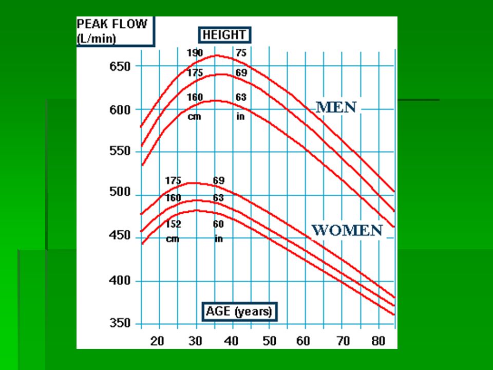
As well as how much air you can get into and out of your lungs, how fast it enters and leaves is also important. We use a Peak Flow Meter to measure the maximum rate of air flow per min Rates will vary depending on your age, gender and height. Maximum rates are normally around age 30 with the rate dropping gradually with age. Males have higher rates than females

33
Gas Exchange Oxygen moves by diffusion into the blood. Here it combines with Haemoglobin giving oxyhaemoglobin. Carbon dioxide diffuses out of the blood and is breathed out, https://www.youtube.com/watch?v=EFCj9STCvdI

34
Diffusion Movement of atoms/molecules from an area of high concentration to one of lower concentration until they are evenly distributed.

35
Fick`s Law Diffusion proportional to: Surface area x concentration difference diffusion distance

36
How are the alveoli adapted for gas exchange?

37
Respiratory Membrane (Air-Blood Barrier) Slide 13.18b Copyright © 2003 Pearson Education, Inc. publishing as Benjamin Cummings Figure 13.6

38
https://www.youtube.com/watch?v=XmuPdYOOrDo The New Living Body-breathing video

39
Control of breathing Slide 13.36 Copyright © 2003 Pearson Education, Inc. publishing as Benjamin Cummings Nerve centres that control rate and depth are located in the medulla of the brain The pons (part of the brain) appears to smooth out respiratory rate Normal breathing rate is 12–15 breaths per minute Breathing rate and depth increases as we exercise
 appears to smooth out respiratory rate Normal breathing rate is 12–15 breaths per minute Breathing rate and depth increases as we exercise.")
40
Factors Influencing Respiratory Rate and Depth Slide 13.39a Copyright © 2003 Pearson Education, Inc. publishing as Benjamin Cummings Chemical factors Carbon dioxide levels Level of carbon dioxide in the blood is the main regulatory chemical for respiration Increased carbon dioxide increases respiration Changes in carbon dioxide act directly on the medulla oblongata

41
Factors Influencing Respiratory Rate and Depth Slide 13.39b Copyright © 2003 Pearson Education, Inc. publishing as Benjamin Cummings Chemical factors (continued) Oxygen levels Changes in oxygen concentration in the blood are detected by chemoreceptors in the aorta and carotid artery Information is sent to the medulla oblongata
 Oxygen levels Changes in oxygen concentration in the blood are detected by chemoreceptors in the aorta and carotid artery Information is sent to the medulla oblongata.")
42
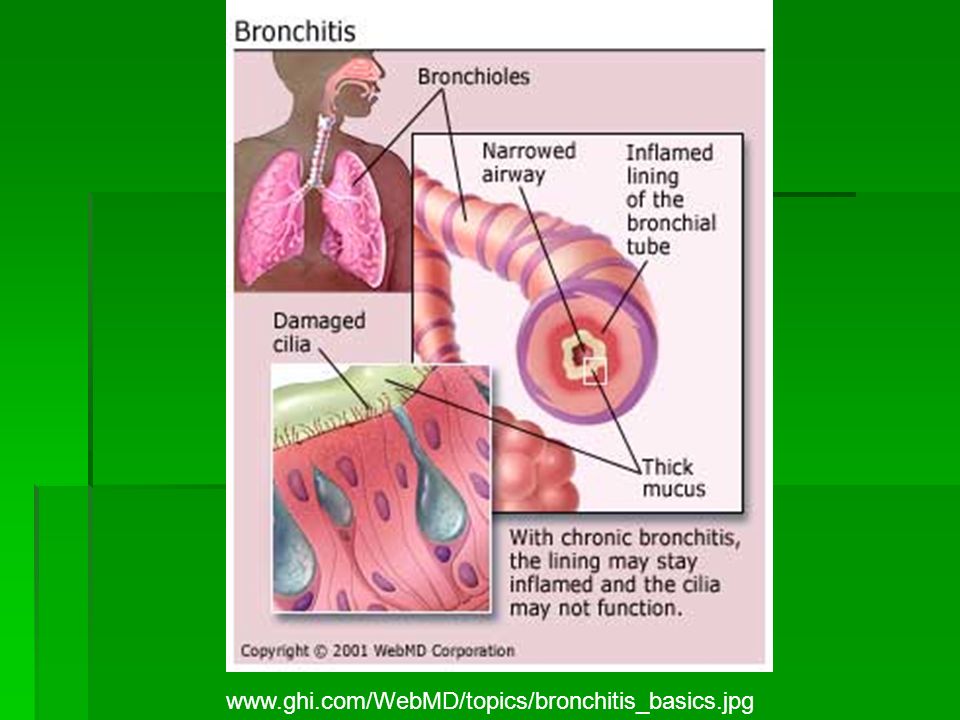
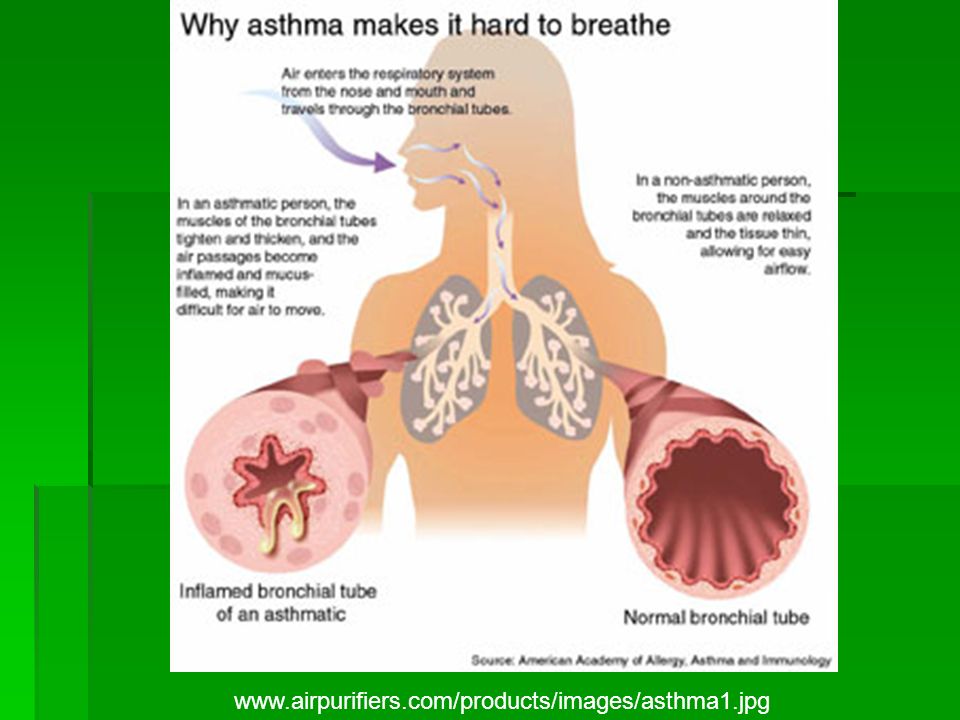
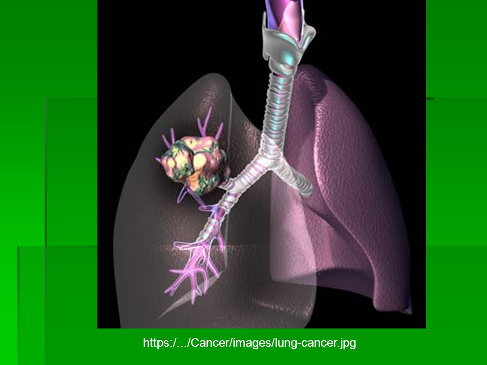
Lung conditions Emphysema Bronchitis Pneumonia Asthma Lung cancer

43
media-2.web.britannica.com/eb-media/04/100104...

44
www.ghi.com/WebMD/topics/bronchitis_basics.jpg
45
www.airpurifiers.com/products/images/asthma1.jpg
46
https:/.../Cancer/images/lung-cancer.jpg
47
Smokers Lung

Similar presentations


. 2.Production of sound (vocal cords). 3.Pulmonary ventilation. 4. Inspiration (intercostals muscles lift.>")


 bronchioles.>")


