Download presentation
Presentation is loading. Please wait.

1
The Lower Limb Sevda LAFCI, MD.PhD.

2
The Lower Limb The bones of the lower limb form the inferior part of the appendicular skeleton the organ of locomotion for bearing the weight of body stronger and heavier than the upper limb for maintaining equilibrium As you know human skeleton divides into two parts: Axial skeleton Appendecular skeleton

3
The Lower Limb 4 parts: The pelvic girdle (coxae) The thigh
The leg (crus) The foot (pes) It consists of four major parts
 The foot (pes) It consists of four major parts.")
4
The Lower Limb The pelvic girdle:
formed by the hip bones (innominate bones-ossa coxae) connection the skeleton of the lower limb to the vertebral column The latin word for the foot
 connection the skeleton of the lower limb to the vertebral column. The latin word for the foot.")
5
The Lower Limb The thigh the femur connecting the hip and knee

6
The Lower Limb The leg the tibia and fibula
connecting the knee and ankle

7
The Lower Limb The foot distal part of the ankle
the tarsal bones, metatarsal bones, phalanges

8
The Lower Limb the organ of locomotion for bearing the weight of body
for maintaining equilibrium

9
The Lower Limb 4 parts: The pelvic girdle The thigh The leg The foot

10
The pelvic girdle Hip the area from the iliac crest to the thigh
the region between the iliac crest and the greater torachanter of the femur formed by the innominate bones-ossa coxae

11
The hip bone os coxae large and irregular shaped
consists of three bones in childhood: ilium ischium pubis fuse at years joined in adult

12
The hip bone the ilium forms the superior 2/3 of the hip bone
has ala (wing), is fan-shaped its body representing the handle iliac crest: superior margin of ilium The first bone of the hip is the ilium
, is fan-shaped. its body representing the handle. iliac crest: superior margin of ilium. The first bone of the hip is the ilium.")
13
The hip bone the ilium iliac crest internal lip (labium internum)
external lips (labium externum)
")
14
The hip bone the ilium iliac crest end posteriorly “posterior superior iliac spine” at the level of the fourth lumbar vertebra bilat.* iliac crest end anteriorly “anterior superior iliac spine easily felt visible if you are not fatty *: it is important for lumbar puncture

15
The hip bone the ilium iliac crest can always be determined
Bilateral posterior superior iliac spina lies beneath a skin dimple* The line connecting the right-l eft skin dimple is at the level of 2. sacral vertebra and the middle of the sacroiliac joint. *: skin dimples formed by the skin and underlying fascia attached to the PSIS This location is important for to obtain bone marrow from the iliac crest.

16
The hip bone the ilium Tubercle of the crest is located 5cm posterior to the anterior superior iliac spine ant. inf. iliac spine post. inf. iliac spine difficult to identfy by palpation

17
The hip bone the ilium greater sciatic notch Furthermore Moreover
it is located in lower part of ilium

18
The hip bone the ilium At the medial side
auricular surface for the sacroiliac joint

19
The hip bone the ischium
it forms the posteroinferior part of hip L-shaped which passes inferiorly from the acetabulum turns anteriorly to join the pubis body ramus

20
The hip bone the ischium
at the inferior end of the body ischial tuberosity is covered by gluteus maximus muscle when the thigh is extended He body of ischium fused with ilium and pubis, when you sitting ischial tuberosity is the most distal part of the pelvis

21
The hip bone the ischium
at the posterior part of the ischium ischial spine (spina ischiadica) separates the greater sciatic notch sup. lesser sciatic notch inf. Ischial spine separates the greater sciatic notch superiorly from the lesser sciatic notch inferiorly G L
 separates the. greater sciatic notch sup. lesser sciatic notch inf. Ischial spine separates the greater sciatic notch superiorly from the lesser sciatic notch inferiorly. G. L.")
22
The hip bone the ischium
the greater sciatic notch is converted “greater sciatic foramen” by the sacrospinous ligament pass the the priformis muscle the vessels and nerves of gluteal region G

23
The hip bone the ischium
The lesser sciatic notch is converted “lesser sciatic foramen” by the sacrospinous and sacrotuberous ligament contains: obtrator internus muscle pudendal nerve internal pudendal vessels L

24
The hip bone the ischium
ramus extends medially from the body joins the inf. ramus of the pubis form “ischiopubic ramus” which completes the obturator foramen

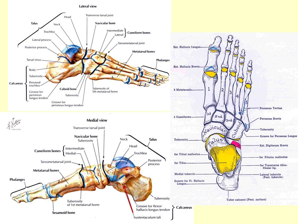
25
The hip bone the pubis forms anterior part of the hip bone
body, lies medially, joins body of the other ones it’s called symphysis pubis (cartilaginous joint) ramus (2) superior ramus passes superiolaterally to the acetabulum inferior ramus passes posteriorly, inferiorly, laterally to join ramus of ischium to form half of the pubic arch (ischiopubic ramus) The pubis consists of three parts.
 ramus (2) superior ramus passes superiolaterally to the acetabulum. inferior ramus passes posteriorly, inferiorly, laterally. to join ramus of ischium. to form half of the pubic arch (ischiopubic ramus) The pubis consists of three parts.")
26
The hip bone the pubis the anterior border of the body is thickened
“pubic crest” its lateral end, pubic tubercule* *: main pubic attachment for the inguinal ligament- bony landmark it provides

27
The hip bone the obturator foramen
oval aperture surrounded by the bodies and rami of the pubis and ischium it lies inferomedial to the acetabulum

28
The hip bone the obturator foramen
is nearly closed by the obturator membrane

29
The hip bone the acetabulum
cup shape cavity articulates with the head of femur it’s names from Roman vinegar cup, it is called acetabulum Until puberty the ilium, ischium and pubis are united by a “Y” shaped hyaline cartilage At years these bones fuse to form the hip bone (cartilage is replaced by bone)
")
30
The Lower Limb 4 parts: The pelvic girdle The thigh The leg The foot

31
The thigh Femur thigh bone is femur
longest strongest heaviest bone articulates with acetabulum and tibia Height ≈ 4xlength of femur A persons height is roughly four times the length of femur

32
The thigh Femur body (shaft) ends (extremities) Proximal end: head
neck greater trochanter lesser trochanter posterior aspect medial aspect

33
The thigh Femur Distal end: broadened
articulates with tibia and patella medial aspect anterior aspect

34
The thigh Femur Proximal end: head neck greater trochanter
lesser trochanter posterior aspect medial aspect

35
The thigh Femur Proximal end: Head forms about 2/3 of a sphere
to fit deeply into the acetabulum sometimes palpable when the thigh is rotated laterally in thin male posterior aspect medial aspect

36
The thigh Femur Proximal end: head neck greater trochanter
lesser trochanter posterior aspect

37
The thigh Femur neck between head and body to meet the body
neck runs inferolaterally with angle of 125˚ limited laterally greater trochanter posterior aspect

38
The thigh Femur Intertrochanteric line between greater and lesser
trochanter (anteriorly) is produced by the attachment of the iliofemoral ligament (massive lig.) anterior aspect
 is produced by. the attachment of. the iliofemoral ligament. (massive lig.) anterior aspect.")
39
The thigh Femur Intertrochanteric crest unites greater and lesser
trochanter, posteriorly posterior aspect

40
The thigh Femur Proximal end: head neck greater trochanter
lesser trochanter posterior aspect

41
The thigh Femur greater trochanter is large, rectangular projection
from the junction of the neck and the body. posterior aspect

42
The thigh Femur greater trochanter is insertion for muscle
of gluteal region the most lateral point of the hip region posterior aspect

43
The thigh Femur greater trochanter
can be easily palpated on the lateral side of the thigh the most lateral point of the hip region

44
The thigh Femur Proximal end: head neck greater trochanter
lesser trochanter posterior aspect

45
The thigh Femur lesser trochanter is located in the
posteromedial surface at the inferior end of the intertorachanteric crest in the angle between the neck and body of the femur posterior aspect

46
The thigh Femur Body (shaft) Linea aspera
in the middle of its posteriorly has medial and lateral lips Diverge inferiorly to form the supracondylar lines not palpable, covered with large muscle

47
The thigh Femur Body (shaft) Pectineal line runs from the
lesser torachanter to the medial lip tendon of the pectineal muscle inserts into it

48
The thigh Femur Distal end:

49
The thigh Femur Distal end: Condyle, epicondyle intercondylar notch
patellar surface adductor tubercle

50
The thigh Femur Distal end: broadened for articulation with tibia
2 large “condyle” project posteriorly are subcutaneous easily palpable separated by a deep U-shaped “intercondylar notch”

51
The thigh Femur Distal end: at the center of the each condyle is a
prominent “epicondyle” tibial and fibular collateral ligaments are attached to the epicondyles

52
The thigh Femur Distal end: articular surfaces of
condyle are confluent anteriorly patellar surface

53
The thigh Femur each condyle is separated from the
patellar surface by a slight groove Patellar surface can be palpated when the leg is flexed. Patella (kneecap) slides during flexion and extension of the leg İt supplies eassily mevement for knee joint.
 slides during flexion and extension of the leg. İt supplies eassily mevement for knee joint.")
54
The thigh Femur The adductor tubercle located in the medial side
A small prominence of bone. The adductor muscle of thigh attaches to it.

55
The Lower Limb 4 parts: The pelvic girdle The thigh The leg The foot

56
The Lower Limb The leg (crus) Between knee and ankle tibia fibula
it is composed of strong oblique fibers are connected by an “interosseous membrane” The bones of leg are tibia and fibula Compose meydana getirmek generate

57
The Lower Limb Tibia (shine bone) proximal end is large
supports most of the weight articulates with the condyle of femur superiorly and the talus inferiorly proximal end is large superior surface of tibia almost flat Medial-lateral condyles articulate with the condyle of femur

58
The Lower Limb sup. surface is flat
consists of med-lat. tibial plateaus

59
The Lower Limb lat. condyle has facet inferiorly
for the head of fibula

60
The Lower Limb Tibial tuberosity is located superior
part of anterior surface patellar ligament is attached to the tibial tuberosity

61
The Lower Limb distal end of tibia is small
facet for the fibula and talus project medially and inferiorly “medial malleolus”

62
The Lower Limb “medial malleolus”
has facet for articulation with talus

63
The Lower Limb body (corpus) medial lateral posterior surface
lateral (interosseous border)* anterior border anterior aspect
* anterior border. anterior aspect.")
64
The Lower Limb body (corpus) *:lat. border is sharp
it gives attachment to the “interosseous membrane” uniting the tibia and fibula

65
The Lower Limb At the posterior surface of tibia
observe a rough diagonal ridge known as the “soleal line” (soleus muscle is attached) Known as posterior aspect
 Known as. posterior aspect.")
66
The Lower Limb At the posterior surface of tibia
observe a rough diagonal ridge known as the “soleal line” (soleus muscle is attached) runs inferioromedially to the medial border posterior aspect
 runs inferioromedially to the medial border. posterior aspect.")
67
The Lower Limb At the posterior surface of tibia
Observe a rough diagonal ridge known as the “soleal line” (soleus muscle is attached) runs inferioromedially to the medial border The nutritient foramen is located posterior aspect
 runs inferioromedially to the medial border. The nutritient foramen is located. posterior aspect.")
68
The Lower Limb At the posterior surface of tibia
Observe a rough diagonal ridge known as the “soleal line” (soleus muscle is attached) runs inferioromedially to the medial border The nutritient foramen is located posterior aspect
 runs inferioromedially to the medial border. The nutritient foramen is located. posterior aspect.")
69
The Lower Limb Fibula (calf bone) Pin-like bone
lies posterolateral to the tibia little /no function in weight hearing providing support for tibia also provides stability to the ankle joint mainly for the attachment of muscle Lateral bone leg is the fibula Without fibular support tibial fractures would occur more frequently

70
The Lower Limb Fibula (calf bone) neck is constricted
interosseous border for attacment to the interosseous memb. nutricient foramen is usually present at the post. side head of fibula is irregular he head of facet on its for articulation with the lat. tibial condyle of tibia Lateral bone leg is the fibula

71
The Lower Limb on the distal end project medially and
inferiorly forms “lateral malleolus” lies more inferior and posterior than does medial malleolus Lateral bone leg is the fibula Without fibular support tibial fractures would occur more frequently So the distal end of fibula forms lateral malleolus Knob-like subcutaneous prominence on the lat surface of the ankle.

72
The Lower Limb 4 parts: The pelvic girdle The thigh The leg The foot

73
The Lower Limb The foot comprise the tarsus metatarsus phalanges

74
The Lower Limb The foot comprise the tarsus metatarsus phalanges
The tarsus consist of seven tarsal bones

75
The Lower Limb tarsus talus* calcaneus cuboid navicular 3 cuneiforms
*:articulates with the leg bones The tarsus consist of seven tarsal bones Only one of them the talus *

76
The Lower Limb talus body-cuboidal shape on the superior side
it has “trochlea” it is pulley shaped part of talus The inferior surface of the body of talus has an oval area for the articulation with the calcaneus Talus has a body It seems like a saddle when viewed from its dorsal aspect It rests on the anterior two thirds of calcaneus, it is inferior bone The saddle shaped superior surface of talus bears the weight of the body transmitted the via tibia It has three articular faces: -one for the facet on the inferior surface of the tibia -one for the facet on the lateral surface of the medial malleus -one for the facet on the medial surface of the lateral malleus

77
The Lower Limb talus posterior part of body has posterior process
has med-lat tubercle 2 tubercle to consist of the groove for the tendon of the flexor hallucis longus muscle Talus has a body It seems like a saddle when viewed from its dorsal aspect It rests on the anterior two thirds of calcaneus, it is inferior bone The saddle shapedsuperior surface of talus bears the weight of the bodytransmitted the via tibia It has three articular faces: one for tha facet on the inferior surface of the tibia one for the facet on the lateral surface of the medial malleus one for the facet on the medial surface of the lateral malleus

78
The Lower Limb talus head of talus has articular surface for
naviculare bone Talus has a body It seems like a saddle when viewed from its dorsal aspect It rests on the anterior two thirds of calcaneus, it is inferior bone The saddle shapedsuperior surface of talus bears the weight of the bodytransmitted the via tibia It has three articular faces: one for tha facet on the inferior surface of the tibia one for the facet on the lateral surface of the medial malleus one for the facet on the medial surface of the lateral malleus

79
The Lower Limb talus at the medial side of the calcaneus shelf-like
projection of calcaneus “Sustentaculum tali” Talus has a body It seems like a saddle when viewed from its dorsal aspect It rests on the anterior two thirds of calcaneus, it is inferior bone The saddle shapedsuperior surface of talus bears the weight of the bodytransmitted the via tibia It has three articular faces: one for tha facet on the inferior surface of the tibia one for the facet on the lateral surface of the medial malleus one for the facet on the medial surface of the lateral malleus

80
The Lower Limb talus the neck is slightly constricted
inferiorly there is a groove called the “sulcus tali” for the interosseous lig. Talus has a body It seems like a saddle when viewed from its dorsal aspect It rests on the anterior two thirds of calcaneus, it is inferior bone The saddle shapedsuperior surface of talus bears the weight of the bodytransmitted the via tibia It has three articular faces: one for tha facet on the inferior surface of the tibia one for the facet on the lateral surface of the medial malleus one for the facet on the medial surface of the lateral malleus

82
Calcaneus Largest-strongest 6 surfaces Sup :joins talus
Inf :calcaneal tuber Ant :joins cuboid Post :forms heel Lat :fibular trochlea Med :sustentaculum tali The posterior end of inferior surface there is a tuber calcanei, it has med-lat processes for the attachment of the muscle. On the medial surface of the calcaneus there is a groove,inf. Part of the sustentaculum tali, flexor hallucis longus muscle On its lateral surface there is atubercle the peroneal (fibular) trochlea
 trochlea.")
83
Navicular 3 facets Ant cuneiform Post talus Lat cuboid
Med tuberosity of navicular It is located between the head of talus and the 3 cuneiform bones. It has facet for articulation with each of them

84
Cuboid Most lat. bone distal tarsus Ant base of metatarsals 4-5
Post calcaneus Med lat cuneiform & navicular Inf groove for fibularis longus Post: İt presents an articular surface for the calcaneus Anteriorly: There is 2 facets for the mt On its medial surface there are facets for lat cuneiform and nav. bones

85
Cunieform Medial (largest) Lateral Intermedium (smallest)
Ant base of metatarsals 1-4 Post Navicular Each cuneiform articulates with the navicular bones posteriorly, and with the base of its articulates with mt anteriorly. Prof. Dr. H. Selçuk Sürücü

86
Metatarsal 5 bones Base Head Body I shortest & thickest II longest
Mt consists of five nt bones Prof. Dr. H. Selçuk Sürücü

87
Digital 14 bones Base Head Body Proximal, middle & distal phalanges
There are 14 phalanges The big one is toe (hallux) has 2 phalanges (proximal-distal) And the other 4 digits have 3 each (proximal-middle-distal) Each phalanx consists of a base proximally, a body and a head distally.
 has 2 phalanges (proximal-distal) And the other 4 digits have 3 each (proximal-middle-distal) Each phalanx consists of a base proximally, a body and a head distally.")
Similar presentations


 (Lower) Leg Foot The lower appendages are attached to the axial skeleton via the pelvic girdle.>")


 Coxae have 3 distinct parts: – Ilium – Ischium – Pubis.>")