Download presentation
Presentation is loading. Please wait.

1
Thinking About Psychology: The Science of Mind and Behavior 2e
As usual Charles T. Blair-Broeker Randal M. Ernst

2
How do scientists study the brain?
Case studies, those who have suffered damage from injury or illness. Provides great info on memory, speech, emotion, movement, personality. It’s hard to make generalizations. Injuries are usually not contained to one area. Lesions, surgically altering, removing, or destroying specific portions of the brain. Severing the corpus callosum for seizures Electrical stimulation, usually produces the opposite behavior.

3
Electroencephalograph
Although not a "brain scan" as the term is usually used. An EEG is a recording of electrical signals from the brain made by hooking up electrodes to the subject's scalp. These electrodes pick up electric signals naturally produced by the brain and send them to galvanometers (instruments that detect and measure small electric currents) that are in turn hooked up to pens, under which graph paper moves continuously. The pens trace the signals onto the graph paper. Although it was known as early as the nineteenth century that living brains have electrical activity, an Austrian psychiatrist named Hans Berger was the first to record this activity in humans, in the late 1920s. EEGs allow researchers to follow electrical impulses across the surface of the brain and observe changes over split seconds of time. An EEG can show what state a person is in -- asleep, awake, anaesthetized -- because the characteristic patterns of current differ for each of these states. One important use of EEGs has been to show how long it takes the brain to process various stimuli. A major drawback of EEGs, however, is that they cannot show us the structures and anatomy of the brain or really tell us which specific regions of the brain do what.
 that are in turn hooked up to pens, under which graph paper moves continuously. The pens trace the signals onto the graph paper. Although it was known as early as the nineteenth century that living brains have electrical activity, an Austrian psychiatrist named Hans Berger was the first to record this activity in humans, in the late 1920s. EEGs allow researchers to follow electrical impulses across the surface of the brain and observe changes over split seconds of time. An EEG can show what state a person is in -- asleep, awake, anaesthetized -- because the characteristic patterns of current differ for each of these states. One important use of EEGs has been to show how long it takes the brain to process various stimuli. A major drawback of EEGs, however, is that they cannot show us the structures and anatomy of the brain or really tell us which specific regions of the brain do what.")
4
CAT - Computerized Axial Tomography
Developed in the 1970s, CAT (or CT) scanning is a process that combines many 2-dimensional x-ray images to generate cross-sections or 3-dimensional images of internal organs and body structures (including the brain). Doing a CAT scan involves putting the subject in a special, donut-shaped x-ray machine that moves around the person and takes many x-rays. Then, a computer combines the 2-dimensional x-ray images to make the cross-sections or 3-dimensional images. CAT scans of the brain can detect brain damage and also highlight local changes in cerebral blood flow (a measure of brain activity) as the subjects perform a task. CAT - Computerized Axial Tomography
 scanning is a process that combines many 2-dimensional x-ray images to generate cross-sections or 3-dimensional images of internal organs and body structures (including the brain). Doing a CAT scan involves putting the subject in a special, donut-shaped x-ray machine that moves around the person and takes many x-rays. Then, a computer combines the 2-dimensional x-ray images to make the cross-sections or 3-dimensional images. CAT scans of the brain can detect brain damage and also highlight local changes in cerebral blood flow (a measure of brain activity) as the subjects perform a task. CAT - Computerized Axial Tomography.")
5
Positron Emission Tomography
Also developed in the 1970s, PET scans allow one to observe blood flow or metabolism in any part of the brain. In a PET scan, the subject is injected with a very small quantity of radioactive glucose. The PET then scans the absorption of the radioactivity from outside the scalp. Brain cells use glucose as fuel, and PET works on the theory that if brain cells are more active, they will consume more of the radioactive glucose, and if less active, they will consume less of it. A computer uses the absorption data to show the levels of activity as a color-coded brain map, with one color (usually red) indicating more active brain areas, and another color (usually blue) indicating the less active areas. The gray outer surface is the surface of the brain from MRI and the inner colored structure is cingulate gyrus, part of the brain's emotional system visualized with PET.
 indicating more active brain areas, and another color (usually blue) indicating the less active areas. The gray outer surface is the surface of the brain from MRI and the inner colored structure is cingulate gyrus, part of the brain s emotional system visualized with PET.")
6
Magnetic Resonance Imaging
Magnetic resonance imaging (MRI) is a test that uses a magnetic field and pulses of radio wave energy to make pictures of organs and structures. Magnetic resonance imaging, or MRI, is a machine used for brain structure imaging. When in some cases a CT scan cannot detect the existing problem, an MRI is helpful for discovering unnoticed anatomical anomalies caused by a disease process or traumatic event. It is a utility used for grand research in determining structural differences and a behavior correlation
 is a test that uses a magnetic field and pulses of radio wave energy to make pictures of organs and structures. Magnetic resonance imaging, or MRI, is a machine used for brain structure imaging. When in some cases a CT scan cannot detect the existing problem, an MRI is helpful for discovering unnoticed anatomical anomalies caused by a disease process or traumatic event. It is a utility used for grand research in determining structural differences and a behavior correlation.")
7
Functional magnetic resonance imaging (fMRI)
It works by detecting the changes in blood oxygenation and flow that occur in response to neural activity – when a brain area is more active it consumes more oxygen and to meet this increased demand blood flow increases to the active area. fMRI can be used to produce activation maps showing which parts of the brain are involved in a particular mental process.

8
FMRI functions through blood flow or blood oxygen level measurements to achieve the brain’s functional image. It is primarily used to gather relevant data as to the consumption of oxygen by the tissues. Through its modernization, fMRI sequences will view a picture of the brain’s active region by picking up the excess blood supply called Blood Oxygen Level Dependence (BOLD). In general, an MRI and fMRI differ from each other in a way that an MRI views the anatomical structure while an fMRI views the metabolic function.
. In general, an MRI and fMRI differ from each other in a way that an MRI views the anatomical structure while an fMRI views the metabolic function.")
9
Brain Development Neural tube (2 wks after conception) ventricles which create CSF Intense growth: at week 4, ½ million neurons created per minute and creation of 2 million synaptic connections per second By 24 weeks of prenatal age the brain is reached full development.

10
Brain develops progressively: hindbrain, midbrain, forebrain
Brain develops progressively: hindbrain, midbrain, forebrain. Eventually the forebrain envelops the hindbrain and midbrain. At birth brain weighs less than a pound. Continues to develop dendrites, myelin forms, axons grow longer, and the branching ends of the axons become more dense. For the longest time scientists believed that people and animals did not experience neurogenesis.

11
Neurogenesis Gould started study in 1998 examining
marmosets’ hippocampus; and concluded they were developing new neurons everyday.

12
Erikson, Gage,et al. applied studies to humans
Erikson, Gage,et al. applied studies to humans. Gould then looked at Macaques’ whole brain in 1999 and found it’s not restricted to just one area. Erikson & Gage used 5 cancer patients who were treated with a drug to determine whether tumor cells were multiplying. The drug would color new cells. Using fluorescent light the chemical tracer could be detected in new cells. After the patients died the hippocampus was removed and scientists found 100s of new neurons had been generated since the drug was administered. All patients were over age 50!

13
Gould has found that stressful experiences inhibit the production of new neurons in the hippocampus of adult rodents and primates. Moreover, developmental stress (either prenatal or early postnatal) persistently diminishes the production of new neurons, even into adulthood.
 persistently diminishes the production of new neurons, even into adulthood.")
14
UCLA “How the Brain Works” Episode 1

15
Lower-Level Brain Structures: The Brainstem
Module 7: The Brain

16
Brainstem The oldest part and central core of the brain
It begins where the spinal cord swells as it enters the skull Is responsible for automatic survival functions comprised of the hindbrain and the midbrain

17
Brainstem

18
Hindbrain Connects the spinal cord with the rest of the brain
Sensory and motor pathways pass through the hindbrain to and from regions that are situated higher up in the brain. Comes in one side of the body and projects to the opposite side of the brain. Strokes affect the opposite side of the body 3 structures: medulla, pons & cerebellum

20
Medulla Located at the base of the brainstem, directly above the spinal cord. Controls life-supporting functions like heartbeat and breathing Also controls some reflexes: swallowing, vomiting, sneezing, coughing Damage to this area can lead to death.

21
Medulla

22
Pons Bulging area above medulla
Helps coordinate and integrate movements on each side of the body. “Bridge” acts as a bridge between higher brain regions and the cerebellum

23
Cerebellum Located above the pons Latin for the “little brain”
Attached to the rear of the brain Helps coordinate voluntary movements, muscle tone, and balance (equilibrium) Involved in learning automated movements: typing, writing, playing tennis, dunking a basketball If damaged, the person could perform basic movements but would lose fine coordination skills. Alcohol affects the cerebellum, causing difficulty holding balance and walking straight.
 Involved in learning automated movements: typing, writing, playing tennis, dunking a basketball. If damaged, the person could perform basic movements but would lose fine coordination skills. Alcohol affects the cerebellum, causing difficulty holding balance and walking straight.")
24
Cerebellum

25
Reticular Formation A nerve network in the brainstem that plays an important role in controlling wakefulness and arousal Extending up and down the spinal cord into the brain Controls level of alertness Damage to this area can cause a coma.

26
Reticular Formation

27
Midbrain (middle and smallest)
Relay station for auditory and visual information After passing through the midbrain level, the info is passed to sensory processors in the forebrain Substantia nigra – large neural pathway involved in motor control and produces dopamine. Parkinson’s disease symptoms are associated with degenerations of dopamine producing neurons in the substantia nigra.

28
The Forebrain The largest aka the cerebrum 90% of the brain

29
Cerebral Cortex cortex means bark
Outer portion of the forebrain Divided into two hemispheres Corpus Callosum, the large band of neural fibers that connects the two brain hemispheres and allows them to communicate with each other Covers the brain’s lower level structures The grooves and bulges of the cerebral cortex allows about 3 ft of surface area to be packed into a human skull. Divided into four lobes

30
Corpus Callosum

31
Longitudinal Fissure The long crevice that divides the cerebral cortex into left and right hemispheres This and other fissures in the brain create major divisions in the brain called lobes (temporal, occipital, parietal, and frontal)
")
33
Frontal Lobes The portion of the cerebral cortex lying just behind the forehead Is involved in planning and judgments Includes the primary motor cortex, movements of different body parts are represented here. 1/3 devoted to hand movements and 1/3 to facial muscles.

34
The Frontal Lobe Play “The Frontal Lobes: Cognition and Awareness” (9:05) Segment #7 from The Mind: Psychology Teaching Modules (2nd edition).
 Segment #7 from The Mind: Psychology Teaching Modules (2nd edition).")
35
Phineas Gage Play “The Frontal Lobes and Behavior: The Story of Phineas Gage” (12:03) Module #25 from The Brain: Teaching Modules (2nd edition).
 Module #25 from The Brain: Teaching Modules (2nd edition).")
37
Parietal Lobes The portion of the cerebral cortex lying behind the frontal lobes. Includes the somatosensory cortex, processes touch, temperature, pressure, and information from the muscles and joints. All body parts are represented, but some more than others.

39
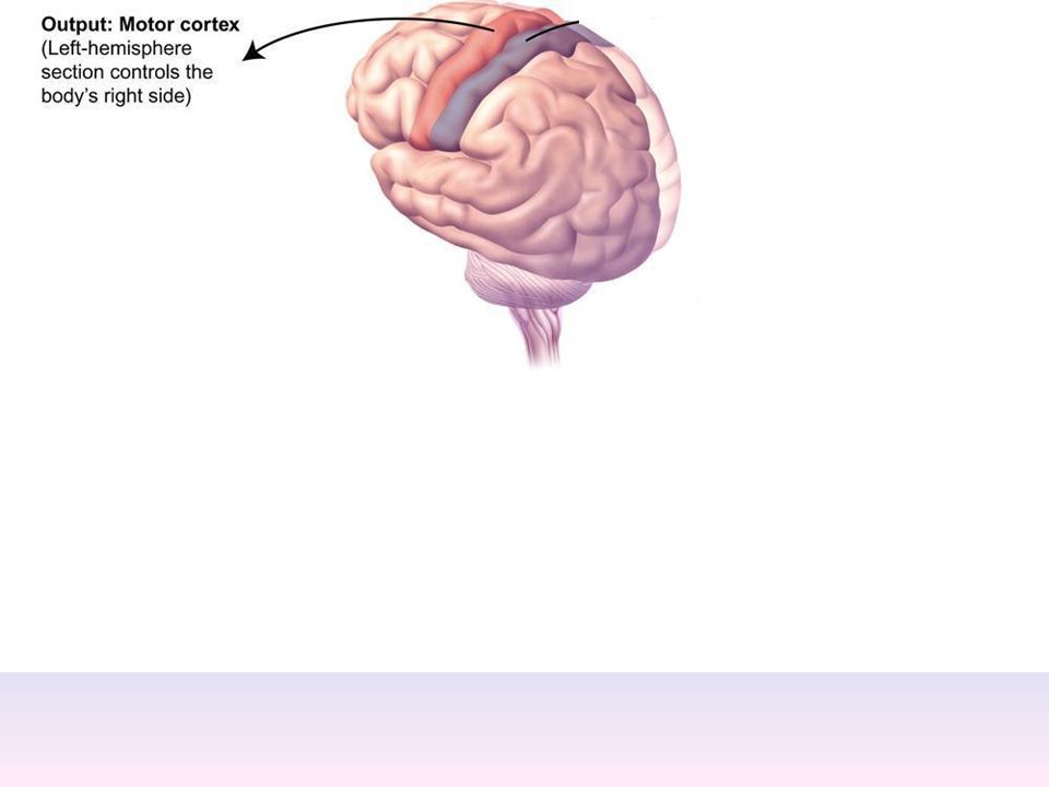
Motor Cortex The strip of brain tissue at the rear of the frontal lobes Controls voluntary movement Different parts of the cortex control different parts of the body. The motor cortex in the left hemisphere controls the right side of the body and visa versa.

43
Somatosensory Cortex The brain are located in the front of the parietal lobes Registers and processes body sensations Soma is Greek for “body.”

45
Occipital Lobe The portion of the cerebral cortex lying at the back of the head It includes the primary visual processing areas of the brain

47
Temporal Lobes Includes the auditory (hearing) areas of the brain
Where sound information is processed Located roughly above the ears An association area thought to help us recognize faces

49
Motor Cortex The strip of brain tissue at the rear of the frontal lobes Controls voluntary movement Different parts of the cortex control different parts of the body. The motor cortex in the left hemisphere controls the right side of the body and visa versa.

53
Somatosensory Cortex The brain are located in the front of the parietal lobes Registers and processes body sensations Soma is Greek for “body.”

55
Lower-Level Brain Structures: The Limbic System
Module 7: The Brain

56
Limbic System (border)
A ring of structures at the border of the brainstem and cerebral cortex, considered apart of the forebrain Helps regulate memory, learning, and emotion Involved and pleasurable and rewarding sensations James Olds discovered rats could be trained to perform a task, such as pressing a bar, in order to receive electrical stimulation. It’s still not completely understood. Includes the hypothalamus, thalamus, hippocampus, and amygdala

57
Hippocampus Embedded in the temporal lobe in each hemisphere.
Helps processing new memories for permanent storage (remember we know that new neurons are created here..) Looks something like a seahorse Hippo is Greek for “horse.”
 Looks something like a seahorse. Hippo is Greek for horse.")
58
Thalamus Sits atop the brainstem
The brain’s sensory switchboard – except for smell Directs messages to the sensory receiving areas in the cortex Thalamus is Greek for “inner chamber.” Thought to be involved in regulating levels of awareness, attention, motivation, and emotional aspects of sensations.

59
Hypothalamus A neural structure lying below the thalamus
Regulates the ANS Regulates the body’s maintenance activities such as; eating, drinking, body temperature, and it linked to emotion One area called the suprachiasmatic nucleus (SCN) is also involved in our sleep/wake cycles. Plays a role in emotions, pleasure, and sexual function The pituitary gland is attached to the hypothalamus. The hypothalamus produces hormones and neurotransmitters that directly affect the pituitary gland. The pituitary gland influences the activity of other glands.
 is also involved in our sleep/wake cycles. Plays a role in emotions, pleasure, and sexual function. The pituitary gland is attached to the hypothalamus. The hypothalamus produces hormones and neurotransmitters that directly affect the pituitary gland. The pituitary gland influences the activity of other glands.")
61
Hypothalamus and Aggression
Play “Aggression, Violence, and the Brain” (7:17) Module #24 from The Brain: Teaching Modules (2nd edition).
 Module #24 from The Brain: Teaching Modules (2nd edition).")
62
Amygdala Almond shaped neural clusters in the limbic system
Controls emotional responses such as fear and anger Research on monkeys showed that destruction of the amygdala made the monkeys lose their fear of predators. Electrical stimulation in humans produced fear and aggression. Involved in learning and forming memories especially those with a strong emotional component.

64
Differences Between the Two Hemispheres
Module 7: The Brain

65
Hemispheric Differences
Brain is divided into two hemispheres but works as a single entity. Both sides continually communicate via the corpus callosum, except in those with split brains. Functional symmetry include the primary motor cortex and the somatosensory cortex Other important processes are not symmetrical

66
Split Brain Patient Play “The Divided Brain” (6:46) Module #5 from The Brain: Teaching Modules (2nd edition).
 Module #5 from The Brain: Teaching Modules (2nd edition).")
67
Differences Between the Two Hemispheres: Language and Spatial Abilities
Module 7: The Brain

68
Left vs. Right Brain?

69
The Brain’s Left Hemisphere
For most people, language functions are in the left hemisphere. (lateralization of function) For a small percentage of people, language functions are in the right hemisphere.
 For a small percentage of people, language functions are in the right hemisphere.")
70
Broca’s Area The brain area of the left frontal lobe
Directs the muscle movements involved in speech If damaged the person can form the ideas but cannot express them as speech, Broca’s Aphasia. Aphasia: partial or complete inability to articulate ideas or understand spoken or written language because of brain injury or damage.

72
Wernicke’s Area A brain area of the left temporal lobe
Involved in language comprehension and expression Our ability to understand what is said to us Wernicke’s Aphasia: can speak, but they have trouble finding the right words and have great difficulty comprehending written or spoken communication. Characterized by nonsensical, meaningless, incoherent words.

74
The Brain’s Right Hemisphere
Houses the brain’s spatial abilities, which allows us to perceive or organize things in a given space, judge distance, etc. Helps in making connections between words (inferences) Seems to help orchestrate our sense of self (sometimes couldn’t recognize themselves in a mirror) Some patients have trouble perceiving who people are in comparison to themselves (nurses as family)
 Seems to help orchestrate our sense of self (sometimes couldn’t recognize themselves in a mirror) Some patients have trouble perceiving who people are in comparison to themselves (nurses as family)")
75
Broca’s and Wernicke’s Areas
Play “Language and Speech: Broca’s and Wernicke’s Areas” (7:44) Module #6 from The Brain: Teaching Modules (2nd edition).
 Module #6 from The Brain: Teaching Modules (2nd edition).")
76
Split Brain Research, Roger Sperry
Read pgs 80-81

77
Split Brain Research

78
Split Brain Research

79
Split Brain Research

80
Split Brain Research

81
Split Brain Research

82
Split Brain Research

Similar presentations


>")
. The more complex.>")


