Download presentation
Presentation is loading. Please wait.

2
Essentials of Human Anatomy & Physiology Seventh Edition Elaine N. Marieb Ch 5 pgs 145-154 and Ch 6 pgs 176-179 The Skeletal System Articulations

3
What is an articulation? Slide 5.43 Copyright © 2003 Pearson Education, Inc. publishing as Benjamin Cummings Functional junction between bones AKA a joint Function? Hold bones together Mobility (movement)
.")
4
Classification of Articulations Slide 5.43 Copyright © 2003 Pearson Education, Inc. publishing as Benjamin Cummings BB y: FF unction- immovable, slightly movable, OR freely movable SS tructure- fibrous, cartilaginous, OR synovial

5
1. Fibrous (Immovable) Articulations Slide 5.46 Copyright © 2003 Pearson Education, Inc. publishing as Benjamin Cummings Bones united by fibrous tissue No movement Ex. Sutures of skull Distal tibiofibular joint Figure 5.27d, h

6
2. Cartilaginous (slightly movable) Articulations Slide 5.47 Copyright © 2003 Pearson Education, Inc. publishing as Benjamin Cummings Cartilage in between No movement or slight movement Exs. symphysis pubis intervertebral joints Costal cartilage Figure 5.27b, c
 Articulations Slide 5.47 Copyright © 2003 Pearson Education, Inc. publishing as Benjamin Cummings Cartilage in between No movement or slight movement Exs. symphysis pubis intervertebral joints Costal cartilage Figure 5.27b, c.")
7
More Cartilaginous Articulations Slide 5.47 Copyright © 2003 Pearson Education, Inc. publishing as Benjamin Cummings

8
3. Synovial (freely movable) Articulations Slide 5.48 Copyright © 2003 Pearson Education, Inc. publishing as Benjamin Cummings Most are freely movable Many types Separated by a joint cavity Contains synovial fluid Figure 5.27f–g Ex: tibiofemoral, Coxal, humeroscapular

9
Structures of Synovial Joints Slide 5.49 Copyright © 2003 Pearson Education, Inc. publishing as Benjamin Cummings epiphyses

10
Special Structures of Synovial Joints Slide 5.51 Copyright © 2003 Pearson Education, Inc. publishing as Benjamin Cummings Tibiofemoral Cartilage to prevent friction, shock absorption, cushioning in joint

11
Structures of Synovial Joints Slide 5.51 Copyright © 2003 Pearson Education, Inc. publishing as Benjamin Cummings Figure 5.28 Fluid filled sac to prevent friction between bone & ligament

12
Homeostatic imbalances/disorders Slide 5.50 Copyright © 2003 Pearson Education, Inc. publishing as Benjamin Cummings Bursitis

13
Homeostatic imbalances/disorders Slide 5.50 Copyright © 2003 Pearson Education, Inc. publishing as Benjamin Cummings Arthritis Rheumatoid arthritis gout osteoarthritis

16
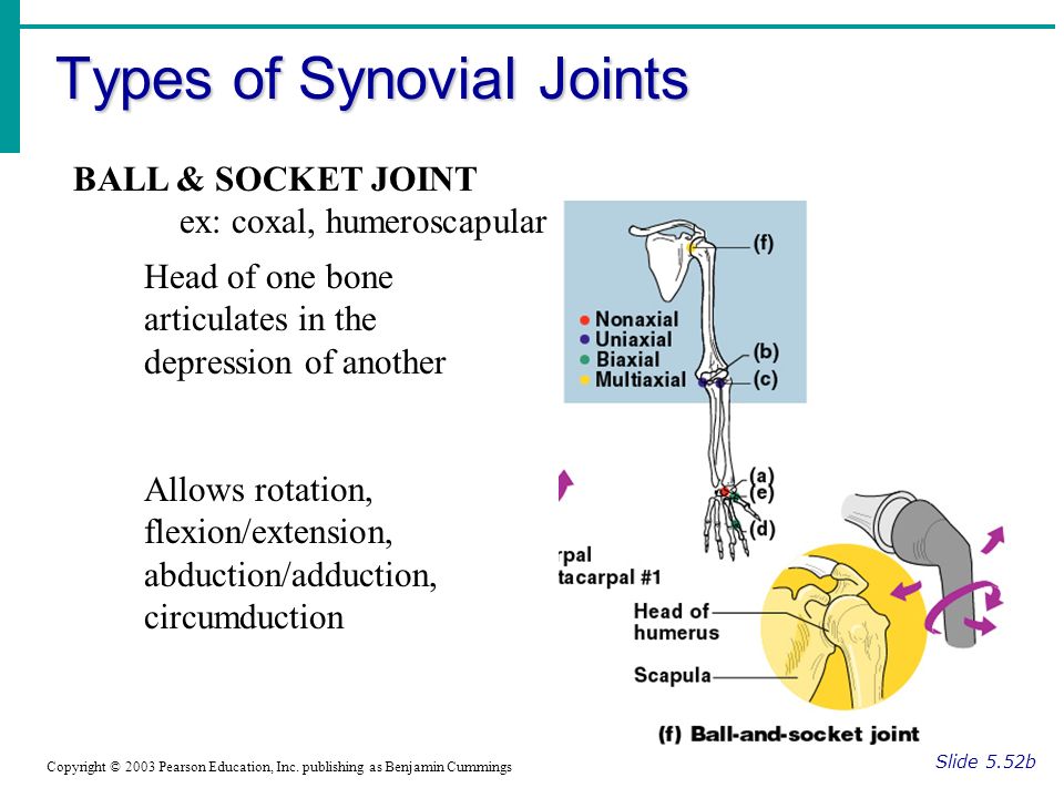
Types of Synovial Joints Slide 5.52b Copyright © 2003 Pearson Education, Inc. publishing as Benjamin Cummings Figure 5.29d–f Head of one bone articulates in the depression of another Allows rotation, flexion/extension, abduction/adduction, circumduction BALL & SOCKET JOINT ex: coxal, humeroscapular

17
Types of Synovial Joints Slide 5.52a Copyright © 2003 Pearson Education, Inc. publishing as Benjamin Cummings Figure 5.29a–c gliding Slightly curved bones articulate with one another Sliding Concave bone and convex bone articulate together Flexion/extension Cylindrical surface of one bone articulates around its axis with ring of another Rotation Sternoclavicular Acromioclavicular Sternoclavicular Acromioclavicular tibiofemoral Proximal Radioulnar

18
Types of Synovial Joints Slide 5.52b Copyright © 2003 Pearson Education, Inc. publishing as Benjamin Cummings Figure 5.29d–f oval shaped bone articulates with oval concavity of another Flexion/extension, abduction/adduction, no rotation 2 bones each with concave and convex surfaces articulate Same mvmts as condyloid Knuckles metacarpals to phalanges Twiddling your thumbs

19
Muscles and Body Movements Slide 6.30b Copyright © 2003 Pearson Education, Inc. publishing as Benjamin Cummings Muscles attached to at least two points : Origin – end attached to an immovable bone Insertion – end attached to a movable bone Figure 6.12 Insertion always moves toward origin!!!!

20
Body Movements at synovial joints Slide 6.34 Copyright © 2003 Pearson Education, Inc. publishing as Benjamin Cummings

21
Body Movements at synovial joints Slide 6.33 Copyright © 2003 Pearson Education, Inc. publishing as Benjamin Cummings Figure 6.13 Flexion- decrease of angle between bones Extension- increase of angle between bones Hyperextension- increase of angle between bones beyond anatomical position

22
Body Movements at synovial joints Slide 6.32 Copyright © 2003 Pearson Education, Inc. publishing as Benjamin Cummings Abduction- movement of body part away from midline Adduction- movement of body part towards the midline

23
Slide 6.33 Copyright © 2003 Pearson Education, Inc. publishing as Benjamin Cummings Figure 6.13 Body Movements at synovial joints Rotation- movement of body part about its axis

24
Slide 6.32 Copyright © 2003 Pearson Education, Inc. publishing as Benjamin Cummings Circumduction- movement of body part so that one end follows a circular path

25
Body Movements at synovial joints Slide 6.34 Copyright © 2003 Pearson Education, Inc. publishing as Benjamin Cummings

26
Special Movements at specific synovial joints Slide 6.34 Copyright © 2003 Pearson Education, Inc. publishing as Benjamin Cummings Supination- rotation at forearm so palms are up, or front Pronation- rotation at forearm so palms are down, or back

27
Special Movements at specific synovial joints Slide 6.34 Copyright © 2003 Pearson Education, Inc. publishing as Benjamin Cummings Plantar flexion- extension of foot at ankle, point toe Dorsiflexion- flexion of foot at ankle, pull toes to shin

28
Special Movements at specific synovial joints Slide 6.34 Copyright © 2003 Pearson Education, Inc. publishing as Benjamin Cummings Inversion- rotation of foot at ankle, roll onto outside of ankle so bottom of foot faces in Eversion- rotation of foot at ankle, roll onto inside of ankle so bottom of foot faces out

29
Special Movements at specific synovial joints Slide 6.34 Copyright © 2003 Pearson Education, Inc. publishing as Benjamin Cummings Elevation- raise a body part Depression- lower a body part

30
Special Synovial Joint- humeroscapular Slide 5.51 Copyright © 2003 Pearson Education, Inc. publishing as Benjamin Cummings Figure 5.28

31
Special Synovial Joint- coxal Slide 5.51 Copyright © 2003 Pearson Education, Inc. publishing as Benjamin Cummings

32
Special Joint- tibiofemoral (knee) Slide 6.34 Copyright © 2003 Pearson Education, Inc. publishing as Benjamin Cummings Patellar surface femur Head of fibula Tibial tuberosity Condyles of tibia Condyles of femur tibia

33
Special Joints- tibiofemoral- label diagram Slide 6.34 Copyright © 2003 Pearson Education, Inc. publishing as Benjamin Cummings Injury to Knees and other joints can be treated with RICE: R: rest I: ice C: compression E: elevation Injury to Knees and other joints can be treated with RICE: R: rest I: ice C: compression E: elevation

34
Homeostatic imbalances/disorders Slide 5.50 3 C’s of most common knee injuries- 1.Collateral (MCL)- torn ligament 2.Cruciate (ACL)- torn ligament 3.Cartilage (menisci)- torn cartilage * Injuries are due to a hit to the knee or twists with rapid shift in weight
- torn ligament 2.Cruciate (ACL)- torn ligament 3.Cartilage (menisci)- torn cartilage * Injuries are due to a hit to the knee or twists with rapid shift in weight")
35
Application- arthroscopy Slide 5.50 Copyright © 2003 Pearson Education, Inc. publishing as Benjamin Cummings -noninvasive 2-3 incisions -lighted camera -diagnosis & repair

36
3 C’s of knee injuries Slide 5.50 Torn Meniscus: transplant with artificial meniscus (plastic) or sewn Torn Ligaments: transplant autographt (from person) or allographt (from organ donor)
 or sewn Torn Ligaments: transplant autographt (from person) or allographt (from organ donor)")
37
Torn rotator cuff injury Slide 5.50 -injury of 1 to 4 of deep muscles or ligaments in the shoulder

39
Homeostatic imbalances/disorders Slide 5.50 Sprain Inversion (rolling in) torn ankle ligaments
 torn ankle ligaments")
40
Homeostatic imbalances/disorders Slide 5.50 Torn ligaments Strains or tears due to twisting or over stretching

41
Homeostatic imbalances/disorders Slide 5.50 Torn tendons Strains or tears due to twisting or overstretching

42
Homeostatic imbalances/disorders Slide 5.50 Separation dislocation

43
Tendonitis When normal smooth gliding motion os tendon is impaired; inflammation & pain

44
Lyme disease -inflammatory disease -deer tick bite with Borrelia burgdorferi (bacteria) -bulls eye rash -neurological problems -inflammatory disease -deer tick bite with Borrelia burgdorferi (bacteria) -bulls eye rash -neurological problems
 -bulls eye rash -neurological problems -inflammatory disease -deer tick bite with Borrelia burgdorferi (bacteria) -bulls eye rash -neurological problems")
45
A.Plane B.Rotation C.Saddle D.Only flexion/extension E.Most movements F.condyloid A.Plantar flexion B.dorsiflexion C.flexion D.extension E.abduction F.Adduction G.Inversion H.Eversion I.Hyperextension J.Supination K.Pronation 1 2 5 4 3

46
A B C D E F

Similar presentations


 and type of substance.>")


 Fibrous Joints 1) connections between adjacent bones 2) syndesmoses to gomphoses 3) ex.suture c) Cartilagenous.>")

