Download presentation
Presentation is loading. Please wait.

1
Joints aka: articulations

2
Classification of Joints
Functional classification (arthr = joints) (Focuses on amount of movement) Synarthroses (immovable joints) – most common in the axial skeleton Amphiarthroses (slightly movable joints) – most common in the axial skeleton Diarthroses (freely movable joints) – most common in the appendicular skeleton
 (Focuses on amount of movement) Synarthroses (immovable joints) – most common in the axial skeleton. Amphiarthroses (slightly movable joints) – most common in the axial skeleton. Diarthroses (freely movable joints) – most common in the appendicular skeleton.")
3
Classification of Joints
Structural classification (Based on the material binding them and presence or absence of a joint cavity) Fibrous Cartilagenous Synovial
 Fibrous. Cartilagenous. Synovial.")
4
Fibrous joints Bones connected by fibrous tissue: dense regular connective tissue No joint cavity Slightly movable or immovable Types Sutures Syndesmoses

5
Sutures Only between bones of skull
Fibrous tissue continuous with periosteum Ossify and fuse in middle age:

6
Syndesmoses In Greek: “ligament” Bones connected by ligaments only
Amount of movement depends on length of the fibers: longer than in sutures

7
Cartilagenous joints Articulating bones united by cartilage
Lack a joint cavity Not highly movable Two types Synchondroses (singular: synchondrosis) Sympheses (singular: symphesis)
 Sympheses (singular: symphesis)")
8
Synchondroses Literally: “junction of cartilage”
Hyaline cartilage unites the bones Immovable (synarthroses) Examples: Epiphyseal plates Joint between rib’s and the costal cartilage and of the sternum
 Examples: Epiphyseal plates. Joint between rib’s and the costal cartilage and of the sternum.")
9
Sympheses Literally “growing together” Fibrocartilage unites the bones
Slightly movable (amphiarthroses) Resilient shock absorber Provide strength and flexibility Hyaline cartilage on articular surfaces of bones to reduce friction Examples Intervertebral discs Pubic symphysis of the pelvis
 Resilient shock absorber. Provide strength and flexibility. Hyaline cartilage on articular surfaces of bones to reduce friction. Examples. Intervertebral discs. Pubic symphysis of the pelvis.")
10
Synchondroses and sympheses
Also pubic symphsis

11
Synovial joints Include most of the body’s joints
All are diarthroses (freely movable) All contain fluid-filled joint cavity
 All contain fluid-filled joint cavity.")
12
General Structure of Synovial Joints
Articular cartilage Hyaline Spongy cushions absorb compression Protects ends of bones from being crushed Joint (synovial) cavity Potential space Small amount of synovial fluid
 cavity. Potential space. Small amount of synovial fluid.")
13
General structure of synovial joints (cont.)
3. Articular (or joint) capsule Two layered Outer*: fibrous capsule of dense irregular connective tissue continuous with periosteum Inner*: synovial membrane of loose connective tissue (makes synovial fluid) Lines all internal joint surfaces not covered by cartilage* * * *
 capsule. Two layered. Outer*: fibrous capsule of dense irregular connective tissue continuous with periosteum. Inner*: synovial membrane of loose connective tissue (makes synovial fluid) Lines all internal joint surfaces not covered by cartilage* * * *")
14
General structure of synovial joints (cont.)
4. Synovial fluid Filtrate of blood Contains special glycoproteins Nourishes cartilage and functions as slippery lubricant “Weeping” lubriication 5. Reinforcing ligaments (some joints) Capsular (most) – thickened parts of capsule Extracapsular Intracapsular
 Capsular (most) – thickened parts of capsule. Extracapsular. Intracapsular.")
15
General structure of synovial joints (cont.)
6. Nerves Detect pain Monitor stretch (one of the ways of sensing posture and body movements) 7. Blood vessels Rich blood supply Extensive capillary beds in synovial membrane (produce the blood filtrate)
 7. Blood vessels. Rich blood supply. Extensive capillary beds in synovial membrane (produce the blood filtrate)")
16
General structure of synovial joints

17
Some joints… Articular disc or meniscus (literally “crescent”)
Only some joints Those with bone ends of different shapes or fitting poorly Some to allow two kinds of movement (e.g. jaw) Made of fibrocartilage Examples: knee TMJ (temporomandibular joint) sternoclavicular joint
 Made of fibrocartilage. Examples: knee. TMJ (temporomandibular joint) sternoclavicular joint.")
18
Bursae and tendon sheaths
Contain synovial fluid Not joints but often associated with them Act like ball bearings Bursa means “purse” in Latin Flattened sac lined by synovial membrane Where ligaments, muscles, tendons, or bones overlie each other and rub together Tendon sheath Only on tendons subjected to friction

19
Bursae and tendon sheaths

20
When you are finished describe the following inflammatory disorders:
Bursitis Arthritis (Osteo, Rhumatoid and Gouty) Tendonitis Synovial joints classified by shape (of their articular surfaces) Working with a book and a partner or alone create a chart that Names, describes and provides examples of the following synovial joints Plane Hinge Pivot Condyloid Saddle Ball-and-socket
 Tendonitis. Synovial joints classified by shape (of their articular surfaces) Working with a book and a partner or alone create a chart that Names, describes and provides examples of the following synovial joints. Plane. Hinge. Pivot. Condyloid. Saddle. Ball-and-socket.")
21
6 synovial joints Classified by the shape of their articular surface
Plane Hinge Pivot Condyloid Saddle Ball-and-socket

22
Types of Joints Hinge-
Rounded process fits into concave surface. Uniaxial.

23
Ball and Socket- Spherical head fits into deep concave surface
Ball and Socket-
Spherical head fits into deep concave surface. Multiaxial. They are found in the hips and shoulders. (Hip, Shoulder)
")
24
Gliding- In a gliding or plane joint flat bones slide past each other
Gliding-
In a gliding or plane joint flat bones slide past each other. Mid-carpal and mid-tarsal joints are gliding joints. Non-axial. (Hands (intercarpal), Feet (intertarsal))
, Feet (intertarsal))")
25
Saddle- Both surfaces of the bones have both concave and convex regions with the shapes of the two bones complementing one other and allowing a wide range of movement. (carpals and metacarpal of the Thumb)
.")
26
Pivot Joint: Round end of bone fits into a sleeve or ring of bones concave. Uniaxial.
Condyloid Joint: oval or egg shaped end fits into a concave shape. Reduced ball and socket joint. Biaxial.

27
The following slides show specific joints
The following slides show specific joints. You are not responsible for this on a quiz or test.

28
Shoulder (glenohumeral) joint
Selected synovial joints Shoulder (glenohumeral) joint Stability sacrificed for mobility Ball and socket: head of humerus with glenoid cavity of scapula Glenoid labrum: rim of fibrocartilage Thin, loose capsule Strongest ligament: coracohumeral Muscle tendons help stability Rotator cuff muscles add to stability Biceps tendon is intra-articular
 joint. Stability sacrificed for mobility. Ball and socket: head of humerus with glenoid cavity of scapula. Glenoid labrum: rim of fibrocartilage. Thin, loose capsule. Strongest ligament: coracohumeral. Muscle tendons help stability. Rotator cuff muscles add to stability. Biceps tendon is intra-articular.")
29
Elbow joint Hinge: allows only flexion and extension
Annular ligament of radius attaches to capsule Capsule thickens into: Radial collateral ligament Ulnar collateral ligament Muscles cross joint

30
Wrist joint Two major joint surfaces Several ligaments stabilize
Radiocarpal joint Between radius and proximal carpals (scaphoid and lunate) Condyloid joint Flexion extension adduction, abduction, circumduction Intercarpal or midcarpal joint Between the proximal and distal rows of carpals
 Condyloid joint. Flexion extension adduction, abduction, circumduction. Intercarpal or midcarpal joint. Between the proximal and distal rows of carpals.")
31
Hip (coxal) joint Ball and socket
Moves in all axes but limited by ligaments and deep socket Three ext. ligaments “screw in” head of femur when standing Iliofemoral Pubofemoral Ischiofemoral

32
Acetabular labrum diameter smaller than head of femur
Dislocations rare Ligament of head of femur supplies artery Muscle tendons cross joint Hip fractures common in elderly because of osteoporosis

33
Knee joint Largest and most complex joint Primarily a hinge
Compound and bicondyloid: femur and tibia both have 2 condyles Femoropatellar joint shares joint cavity At least a dozen bursae Prepatellar Suprapatellar

34
Lateral and medial menisci
“torn cartilage” Capsule absent anteriorly Capsular and extracapsular ligaments Taut when knee extended to prevent hyperextension

35
Fibular and tibial collateral ligaments
Patellar ligament Continuation of quad tendon Medial and lateral retinacula Fibular and tibial collateral ligaments Called medial and lateral Extracapsular

36
Cruciate ligaments Cross each other (cruciate means cross)
Anterior cruciate (ACL) Anterior intercondylar area of tibia to medial side of lateral condyl of femur Posterior cruciate Posterior intercondylar area of tibia to lateral side of medial condoyle
 Anterior intercondylar area of tibia to medial side of lateral condyl of femur. Posterior cruciate. Posterior intercondylar area of tibia to lateral side of medial condoyle.")
37
Cruciate ligaments

38
Knee injuries Flat tibial surface predisposes to horizontal injuries
Lateral blow: multiple tears ACL injuries Stop and twist Commoner in women athletes Heal poorly Require surgery

39
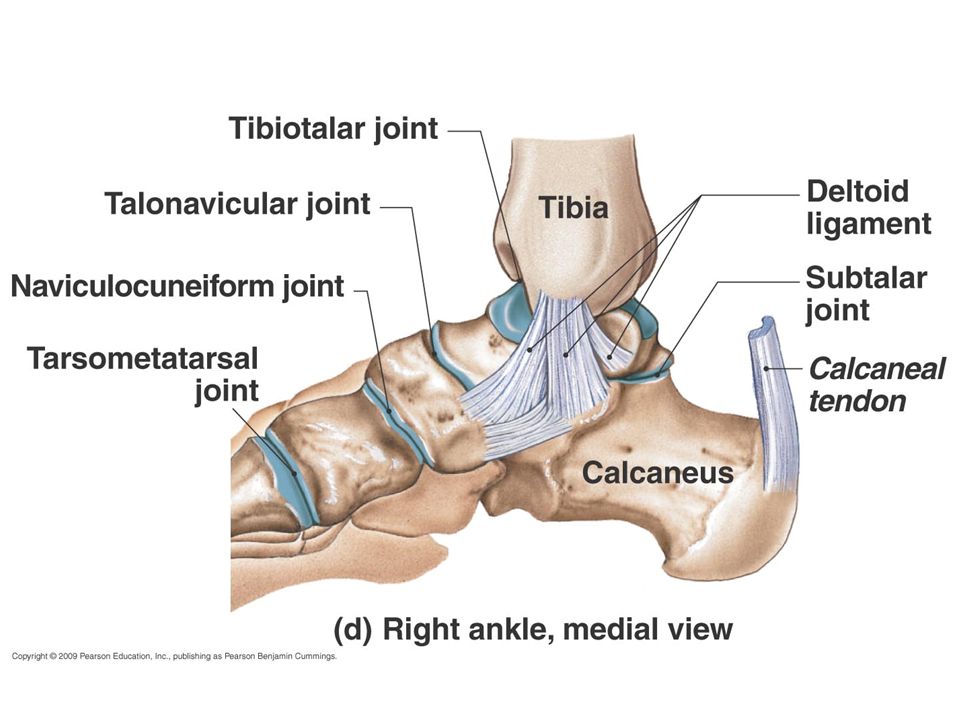
Ankle joint Hinge joint Distal tibia and fibula to talus
Dorsiflexion and plantar flexion only Medial deltoid ligament Lateral ligaments: 3 bands Anterior talofibular Posterior talofibular Calcaneofibular Anterior and posterior tibiofibular (syndesmoses)
")
40
Right ankle, lateral view

42
Temporomandibular joint (TMJ)
Head of mandible articulates with temporal bone Disc protects thin mandibular fossa of temporal bone

43
Sternoclavicular joint
Saddle joint Only other example is trapezium and metacarpal 1 (thumb), allowing opposion Sternum and 1st costal (rib) cartilage articulate with clavicle Very stable: clavicle usually breaks before dislocation of joint Only bony attachment of axial skeleton to pectoral girdle
, allowing opposion. Sternum and 1st costal (rib) cartilage articulate with clavicle. Very stable: clavicle usually breaks before dislocation of joint. Only bony attachment of axial skeleton to pectoral girdle.")
44
Disorders of joints Injuries Inflammatory and degenerative conditions
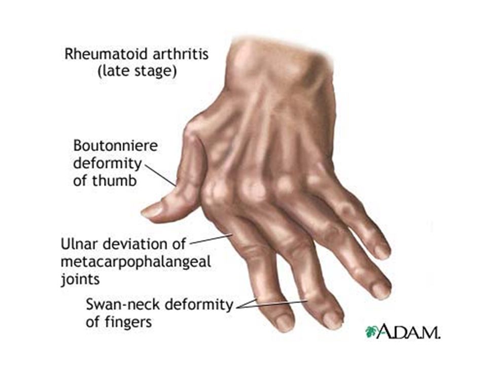
Sprains Dislocations Torn cartilage Inflammatory and degenerative conditions Bursitis Tendonitis Arthritis Osteoarthritis (“DJD” – degenerative joint disease) Rheumatoid arthritis (one of many “autoimmune” joint disorders) Gout (crystal arthropathy)
 Rheumatoid arthritis (one of many autoimmune joint disorders) Gout (crystal arthropathy)")
45
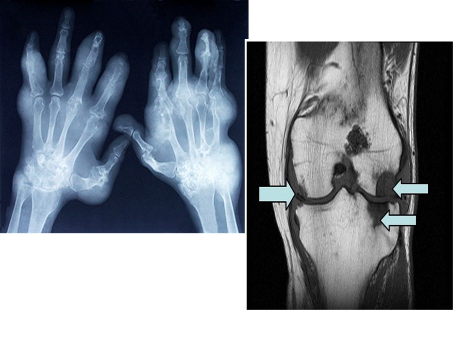
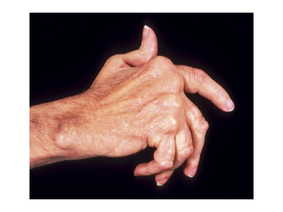
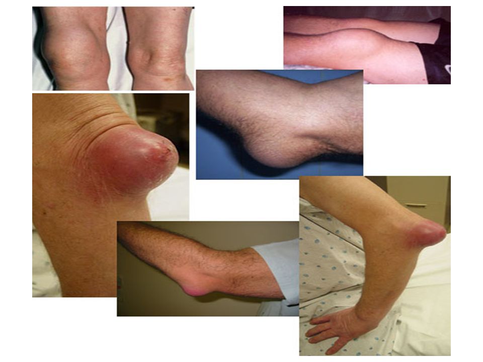
Arthritis

49
Bursitis Inflammation of the Bursa (fluid filled sac surrounding the joint). A bursa can become inflamed from injury, infection (rare in the shoulder), or due to an underlying rheumatic condition. Bursitis is typically identified by localized pain or swelling, tenderness, and pain with motion of the tissues in the affected area.
, or due to an underlying rheumatic condition. Bursitis is typically identified by localized pain or swelling, tenderness, and pain with motion of the tissues in the affected area.")
51
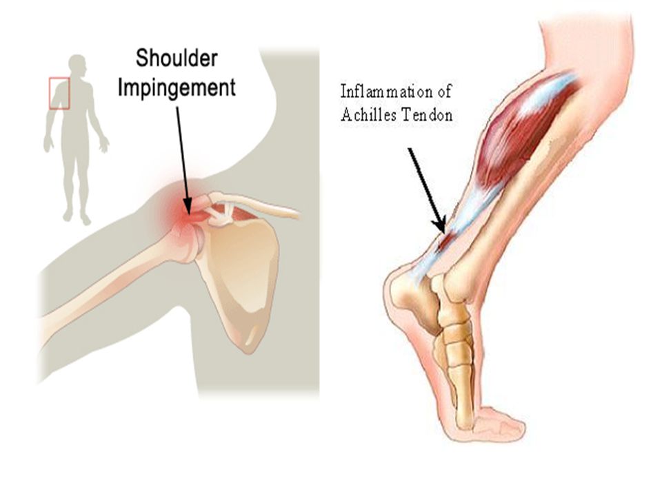
Tendonitis Sometimes the tendons become inflamed for a variety of reasons, and the action of pulling the muscle becomes irritating. If the normal smooth gliding motion of your tendon is impaired, the tendon will become inflamed and movement will become painful. This is called tendonitis, and literally means inflammation of the tendon. The most common cause of tendonitis is overuse.

Similar presentations


>")
: a point of contact between bones. Some allow movement, others are immovable (sutures). Most joints.>")


 and type of substance.>")


 Weakest parts of the skeleton Weakest parts of the skeleton Articulation – site where two or more bones meet Articulation – site.>")
