Download presentation
Presentation is loading. Please wait.

17
Figure 7.20c The first and second cervical vertebrae.

18
Figure 7.27 The humerus of the right arm and detailed views of articulation at the elbow.

19
Figure 7.26b The scapula.

20
Figure 7.28a Radius and ulna of the right forearm.

22
Figure 7.29 Bones of the right hand.

23
Figure 7.31a The hip (coxal) bones.
 bones.")
24
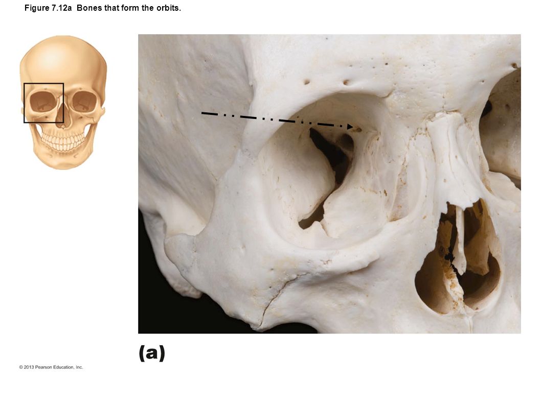
Figure 7.12a Bones that form the orbits.

26
Figure 7.34a Bones of the right foot.

27
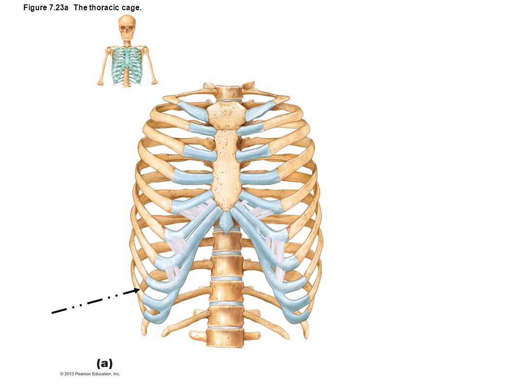
Figure 7.23a The thoracic cage.

29
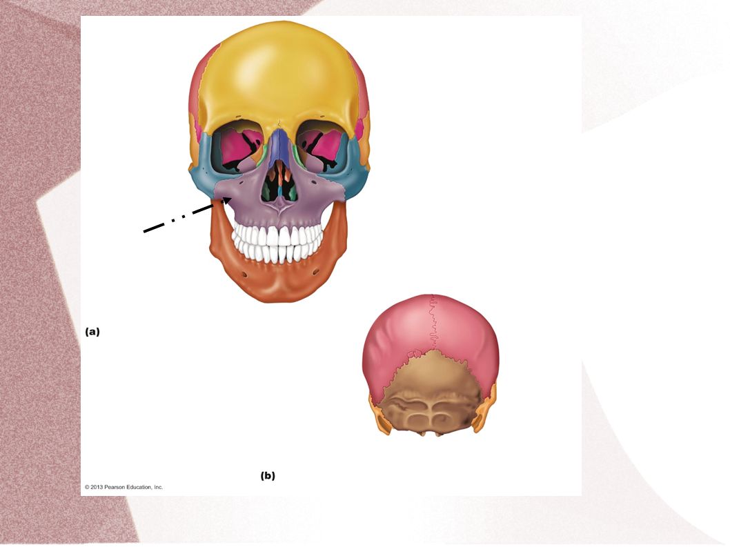
Figure 7.4 Anatomy of the anterior and posterior aspects of the skull.

30
Figure 7.30 Pelvis.

31
Figure 7.29b Bones of the right hand.

32
Figure 7.23a The thoracic cage.

33
Figure 8.1b Fibrous joints.

34
Figure 8.5e Movements allowed by synovial joints.

35
Figure 8.4a Bursae and tendon sheaths.

38
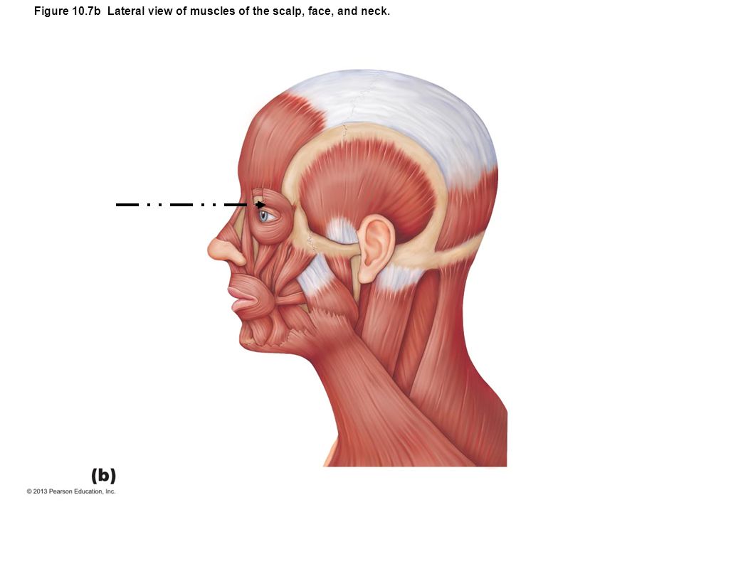
Figure 10.7b Lateral view of muscles of the scalp, face, and neck.

40
Figure 10.24a–b Muscles of the posterior compartment of the right leg.

41
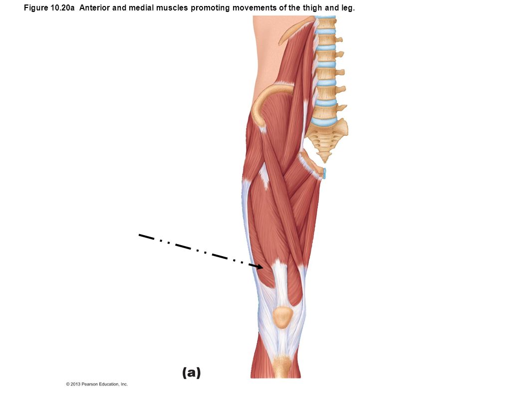
Figure 10.20a Anterior and medial muscles promoting movements of the thigh and leg.

43
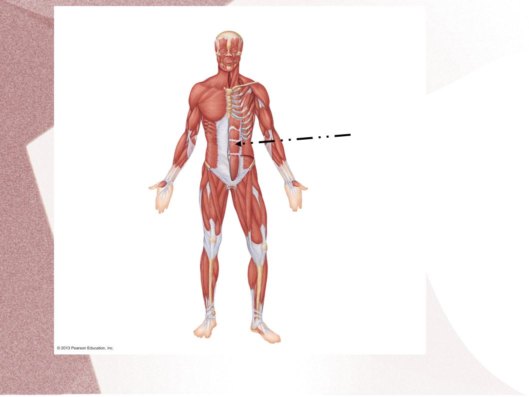
Figure 10.12a Muscles of the abdominal wall.

45
Figure 10.15b Muscles crossing the shoulder and elbow joints, causing movements of the arm and forearm, respectively.

46
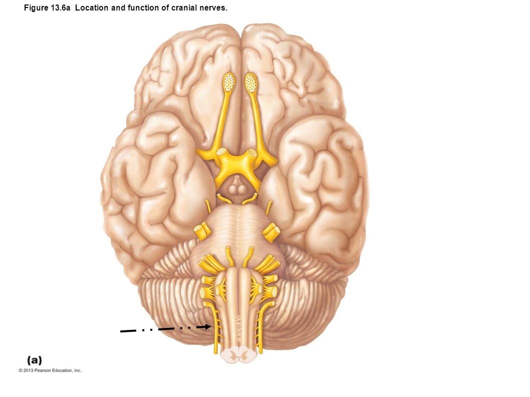
Figure 13.6a Location and function of cranial nerves.

48
Figure 12.10a Midsagittal section of the brain.

49
Figure 12.22 Meninges: dura mater, arachnoid mater, and pia mater.

50
Figure 12.28b Anatomy of the spinal cord.

52
Figure 12.3 Ventricles of the brain.

53
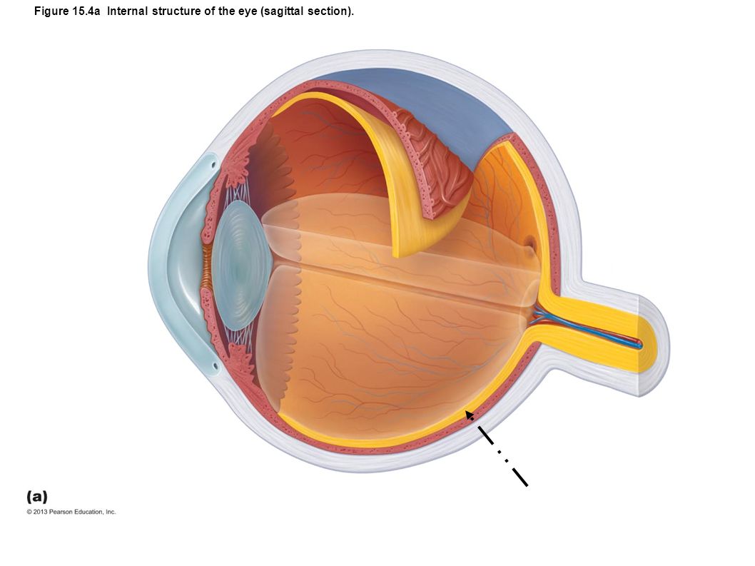
Figure 15.4a Internal structure of the eye (sagittal section).
.")
55
Figure 15.3a Extrinsic eye muscles.

56
Figure 15.2 The lacrimal apparatus.

57
Figure 15.24b Structure of the ear.

58
Figure 15.26 Membranous labyrinth of the internal ear.

59
Figure 15.30a Pathway of sound waves and resonance of the basilar membrane.

60
Figure 15.27b Anatomy of the cochlea.

Similar presentations