Download presentation
Presentation is loading. Please wait.

1
Development of the vertebral column, the locomotor system and the skull
Dr. Károly Altdorfer

2
W.J. Larsen, Human embryology, Churchil Livingstone
K.L. Moore, T.V.N. Persaud, The developing human, Saunders J. Szentágothai, M. Réthelyi, Funkcionális anatómia, Medicina Prof. Mihály Kálmán’s lectures

4
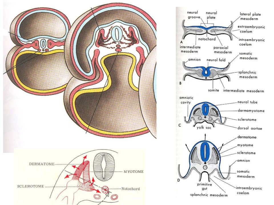
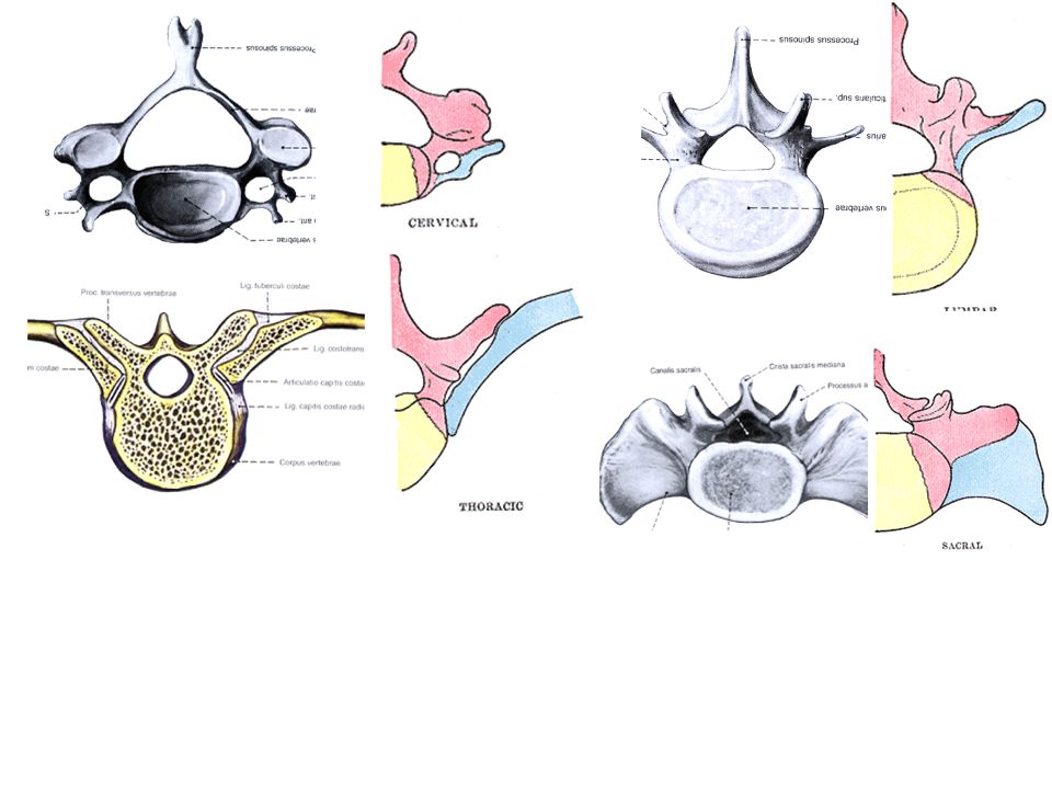
Development of the vertebral column
rearrangement

7
Vertebrae + ribs, sternum (2 sternal bars)
")
10
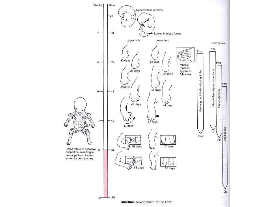
Development of the limbs
From somatopleura

11
Ectoderm: Epithelium, glands of the skin: ectoderm Mesoderm: Bones, joints, vessels, connective tissue: from somatopleura Muscles: somatopleura+myotomes Nerves: neural tube and neural crest

16
Development: innervation shows the origin

17
Thalidomid (Contergan ) – sedative: 1957 – especially recommended for pregnant women…
 – sedative: 1957 – especially recommended for pregnant women…")
18
Amelia, meromelia

19
syndaktylia

20
polydaktylia

21
Development of muscles
Branchial arches Sources: Myotome (trunk muscles with diaphragm, tongue + eye muscles) Somatopleura (limb muscles) Brancial arches (mastication, facial expr.) Tail mesoderm (pelvic floor)
 Somatopleura (limb muscles) Brancial arches (mastication, facial expr.) Tail mesoderm (pelvic floor)")
22
Differentiation of the myotome
(Epimere, hypomere) 1 2 3 4
")
23
Differentiation of the myotome
(Epimere, hypomere) 1 2 3 4
")
24
Differentiation of the myotome
(Epimere, hypomere) 1 2 3 4
")
25
Differentiation of the myotome
(Epimere, hypomere) 1 2 3 4
")
26
Differentiation of the myotome
(Epimere, hypomere) 1 2 3 4
")
28
Development of the diaphragm
Muscle: from C4 myotome (phrenic nerve!) Development: innervation shows the origin…
 Development: innervation shows the origin…")
29
Development of the skull
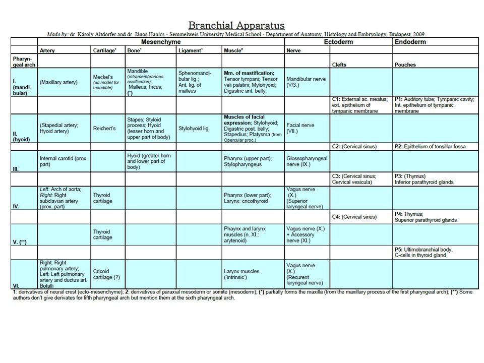
The development of the skull - Viscerocranium (Splanchnocranium = visceral skull, facial skeleton) - Neurocranium (around the brain) Bones can develop by - enchondral ossification (base of skull + some facial bones): chondrocranium intramembranous (calvaria + mandible, maxilla etc.): desmocranium Sources: the embryonic tissue around the developing brain: The first 4 occipital somites lat. parts of occipital bone non-segmented paraxial mesoderm: somitomeres base of the skull (partly) neural crest (ecto-mesenchyme) first two branchial arches (gill arch in fish) splanchnocranium (facial skeleton), bones of calvaria Development of the skull + structures from branchial arches I-II.
 - Neurocranium (around the brain) Bones can develop by. - enchondral ossification (base of skull + some facial bones): chondrocranium. intramembranous (calvaria + mandible, maxilla etc.): desmocranium. Sources: the embryonic tissue around the developing brain: The first 4 occipital somites lat. parts of occipital bone. non-segmented paraxial mesoderm: somitomeres base of the skull (partly) neural crest (ecto-mesenchyme) first two branchial arches (gill arch in fish) splanchnocranium (facial skeleton), bones of calvaria. Development of the skull. + structures from branchial arches I-II.")
30
The development of the skull
- Viscerocranium (Splanchnocranium = visceral skull, facial skeleton) - Neurocranium (around the brain) Bones can develop by - enchondral ossification (base of skull + some facial bones) intramembranous (calvaria + mandible, maxilla etc.) Sources: the embryonic tissue around the developing brain: The first 4 occipital somites lat. parts of occipital bone non-segmented paraxial mesoderm: somitomeres base of the skull (partly) neural crest (ecto-mesenchyme) first two branchial arches (gill arch in fish) splanchnocranium (facial skeleton), bones of calvaria Prechordal or
 - Neurocranium (around the brain) Bones can develop by. - enchondral ossification (base of skull + some facial bones) intramembranous (calvaria + mandible, maxilla etc.) Sources: the embryonic tissue around the developing brain: The first 4 occipital somites lat. parts of occipital bone. non-segmented paraxial mesoderm: somitomeres base of the skull (partly) neural crest (ecto-mesenchyme) first two branchial arches (gill arch in fish) splanchnocranium (facial skeleton), bones of calvaria. Prechordal or.")
31
- at the base of the skull and - at the face (from the nasal capsule).
Chondrocranium - enchondral ossification: cartilage model turns into bone - at the base of the skull and - at the face (from the nasal capsule). The base of the skull: occipital bone (except upper part of its squama), petrous part of temporal (around the ectodermal acoustic placod -organ of hearing), sphenoid: body, wings and lateral (!) plates of pterygoid process. The nasal capsule ethmoid, inferior nasal concha. The remaining parts of the cartilaginous nasal capsule give the nasal cartilages which persist in adults. + structures from branchial arches I-II.
. The base of the skull: occipital bone (except upper part of its squama), petrous part of temporal (around the ectodermal acoustic placod -organ of hearing), sphenoid: body, wings and lateral (!) plates of pterygoid process. The nasal capsule ethmoid, inferior nasal concha. The remaining parts of the cartilaginous nasal capsule give the nasal cartilages which persist in adults. + structures from branchial arches I-II.")
32
Desmocranium - intramembranous ossification: from the connective tissue covering of the developing brain. The first centers of ossification: tuber frontalia and parietalia ossification of the connective tissue spreads radially. Calvaria: frontal, parietal, squamous part of temporal, tympanic part of temporal bones. On the face: nasal, lacrimal, maxilla, zygomatic, vomer, palatine; medial plate of pterygoid process. Mandible also, but it develops from branchial arch. + structures from branchial arches I-II.

33
The splanchnocranium originally develops from the first and second branchial arches.
1. The first (or mandibular) branchial arch contains the Meckel's cartilage. The Meckel's cartilage in the cartilaginous fish is an ancient form of the jaw which articulates with the cartilagineous part of temporal bone. The posterior part of the Meckel’s cartilage is transformed into the malleus and incus (2 auditory ossicles, the hammer and the anvil). Around (!) the Meckel’s cartilage model: the mandible is ossified via intramembranous ossification. 2. The second (or hyoid) branchial arch contains the Reichert's cartilage. The derivatives: the upper part of the hyoid bone*, the styloid process* and the stapes (the third auditory ossicle, the stirrups). (*Between them the mesechyme forms the stylo-hyoid ligament.)
 branchial arch contains the Meckel s cartilage. The Meckel s cartilage in the cartilaginous fish is an ancient form of the jaw which articulates with the cartilagineous part of temporal bone. The posterior part of the Meckel’s cartilage is transformed into the malleus and incus (2 auditory ossicles, the hammer and the anvil). Around (!) the Meckel’s cartilage model: the mandible is ossified via intramembranous ossification. 2. The second (or hyoid) branchial arch contains the Reichert s cartilage. The derivatives: the upper part of the hyoid bone*, the styloid process* and the stapes (the third auditory ossicle, the stirrups). (*Between them the mesechyme forms the stylo-hyoid ligament.)")
35
Which type of ossification?
Where there is no clear ’model’ (shape) for a body area yet and an expansive development of the body part is needed (e.g. limbs, face): first cartilage appears, later enchondral ossification will generate the model for the body part. Where there is a pre-existing organ with well defined shape and it has an ability for expansive growth (e.g. the brain!), it is enough to ossify its connective tissue capsule (=intramembranous ossification). Malformations: Problems with the endochondral ossification: chondrodystrophia results in a serious problem in limb longitudinal growth and chondral skull bone growth dwarfs with depressed nasal root and huge, balloon-like neurocranium and very prominent facial bones. Problems with the intramembranous ossification: the calvaria of the skull and the clavicle are poorly developed (dysostosis cleidocranialis).
 for a body area yet and an expansive development of the body part is needed (e.g. limbs, face): first cartilage appears, later enchondral ossification will generate the model for the body part. Where there is a pre-existing organ with well defined shape and it has an ability for expansive growth (e.g. the brain!), it is enough to ossify its connective tissue capsule (=intramembranous ossification). Malformations: Problems with the endochondral ossification: chondrodystrophia results in a serious problem in limb longitudinal growth and chondral skull bone growth dwarfs with depressed nasal root and huge, balloon-like neurocranium and very prominent facial bones. Problems with the intramembranous ossification: the calvaria of the skull and the clavicle are poorly developed (dysostosis cleidocranialis).")
Similar presentations