Download presentation
Presentation is loading. Please wait.

1
PTA 120 Pathophysiology Week 5

2
Objectives Discuss anatomic structures and physiologic processes related to the skeletal system. Discuss physical effects of aging on the skeletal system. Define skeletal pathological conditions including signs and symptoms of each. Discuss how skeletal pathologies can adversely affect function requiring the modification of treatment intervention to protect the patient from worsening the condition and optimize treatment outcomes. Discuss the modifications and precaution that may be required for the treatment of patients with skeletal disorders. Demonstrate understanding of the PTA’s role in the disease processes.

3
Textbooks Pathology for the Physical Therapist Assistant, Ch. 1 pp. 23 – 25, Ch. 5, Ch. 6 pp. 213 – 250 Physical Therapy Clinical Handbook for PTAs

6
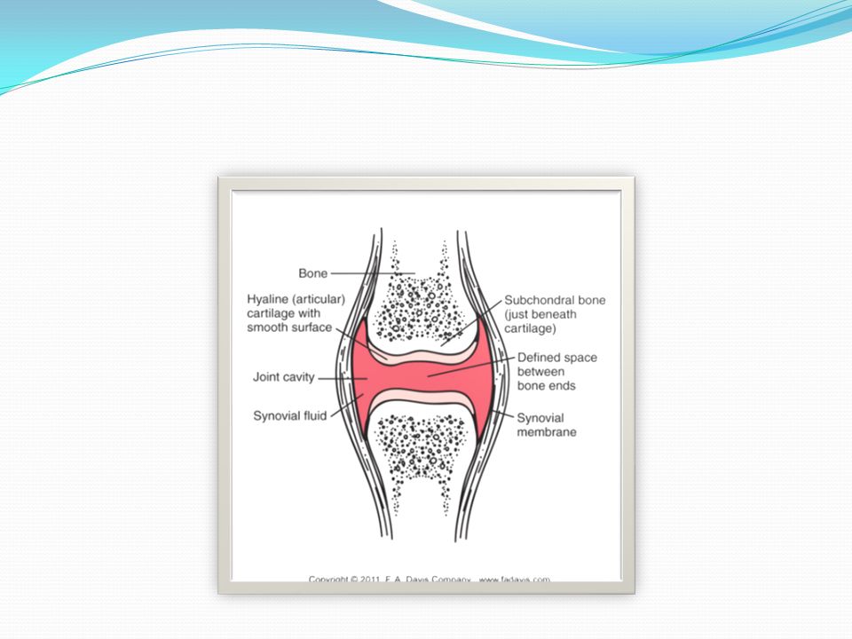
Functions of Bones Support of the body Protection of soft organs Movement due to attached skeletal muscles Storage of minerals and fats Blood cell formation

7
Effects of Aging Physiologic ChangeFunctional Effect Bone mass decreasesBones become more porous, brittle, and fragile Increased instances of osteoporosis and pathologic fractures Changes in joint structuresArticular cartilage erodes, synovial fluid becomes fibrotic Decreased mobility; altered gait Intervertebral discs dehydrate and narrow Spinal changes such as decreased height and kyphosis Altered gait

8
Healing time Bone 3-4days- Fractured edges become necrotic. Osteoclasts mobilize in the area. Up to 4 wks- Osteoclasts form soft callus. 3-4wks- Hard callus develops. 3-4 mos- Fracture is healed, but remodeling continues. Concorde Career College

9
Bone Healing

10
Osteoarthritis Description Also known as Degenerative Joint Disease, Degenerative Arthritis, Osteoarthrosis, OA Progressive damage and loss of articular cartilage Joint spaces narrow Older and elderly adults Most common form of arthritis, most commonly seen in hips, knees, hands, feet

11
Osteoarthritis Progression of OA Cartilage becomes thin and soft, begins to flake off and enter joint cavity Damaged tissues stimulates enzymes that accelerate the degenerative process Surface defects appear and expose underlying bone, which then becomes sclerotic Cysts develop, and new bone growths occur All events lead to inflammation and swelling

12
Osteoarthritis From McCance KL, Heuther SE: Pathophysiology: the biologic basis for disease in adults and children, ed 5, St. Louis, 2006, Mosby.

14
Osteoarthritis Etiology Aging process Can be inherited Trauma and long-term stress can accelerate the process Pain on weight bearing Stiffness after inactivity Reduced range of motion Muscle atrophy, weakness, and spasm Joint deformity and enlargement Joint crepitus Reduced or loss of function

15
Osteoarthritis Joints may enlarge and present with nodes Buochard nodes on proximal IP joints Heberden nodes on distal IP joints From Lemmi FO, Lemmi CAE: Physical assessment findings CD-ROM, Philadelphia, 2000, WB Saunders.

16
Osteoarthritis Treatment NSAIDS and other medications Injections Surgery to remove torn cartilage or small pieces of bone, or to perform joint replacements Dietary changes for weight loss

17
physical therapy intervention Osteoarthritis Improve functional status Reduce pain Modalities Patient education

18
Spondylosis Description Degenerative change in the spine Commonly affects the neck Result in bone spur growths that can put pressure on spinal nerves or the spinal cord Etiology Aging process Laxity in spinal ligaments, herniated discs Previous spinal injury

19
Clinical picture of spondylosis Symptoms begin after a period of worry or stress Headaches, neck aches, as well as shoulder and arm pain Neck weakness Radiologic changes

20
Clinical picture of spondylosis Referred pain into the arm Pain in sacroiliac region, buttocks, and hips Muscle spasms Altered sensation and paresthesias Limited range of motion

21
Spondylosis Treatment Analgesics, anti-inflammatories Decompressive surgery Cortisone injections

22
physical therapy intervention Spondylosis Restore function through postural re-education and exercises Reduce pain with use of heat, massage, relaxation exercises Modalities Patient education

23
Spondylolysis Description Structural deformity in the pars interarticularis of lumbar spine vertebrae Usually occurs at L4/L5 or L5/S1 Incidence is 3-7% of the U.S. population, increases with athletes in contact sports or gymnastics Can cause anterior slipping of the L5 vertebra over the sacrum called spondylolisthesis

24
Spondylolysis

25
Etiology Cause unknown Repeated microtrauma from gymnastics, weight lifting, football Genetic defect Spina bifida occulta

26
Spondylolysis Signs and Symptoms May be clinically absent Discovered on x-ray Treatment Inactivity Analgesics Spinal fusion

27
physical therapy intervention Spondylolysis Abdominal and extensor muscle strengthening Postural re-education, intensive functional re- education Lifestyle adaptation

28
Spondylolisthesis Description Vertebra becomes anteriorly displaced Usually affects L4/L5 or L5/S1 Age of onset usually > 40, women > men (3:1) Etiology Disc degeneration and bone disease (arthritis) Birth defect or trauma to vertebral column
 Etiology Disc degeneration and bone disease (arthritis) Birth defect or trauma to vertebral column")
29
Spondylolisthesis

30
Signs and Symptoms Low back pain, referred pain Muscle spasm Increased lumbar lordosis Treatment Analgesics Exercise Spinal fusion

31
physical therapy intervention Spondylolisthesis Abdominal strengthening Postural re-education Modalities for pain relief Abdominal binder Patient education

32
Infective Arthritis Description Also called Septic Arthritis Infection of the joint Children and elderly most commonly affected Knee is most affected joint

33
Infective Arthritis Etiology Bacteria Staphylococcus, Streptococcus pneumoniae, Neisseria gonorrhoeae, Mycobacterium tuberculosis Viruses and fungi

34
Infective Arthritis Signs and Symptoms Fever, fatigue, irritability, loss of appetite Affected joint becomes red and swollen Severe pain on movement and decreased range of motion

35
Infective Arthritis Treatment Immediate, aggressive antimicrobial treatment Rest and elevation Fluid aspiration

36
physical therapy intervention Infective Arthritis After the infection fully treated by physician Strengthening and stretching to decrease stiffness Endurance activities

37
Diseases of bone Why does the PTA need to know about diseases of the bone?

38
Diseases of Bone … to enable appropriate development of exercise programs and interventions, taking into account comorbid diagnoses for patients, as well as understanding the precautions and contraindications for these patients.

39
Osteoporosis Description Systemic condition and metabolic disease involving a wasting or deterioration of bone in mass and density Combination of decreased bone mass and microdamage to the bone structure that results in a susceptibility to fracture Etiology Bone reabsorption exceeds bone formation

40
Osteoporosis Normal Bone Osteoporotic Bone

41
Osteoporosis Risk factors Non-modifiable Female Age > 50 Caucasian / Asian Thin, small-frame Early menopause ( < age 45) Family history
 Family history")
42
Osteoporosis Modifiable: Sedentary lifestyle Diet and nutrition Low calcium intake Smoking Excessive alcohol intake (> 2 drinks / day) Excessive caffeine intake (equivalent to > 3 cups of caffeinated coffee) Excessive tobacco use Certain medications Low body weight and BMI
 Excessive caffeine intake (equivalent to > 3 cups of caffeinated coffee) Excessive tobacco use Certain medications Low body weight and BMI")
43
Osteoporosis Signs and Symptoms Loss of height, leading to breathing problems and eating difficulties Marked thoracic spine kyphosis and “Dowager’s Hump” Fractures, commonly in the wrist or hip Low back and neck pain

44
Osteoporosis

45
Compression fracture of the vertebra

46
Osteoporosis Treatment Prevention is best Estrogen replacement therapy Calcium and vitamin D supplements Regular weight-bearing exercise Lifestyle changes to decrease risk factors

47
physical therapy intervention Osteoporosis Contraindications include spinal flexion exercises, mobilization techniques, traction Patient education regarding exercises and postural advice Spinal extension exercises Heat, massage, spinal support to reduce pain Balance training if needed

49
PTA 120 Pathophysiology Day 3

51
Abnormal curvatures

52
Kyphosis Description Exaggeration of the normal posterior thoracic curvature Excessive rounding > 45-50 degrees (20-40 degrees is normal) Can have rounded shoulders and dowager’s hump
 Can have rounded shoulders and dowager’s hump")
53
Kyphosis Kamal A, Brockelhurst JC: Color atlas of geriatric medicine, ed 2, St. Louis, 1992, Mosby.

54
Kyphosis Etiology Can be from posture or disease Chronic spasticity of pectoralis major and minor, serratus anterior, weak rhomboid major and minor Osteoporosis in older persons Tuberculosis of vertebral bodies, ankylosing spondylitis, cancer, benign tumors, spina bifida, cerebral palsy, poliomyelitis

55
Kyphosis Signs and Symptoms Asymptomatic until hump becomes obvious Mild back pain and fatigue Decreased mobility of the spine Rounded back and shoulders Chest may cave in, head moves forward

56
Kyphosis Treatment Exercises Bracing Spinal fusion

57
physical therapy intervention Kyphosis Strengthening of the back muscles Stretching of the chest muscles Postural education

58
Lordosis Description Also known as hyperlordosis, saddleback, or swayback Exaggeration of the normal anterior curvature of the lumbar spine Can lead to degenerative disc disease or herniated discs in the lumbar spine

59
Lordosis

60
Etiology Postural compensation for added abdominal mass, girth, as in pregnancy and obesity May accompany spinal disease such as osteoporosis or spondylolisthesis

61
Lordosis Signs and Symptoms Can be asymptomatic Low back pain due to strain on muscles and ligaments Protruding abdomen and buttocks, arched lower back

62
Lordosis Treatment If caused by pregnancy, delivery of the baby resolves the condition Weight loss Spinal fusion

63
physical therapy intervention Lordosis Exercises for weight loss Strengthening of abdominal muscles Pelvic tilt exercises, stretching of back extensors Postural education

64
Scoliosis Description Lateral curvature in the normally straight vertical line of the spine Affects approximately 2% of the population Girls > boys (5:1)
")
65
Scoliosis Etiology Structural Unknown (80% of cases), or a congenital malformation of the spine and diseases Causes a permanent change in the spine Functional Caused by pain or poor posture Appears to change configuration when the patient moves
, or a congenital malformation of the spine and diseases Causes a permanent change in the spine Functional Caused by pain or poor posture Appears to change configuration when the patient moves")
66
Scoliosis Etiology Can result from other disorders such as muscular dystrophy or spinal cord injuries

67
Scoliosis Signs and Symptoms Best observed from posterior view Unilateral fullness in a segment of the paraspinal region Caused by protrusion of transverse process of the rotated vertebrae On the side of curve’s convexity

68
Scoliosis Waistline uneven Hip and shoulder malalignment One scapula is more prominent Leaning to one side Restricted and painful breathing in severe cases

69
Scoliosis From Gould BE: Pathophysiology for the health professions, ed 3, 2006, St. Louis, Saunders.

70
Scoliosis Treatment Monitoring Body / back brace if curve is > 20 degrees Surgery if curve is > 40 -45 degrees Spinal fusion

71
physical therapy intervention Scoliosis Functional scoliosis can be improved by exercises and postural education to restore alignment of the spine PT goal for structural scoliosis is to ensure good functioning of cardiac and respiratory system

72
Slipped Capital Femoral Epiphysis Description SCFE Femoral head slides on the femur at the epiphysis during bone growth Femur becomes laterally rotated Etiology Traumatic twisting injury Boys > girls, between 11 and 16 years old Obesity or growing rapidly

73
Slipped Capital Femoral Epiphysis Signs and Symptoms Pain, restriction of movement at hip Knee pain, referred from the hip Impaired ambulation Treatment Surgery

74
physical therapy intervention Slipped Capital Femoral Epiphysis Strengthening exercises Gait training with crutches or a walker for change in weight- bearing status after surgery Patient education in weight- bearing precautions

75
Paget’s Disease Description Also called osteitis deformans Bone resorption followed by excessive bone formation, resulting in weakened, deformed bones of increased mass Seldom seen before 40 years of age, usually affects those older than 70 Affects men > women

76
Paget’s Disease

77
Etiology Unknown Slowly developing virus of osteoclasts May be genetic link

78
Paget’s Disease Signs and Symptoms Can be asymptomatic Minor skeletal pain, worse at night Kyphosis, bowlegs, larger/misshapen skull Thickened cranial bones may compress nerves and cause headaches, hearing loss or dizziness From Damjanov I: Pathology for the health-related professions, ed 2, Philadelphia, 2000, WB Saunders.

79
Paget’s Disease Treatment Medications to inhibit bone resorption and decrease pain

80
physical therapy intervention Paget’s Disease Fall prevention Ambulation re-training Exercises for strengthening and endurance

82
Bone Cancer Description Malignant tumors that originate in bone, marrow, and cartilage Common bone cancers – characterized by primary tissue type, peak age, bones involved Ostoesarcoma – growing bone Chondrosarcoma – cartilage Ewing sarcoma – marrow of growing bone

83
Bone Cancer Metastatic or secondary bone tumors are common Cancers that metastasize include breast, lung, prostate, thyroid, kidney The bones most affected with metastatic tumors are the pelvis, spine, ribs, hips, femur, humerus

84
Bone Cancer Risk factors Cancer in other parts of the body Paget’s disease History of radiation therapy

85
Bone Cancer Signs and Symptoms Pain increases at night Localized swelling, limited movement Palpable mass May develop fever, fatigue, anemia, pathologic fracture in the later stages Possible leg length discrepancy May mimic arthritis, requiring referral back to the physician

86
Bone Cancer Treatment Surgical excision Chemotherapy and radiation Joint replacement Limb amputation Other medications, such as ones used mostly for arthritis

87
physical therapy intervention Bone Tumors Assist patients to return to functional activity after surgery or chemotherapy Ambulation re-training for non-weight-bearing status Exercises for strengthening and endurance Transfer and Hoyer lift training if appropriate

89
Talipes Equinovarus Description Also called clubfoot Foot and ankle held in plantarflexion and inversion Affects 1 in 1000 live births Males > females Etiology Genetic trait, family history of clubfoot Cerebral palsy or spina bifida

90
Talipes Equinovarus Signs and symptoms Ankle is plantarflexed, subtaloid and mid-tarsal joints are adducted and inverted Can be bilateral or unilateral Can be postural or positional Treatment Splinting Surgery

91
Talipes Equinovarus

92
physical therapy intervention Talipes Equinovarus Positioning and stretching Weight-bearing activities after cast removal

93
Developmental Dysplasia of the Hip Description Congenital hip disorder affecting infants and children Unilateral or bilateral Three stages or types Unstable hip dysplasia Subluxation or incomplete dislocation Complete dislocation

94
Developmental Dysplasia of the Hip Etiology Hereditary Hip ligament laxity, shallow acetabular shape Breech deliveries First born infants

95
Developmental Dysplasia of the Hip Signs and Symptoms Shortening of the affected leg Trendelenburg gait pattern Decreased active movement and external rotation in affected leg Asymmetry in buttock or gluteal folds, extra thigh skin folds Spinal scoliosis and lumbar lordosis associated

96
Developmental Dysplasia of the Hip Treatment Early detection is important Positioning in spica cast or Pavlik harness Surgery

97
Developmental Dysplasia of the Hip

98
physical therapy intervention Developmental Dysplasia of the Hip Positioning and stretching Weight-bearing activities after cast removal

99
Osteogenesis Imperfecta Description Also known as OI or “Brittle Bone Disease” Disorder of bones and connective tissue resulting in extremely fragile bones Staged I through VIII by characteristics, not by severity Etiology Defect in genes that are responsible for collagen synthesis, which results in brittle bones

100
Osteogenesis Imperfecta Signs and Symptoms Bones are easily fractured, often while still in the uterus Triangular-shaped head, thin skin, reduced enamel on the teeth Scoliosis and bone deformities where fractures have healed, osteoporosis Decreased height due to lack of bone growth Weakened heart valves

101
Osteogenesis Imperfecta From J Med Genet 2002;39:23-29

102
Osteogenesis Imperfecta Treatment Casting, bracing, and splinting Try to avoid surgery Medications to increase bone strength

103
physical therapy intervention Osteogenesis Imperfecta Encourage gross motor skills Resistive exercises are contraindicated Positioning and seating systems Gait training with assistive devices Patient, family, and school education for handling and fall prevention Splinting and bracing Pool therapy

104
Spina Bifida Description Neural tube defect Posterior spinous processes of vertebral column do not close properly during development in utero Bony gap can permit meninges, spinal fluids and spinal cord to hernitate, resulting in neurological impairment Caucasian and Hispanic descent > risk than African-American descent

105
Spina Bifida Incidence is 17.96 per 100,000 live births affecting 200,000 people in the US Three types Spina bifida occulta – condition is hidden Meningocele – meninges and spinal fluid protrude through bony defect Myelomeningocele – herniation of spinal cord, nerves, meninges, spinal fluid Most serious form Neurologic impairment

106
Spina Bifida

107
Etiology Unknown Maternal folic-acid deficiency and genetic alterations are associated Maternal diabetes, use of anti-seizure medications or exposure to high temperatures during pregnancy Caucasian and Hispanic descent > risk than African-Americans

108
Spina Bifida Signs and Symptoms Spina bifida occulta Skin depression, dark tuft of hair, hemangioma, subcutaneous mass Meningocele or myelomeningocele Sac on the back Weakness and paralysis possible if nerves involved

109
Spina Bifida Hydrocephalus, often requiring surgery Hypotonia Bowel and bladder dysfunction Scoliosis, dislocated hip joint contractures, talipes equinovarus Pressure ulcers

110
Spina Bifida Treatment Dependent on the degree of severity Spina bifida occulta No treatment Meningocele and myelomeningocele Surgically closed after birth Defects can be seen in ambulation, bowel or bladder function

111
physical therapy intervention Spina Bifida Assessment of and training in adaptive equipment and wheelchairs Precautions surrounding shunt for hydrocephalus Family education including ROM and signs of shunt malfunction

112
Other skeletal disorders Hemophilic Arthritis Lyme Disease Gout Rickets Osteomalacia Legg-Calve-Perthes Disease Osteomyelitis Marfan’s Syndrome

113
Other diseases which can affect the skeletal system HIV / AIDS Syphilis Tuberculosis in the Bone Lyme Disease

115
PTA 120 Pathophysiology Day 3

117
Abnormal curvatures

118
Kyphosis Description Exaggeration of the normal posterior thoracic curvature Excessive rounding > 45-50 degrees (20-40 degrees is normal) Can have rounded shoulders and dowager’s hump
 Can have rounded shoulders and dowager’s hump")
119
Kyphosis Kamal A, Brockelhurst JC: Color atlas of geriatric medicine, ed 2, St. Louis, 1992, Mosby.

120
Kyphosis Etiology Can be from posture or disease Chronic spasticity of pectoralis major and minor, serratus anterior, weak rhomboid major and minor Osteoporosis in older persons Tuberculosis of vertebral bodies, ankylosing spondylitis, cancer, benign tumors, spina bifida, cerebral palsy, poliomyelitis

121
Kyphosis Signs and Symptoms Asymptomatic until hump becomes obvious Mild back pain and fatigue Decreased mobility of the spine Rounded back and shoulders Chest may cave in, head moves forward

122
Kyphosis Treatment Exercises Bracing Spinal fusion

123
physical therapy intervention Kyphosis Strengthening of the back muscles Stretching of the chest muscles Postural education

124
Lordosis Description Also known as hyperlordosis, saddleback, or swayback Exaggeration of the normal anterior curvature of the lumbar spine Can lead to degenerative disc disease or herniated discs in the lumbar spine

125
Lordosis

126
Etiology Postural compensation for added abdominal mass, girth, as in pregnancy and obesity May accompany spinal disease such as osteoporosis or spondylolisthesis

127
Lordosis Signs and Symptoms Can be asymptomatic Low back pain due to strain on muscles and ligaments Protruding abdomen and buttocks, arched lower back

128
Lordosis Treatment If caused by pregnancy, delivery of the baby resolves the condition Weight loss Spinal fusion

129
physical therapy intervention Lordosis Exercises for weight loss Strengthening of abdominal muscles Pelvic tilt exercises, stretching of back extensors Postural education

130
Scoliosis Description Lateral curvature in the normally straight vertical line of the spine Affects approximately 2% of the population Girls > boys (5:1)
")
131
Scoliosis Etiology Structural Unknown (80% of cases), or a congenital malformation of the spine and diseases Causes a permanent change in the spine Functional Caused by pain or poor posture Appears to change configuration when the patient moves
, or a congenital malformation of the spine and diseases Causes a permanent change in the spine Functional Caused by pain or poor posture Appears to change configuration when the patient moves")
132
Scoliosis Etiology Can result from other disorders such as muscular dystrophy or spinal cord injuries

133
Scoliosis Signs and Symptoms Best observed from posterior view Unilateral fullness in a segment of the paraspinal region Caused by protrusion of transverse process of the rotated vertebrae On the side of curve’s convexity

134
Scoliosis Waistline uneven Hip and shoulder malalignment One scapula is more prominent Leaning to one side Restricted and painful breathing in severe cases

135
Scoliosis From Gould BE: Pathophysiology for the health professions, ed 3, 2006, St. Louis, Saunders.

136
Scoliosis Treatment Monitoring Body / back brace if curve is > 20 degrees Surgery if curve is > 40 -45 degrees Spinal fusion

137
physical therapy intervention Scoliosis Functional scoliosis can be improved by exercises and postural education to restore alignment of the spine PT goal for structural scoliosis is to ensure good functioning of cardiac and respiratory system

138
Slipped Capital Femoral Epiphysis Description SCFE Femoral head slides on the femur at the epiphysis during bone growth Femur becomes laterally rotated Etiology Traumatic twisting injury Boys > girls, between 11 and 16 years old Obesity or growing rapidly

139
Slipped Capital Femoral Epiphysis Signs and Symptoms Pain, restriction of movement at hip Knee pain, referred from the hip Impaired ambulation Treatment Surgery

140
physical therapy intervention Slipped Capital Femoral Epiphysis Strengthening exercises Gait training with crutches or a walker for change in weight- bearing status after surgery Patient education in weight- bearing precautions

141
Paget’s Disease Description Also called osteitis deformans Bone resorption followed by excessive bone formation, resulting in weakened, deformed bones of increased mass Seldom seen before 40 years of age, usually affects those older than 70 Affects men > women

142
Paget’s Disease

143
Etiology Unknown Slowly developing virus of osteoclasts May be genetic link

144
Paget’s Disease Signs and Symptoms Can be asymptomatic Minor skeletal pain, worse at night Kyphosis, bowlegs, larger/misshapen skull Thickened cranial bones may compress nerves and cause headaches, hearing loss or dizziness From Damjanov I: Pathology for the health-related professions, ed 2, Philadelphia, 2000, WB Saunders.

145
Paget’s Disease Treatment Medications to inhibit bone resorption and decrease pain

146
physical therapy intervention Paget’s Disease Fall prevention Ambulation re-training Exercises for strengthening and endurance

148
Bone Cancer Description Malignant tumors that originate in bone, marrow, and cartilage Common bone cancers – characterized by primary tissue type, peak age, bones involved Ostoesarcoma – growing bone Chondrosarcoma – cartilage Ewing sarcoma – marrow of growing bone

149
Bone Cancer Metastatic or secondary bone tumors are common Cancers that metastasize include breast, lung, prostate, thyroid, kidney The bones most affected with metastatic tumors are the pelvis, spine, ribs, hips, femur, humerus

150
Bone Cancer Risk factors Cancer in other parts of the body Paget’s disease History of radiation therapy

151
Bone Cancer Signs and Symptoms Pain increases at night Localized swelling, limited movement Palpable mass May develop fever, fatigue, anemia, pathologic fracture in the later stages Possible leg length discrepancy May mimic arthritis, requiring referral back to the physician

152
Bone Cancer Treatment Surgical excision Chemotherapy and radiation Joint replacement Limb amputation Other medications, such as ones used mostly for arthritis

153
physical therapy intervention Bone Tumors Assist patients to return to functional activity after surgery or chemotherapy Ambulation re-training for non-weight-bearing status Exercises for strengthening and endurance Transfer and Hoyer lift training if appropriate

155
Talipes Equinovarus Description Also called clubfoot Foot and ankle held in plantarflexion and inversion Affects 1 in 1000 live births Males > females Etiology Genetic trait, family history of clubfoot Cerebral palsy or spina bifida

156
Talipes Equinovarus Signs and symptoms Ankle is plantarflexed, subtaloid and mid-tarsal joints are adducted and inverted Can be bilateral or unilateral Can be postural or positional Treatment Splinting Surgery

157
Talipes Equinovarus

158
physical therapy intervention Talipes Equinovarus Positioning and stretching Weight-bearing activities after cast removal

159
Developmental Dysplasia of the Hip Description Congenital hip disorder affecting infants and children Unilateral or bilateral Three stages or types Unstable hip dysplasia Subluxation or incomplete dislocation Complete dislocation

160
Developmental Dysplasia of the Hip Etiology Hereditary Hip ligament laxity, shallow acetabular shape Breech deliveries First born infants

161
Developmental Dysplasia of the Hip Signs and Symptoms Shortening of the affected leg Trendelenburg gait pattern Decreased active movement and external rotation in affected leg Asymmetry in buttock or gluteal folds, extra thigh skin folds Spinal scoliosis and lumbar lordosis associated

162
Developmental Dysplasia of the Hip Treatment Early detection is important Positioning in spica cast or Pavlik harness Surgery

163
Developmental Dysplasia of the Hip

164
physical therapy intervention Developmental Dysplasia of the Hip Positioning and stretching Weight-bearing activities after cast removal

165
Osteogenesis Imperfecta Description Also known as OI or “Brittle Bone Disease” Disorder of bones and connective tissue resulting in extremely fragile bones Staged I through VIII by characteristics, not by severity Etiology Defect in genes that are responsible for collagen synthesis, which results in brittle bones

166
Osteogenesis Imperfecta Signs and Symptoms Bones are easily fractured, often while still in the uterus Triangular-shaped head, thin skin, reduced enamel on the teeth Scoliosis and bone deformities where fractures have healed, osteoporosis Decreased height due to lack of bone growth Weakened heart valves

167
Osteogenesis Imperfecta From J Med Genet 2002;39:23-29

168
Osteogenesis Imperfecta Treatment Casting, bracing, and splinting Try to avoid surgery Medications to increase bone strength

169
physical therapy intervention Osteogenesis Imperfecta Encourage gross motor skills Resistive exercises are contraindicated Positioning and seating systems Gait training with assistive devices Patient, family, and school education for handling and fall prevention Splinting and bracing Pool therapy

170
Spina Bifida Description Neural tube defect Posterior spinous processes of vertebral column do not close properly during development in utero Bony gap can permit meninges, spinal fluids and spinal cord to hernitate, resulting in neurological impairment Caucasian and Hispanic descent > risk than African-American descent

171
Spina Bifida Incidence is 17.96 per 100,000 live births affecting 200,000 people in the US Three types Spina bifida occulta – condition is hidden Meningocele – meninges and spinal fluid protrude through bony defect Myelomeningocele – herniation of spinal cord, nerves, meninges, spinal fluid Most serious form Neurologic impairment

172
Spina Bifida

173
Etiology Unknown Maternal folic-acid deficiency and genetic alterations are associated Maternal diabetes, use of anti-seizure medications or exposure to high temperatures during pregnancy Caucasian and Hispanic descent > risk than African-Americans

174
Spina Bifida Signs and Symptoms Spina bifida occulta Skin depression, dark tuft of hair, hemangioma, subcutaneous mass Meningocele or myelomeningocele Sac on the back Weakness and paralysis possible if nerves involved

175
Spina Bifida Hydrocephalus, often requiring surgery Hypotonia Bowel and bladder dysfunction Scoliosis, dislocated hip joint contractures, talipes equinovarus Pressure ulcers

176
Spina Bifida Treatment Dependent on the degree of severity Spina bifida occulta No treatment Meningocele and myelomeningocele Surgically closed after birth Defects can be seen in ambulation, bowel or bladder function

177
physical therapy intervention Spina Bifida Assessment of and training in adaptive equipment and wheelchairs Precautions surrounding shunt for hydrocephalus Family education including ROM and signs of shunt malfunction

178
Other skeletal disorders Hemophilic Arthritis Lyme Disease Gout Rickets Osteomalacia Legg-Calve-Perthes Disease Osteomyelitis Marfan’s Syndrome

179
Other diseases which can affect the skeletal system HIV / AIDS Syphilis Tuberculosis in the Bone Lyme Disease

Similar presentations