Download presentation
Presentation is loading. Please wait.

1
MUSCULOSKETAL SYSTEM Windsor University School of Medicine
Premed Biology September 2014

2
Pre Med – Biology Chapter 13 Musculoskeletal System
There is more to lectures than the power point slides! Engage your mind

3
SKELETON Animals have a framework similar to the framework of a home. The framework in animals is called the skeleton. In animals or man, the framework has muscles attached to it instead of walls or roof. “Endoskeleton” Protection, support, and movement of the body

4
ENDOSKELETON EXOSKELETON

5
Bones serve the following functions: -
Protect the vital organs inside the body Provide anchor or support to the muscles Produce blood cells

6
The Skeletal System Gives form to the body Protects vital organs
Consists of 206 bones Acts as a framework for attachment of muscles Designed to permit motion of the body

7
Bone cells are living, they can reproduce resulting in the hardening of the bones called “ossification” and bone growth Not only does bone size change, their number also changes. As you grow the number of your bones increases although some bones fuse

9
Skeletal system includes the bones, cartilage, ligaments, and tendons
Skeletal system includes the bones, cartilage, ligaments, and tendons. These are tissues that make up the skeleton. A bone is a hard, living tissue and contains blood vessels, nerves and dividing cells Bones are hollow or spongy inside Hollow portion of the bone is made of “Marrow” The marrow produces red and white blood cells and stores some of the body’s excess fat

10
Cartilage – Tough, flexible tissue that functions as cusioning
Found in lower nose, earlobes, trachea, voicebox Ligaments – Tough, connective tissue that connect one bone to another E.g. Hurt when sprain an ankle Tendons – Connect a bone to a muscle

11
Skeletal System Bone types Bone structure Bone function
Bone growth and metabolism affected by calcium and phosphorous, calcitonin, vitamin D, parathyroid, growth hormone, glucocorticoids, estrogens and androgens, thyroxine, and insulin.

13
There are 3 main parts of the AXIAL skeleton –
Skull Rib cage Back bone or spinal cord Appendages which includes the hands, arms, shoulders and collar bone and the back appendages composed of the feet, legs, knees and hip bone.

14
VERTEBRAE BACKBONE is made up of thirty-three small bones called vertebrae Neck to tailbone 33 bones Total: 1. Cervical = 7 2. Thoracic = 12 3. Lumbar = 5 4. Sacral = 5 5. Coccyx = 4

15
Musculoskeletal Anatomy and Physiology
Flat, Short, Long, Irregular bones Muscles – visceral, cardiac, skeletal Joints – freely & slight moveable, synovial fluid Cartilage,Ligaments, Tendons, Fascia, Bursae Physiology Structure, shape, movement, protection, support, hematopoiesis

16
Joints Types include synarthrodial, amphiarthrodial, diarthrodial
Structure and function of the diarthrodial or synovial joint Subtyped by anatomic structure Ball-and-socket Hinge Condylar Biaxial Pivot

17
Functions of the Musculoskeletal System
Gives the body shape Protects internal organs Provides for movement Consists of more than 600 muscles

18
Anatomy Muscles - provide movement & generate heat.
Ligaments - connect bone to bone injury = sprain Tendons - connect bone to muscle injury = strain Bones - protection & shape

19
The Skull Note that text on graphic may be difficult to read.

20
The Neck

21
The Spinal Column Note that text on graphic may be difficult to read

22
The Thorax Note that text on graphic may be difficult to read

23
The Pelvis Note that text on graphic may be difficult to read

24
The Lower Extremity Hip Thigh Knee Leg Ankle Foot
Note that text on graphic may be difficult to read

25
The Upper Extremity Shoulder girdle Arm Elbow Forearm Wrist Hand
Note that text on graphic may be difficult to read

26
Joints Note that images are missing. Will any text accompany the images?

27
Types of Muscle (1 of 2) Skeletal (voluntary) muscle
Attached to the bones of the body Smooth (involuntary) muscle Carry out the automatic muscular functions of the body
 muscle. Carry out the automatic muscular functions of the body.")
28
Types of Muscle (2 of 2) Cardiac muscle Involuntary muscle
Has own blood supply and electrical system Can tolerate interruptions of blood supply for only very short periods

29
THE MUSCULOSKELETAL SYSTEM
The musculoskeletal system includes all of the bones in a body and the muscles that make them move. It supports the body and protects delicate organs.

30
THE MUSCULOSKELETAL SYSTEM
The skeleton consists of bones, ligaments, and cartilage, which are three types of connective tissue.

31
THE MUSCULOSKELETAL SYSTEM
The other component of the musculoskeletal system is muscles. Muscle tissue consists of bundles of long cells called muscle fibres, which contain specialized proteins. These proteins cause muscles to contract when signalled by nerve cells.

32
THE MUSCULOSKELETAL SYSTEM
Skeletal muscles are attached to the bone by tendons. The other muscle types include smooth muscles (generally located in the intestines) and cardiac muscles in the heart.
 and cardiac muscles in the heart.")
33
THE MUSCULOSKELETAL SYSTEM
Similar to other organ systems, the musculoskeletal system is susceptible to disease. Osteoporosis is a common disease that involves the loss of bone tissue, which makes bones weak and brittle. It is common among older women. As well, extreme movements can fracture bones and damage muscles, ligaments, and cartilage. Some invertebrates have no rigid frame to give them structure, while others have their skeletal system on the outside (exoskeleton).
.")
34
Structure and function of bone Organization of the skeleton Joints
The skeletal system Structure and function of bone Organization of the skeleton Joints Functions of bone (skeleton) Support and protection Blood cell formation Mineral storage (calcium especially) Site for muscle attachment body movement
 Support and protection. Blood cell formation. Mineral storage (calcium especially) Site for muscle attachment body movement.")
35
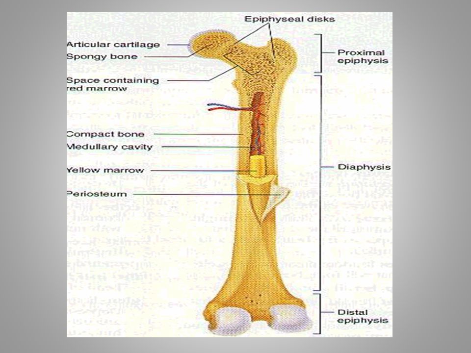
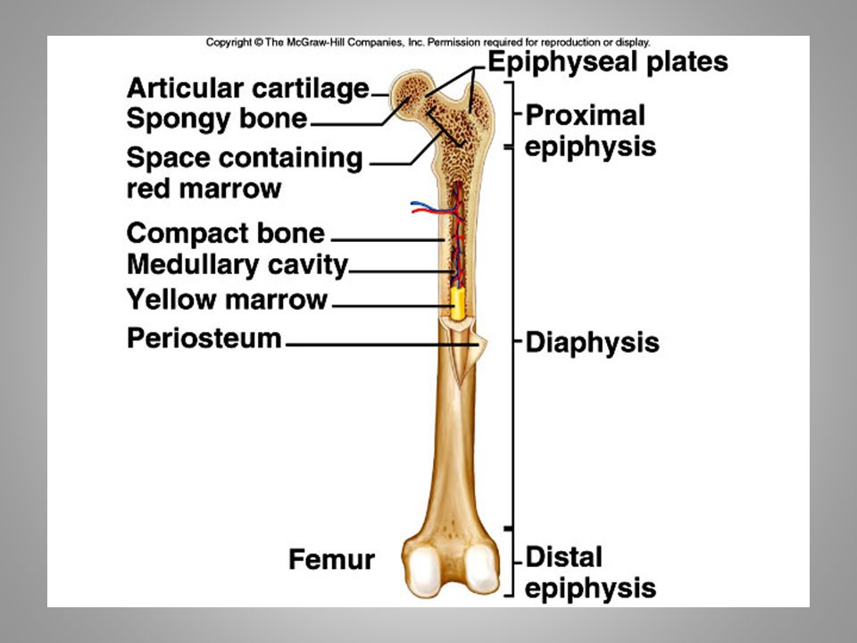
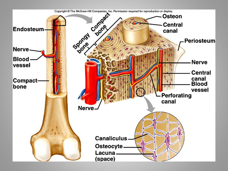
Bones classified by shape: long, short, flat,
irregular, round Bone enclosed in periosteum, which is continuous with tendons and ligaments blood vessels in periosteum Epiphysis- ends spongy bone contains red marrow compact bone, articular cartilage Diaphysis- middle compact bone medullary cavity- contains yellow marrow (fat) lined with endosteum (squamous epithelium)
 lined with endosteum (squamous epithelium)")
37
Compact bone osteocytes within lacunae arranged in concentric circles called lamellae This surround a central canal; complex is called Haversian system Canaliculi connect osteocytes to central canal and to each other

39
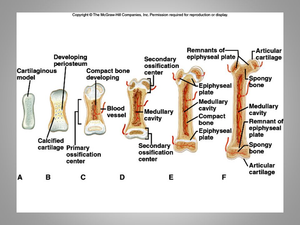
Prenatal development skeleton is mostly cartilaginous Cartilage cells and then osteoblasts start to deposit minerals Cartilaginous disk (epiphyseal disk) remains in epiphysis Cells eventually stop dividing
 remains. in epiphysis. Cells eventually stop dividing.")
41
Adults continually break down and build up bone
Osteoclasts remove damaged cells and release calcium into blood Osteoblasts remove calcium from blood and build new matrix. They become trapped osteoclasts

42
Types of bone breaks Simple- skin is not pierced Compound- skin is pierced Complete- bone is broken in half Partial- broken lengthwise but not into two parts Greenstick- incomplete break on outer arc Comminuted- broken into several pieces Spiral- twisted

43
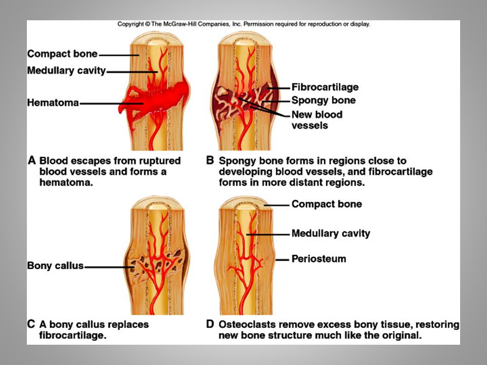
Fracture repair Hematoma- blood clot in space between edges of break Fibrocartilage callus- begins tissue repair Bony callus- osteoblasts produce trabeculae (structural support) of spongy bone and replace fibrocartilage Remodeling- osteoblasts build new compact bone, osteoclasts build new medullary cavity
 of spongy bone and. replace fibrocartilage. Remodeling- osteoblasts build new compact bone, osteoclasts build new medullary cavity.")
45
Axial skeleton skull (cranium and facial bones) hyoid bone (anchors tongue and muscles associated with swallowing) vertebral column (vertebrae and disks) thoracic cage (ribs and sternum) Appendicular skeleton pectoral girdle (clavicles and scapulae) upper limbs (arms) pelvic girdle (coxal bones, sacrum, coccyx) lower limbs (legs)
 thoracic cage (ribs and sternum) Appendicular skeleton. pectoral girdle (clavicles and scapulae) upper limbs (arms) pelvic girdle (coxal bones, sacrum, coccyx) lower limbs (legs)")
46
posterior view p. 135

47
Axial skeleton supports and protects organs
of head, neck and trunk Appendicular skeleton- bones of limbs and bones that anchor them to the axial skeleton Articulation- where joints are formed

48
22 bones in skull 6 in middle ears 1 hyoid bone 26 in vertebral column 25 in thoracic cage 4 in pectoral girdle 60 in upper limbs 60 in lower limbs 2 in pelvic girdle 206 bones in all

49
The skull 8 sutured bones in cranium Facial bones: 13 sutured bones, 1 mandible Cranium encases brain attachments for muscles sinuses

51
Allows for growth

52
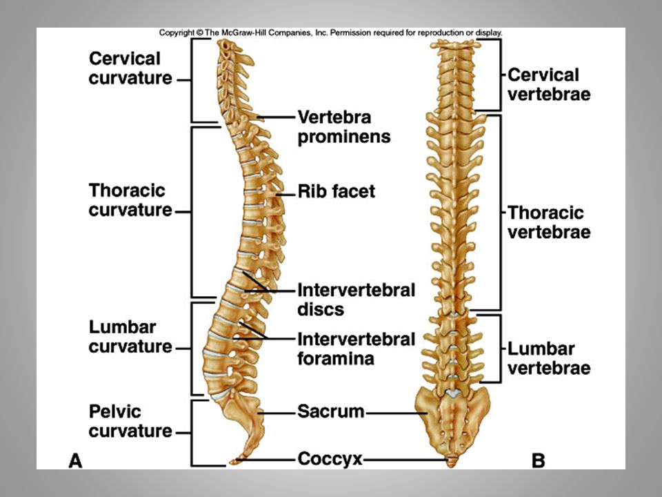
Vertebral column 7 cervial vertebrae 12 thoracic 5 lumbar 1 sacrum (5 fused 1 coccyx (4 fused) Vertebrae vary in size and morphology

54
Thoracic cage ribs thoracic vertebrae sternum costal cartilages True ribs are directly attached to the sternum (first seven pairs) Three false ribs are joined to the 7th rib Two pairs of floating ribs
 Three false ribs are joined to the 7th rib. Two pairs of floating ribs.")
56
Clavicles and scapulae
Help brace shoulders Attachment sites for muscles

58
Bones of upper limb Humerus (upper arm) Radius; ulna Carpals, metacarpals, phalanges Bones of lower limb Femur Patella Tibia, fibula Tarsals, metatarslas, phalanges

59
Joints Immovable (synarthoses) bones sutured together by connective tissue: skull Slightly movable (amphiarthoses) connected by fibrocartilage or hyaline cartilage: vertebrae, rib/sternum joint, pubic symphysis Freely movable (diarthroses)- separated ligaments- hold bones together tendons- muscle to bone lined by synovial membrane
- separated. ligaments- hold bones together. tendons- muscle to bone. lined by synovial membrane.")
61
Types of freely movable joints
Saddle: carpal and metacarpal bones of thumb Ball and socket: shoulder and hip joints Pivot- rotation only: proximal end of radius and ulna Hinge- up and own movement in one plane: knee and elbow Gliding- sliding and twisting: wrist and ankle Condyloid- movement in different planes but not rotations: btw metacarpals and phalanges

62
Types of movement and examples (with muscles)
flexion- move lower leg toward upper extension- straightening the leg abduction- moving leg away from body adduction- movong leg toward the body rotation- around its axis supination- rotation of arm to palm-up position pronation- palm down circumduction- swinging arms in circles inversion- turning foot so sole is inward eversion- sole is out

66
Elevation and depression- raising body part up
or down Aging and bones both bone and cartilage tend to deteriorate cartilage: chondrocytes die, cartilage becomes calcified osteoporosis; bone is broken down faster than it can be built bones get weak and brittle; tend to fracture easily

67
Risk factors for osteoporosis
Inadequate calcium Little weight-bearing exercise Drinking alcohol, smoking Being female: decreased estrogen secretion after menopause Small frame Caucasian or Asian ethnicity

68
Skeleton and other systems
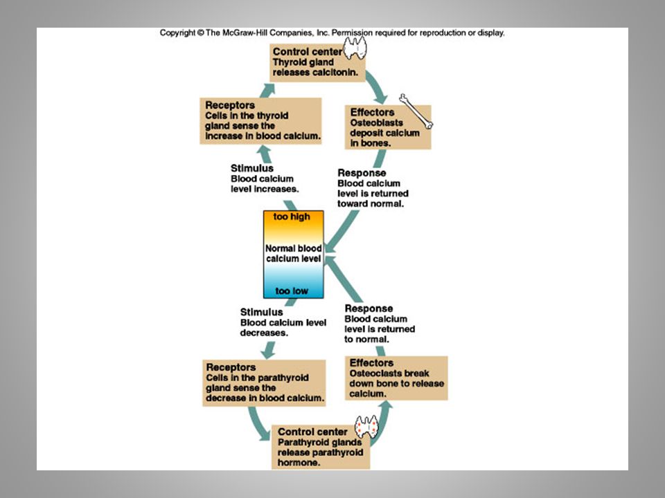
Skin makes vitamin D which enhances calcium absorption Skeleton stores calcium for muscle contraction, nervous stimulation, blood clot formation Red marrow- site of blood cell formation Calcium levels regulated by parathyroid hormone and calcitonin kidneys (can help provide vitamin D) digestive system (can release calcium into blood
 digestive system (can release calcium. into blood.")
70
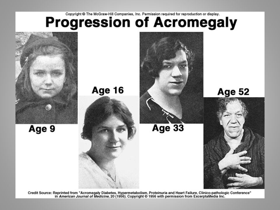
Growth hormone regulates skeletal growth
stimulates cell division in epiphyseal disks in long bones Growth stops when epiphyseal disks are converted to bone When excess growth hormone is produced in childhoodgigantism In adulthood- acromegaly. Bones can’t grow but soft tissue can

72
When muscle contracts, it shortens and causes
movement Skeletal muscles attached to bones by tendons Insertion- attachment to more movable bone Origin- less movable Flexors and extensors act on the same joint to produce opposite actions

Similar presentations