Download presentation
Presentation is loading. Please wait.

1
General principles of fractures

2
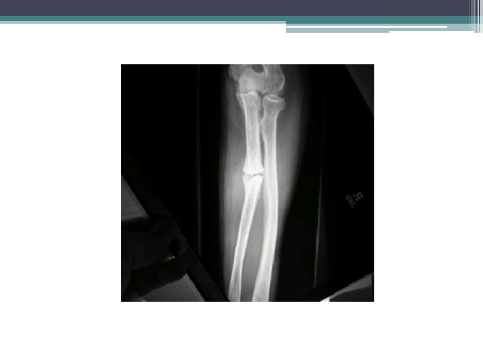
A fracture is a break in the structural continuity of bone. It my be no more than a crack, a crumpling or a splintering of the cortex; more often the break is complete and the bone fragments are displaced

3
If the overlying skin remains intact it is a closed (or simple) fracture, if the skin or one of the body cavities is breached it is an open (or compound) fracture liable to contamination and infection.
 fracture, if the skin or one of the body cavities is breached it is an open (or compound) fracture liable to contamination and infection.")
8
How fractures happen Bone is relatively brittle; it has sufficient strength and resilience to withstand considerable stress. Fractures result from: (1) a single traumatic (injury). (2) repetitive stress. (3) abnormal weakening of the bone (a 'pathological' fracture).
 a single traumatic (injury). (2) repetitive stress. (3) abnormal weakening of the bone (a pathological fracture)..")
9
Fractures due to injury Most fractures are caused by sudden and excessive force, which may be tapping, crushing, bending, twisting or pulling.

11
With a direct force the bone breaks at the point of impact; the soft tissues also must be damaged. Tapping (a momentary blow) usually causes a transverse fracture and damage to the overlying skin; crushing is more likely to cause a comminuted fracture with extensive soft-tissue damage.
 usually causes a transverse fracture and damage to the overlying skin; crushing is more likely to cause a comminuted fracture with extensive soft-tissue damage..")
12
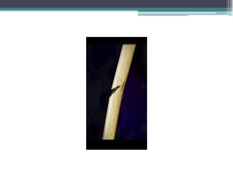
With an indirect force the bone breaks at a distance from where the force is applied; soft- tissue damage at the fracture site is not inevitable. The force may be: (1) twisting, which causes a spiral fracture; (2) bending, which causes a transverse fracture;
 twisting, which causes a spiral fracture; (2) bending, which causes a transverse fracture;.")
13
(3) bending and compressing, which results in a fracture that is partly transverse but with a separate triangular 'butterfly fragment; (4) a combination of twisting, bending and compressing, which causes a short oblique fracture. (5) pulling in which a tendon or ligament pulls the bone apart.
 pulling in which a tendon or ligament pulls the bone apart..")
14
The above description applies mainly to the long bones. A cancellous bone, such as a vertebra or the calcaneum, when subjected to sufficient force, sustains a comminuted crush fracture. At the knee or elbow resisted extension may cause an avulsion fracture of the patella or olecranon.

17
Fatigue or stress fractures Cracks can occur in bone, as in metal and other materials. Due to repetitive stress. This is most often seen in the tibia or fibula or metatarsals, especially in athletes, dancers and army recruits who go on long route marches.

19
Pathological fractures Fractures may occur even with normal stresses if the bone has been weakened (e.g. by a tumour) or if it is excessively brittle (e.g. in Paget's disease).
 or if it is excessively brittle (e.g. in Paget s disease)..")
21
Types of fracture Fractures are infinitely variable in appearance but for practical reasons they are divided into a few well- defined groups.

22
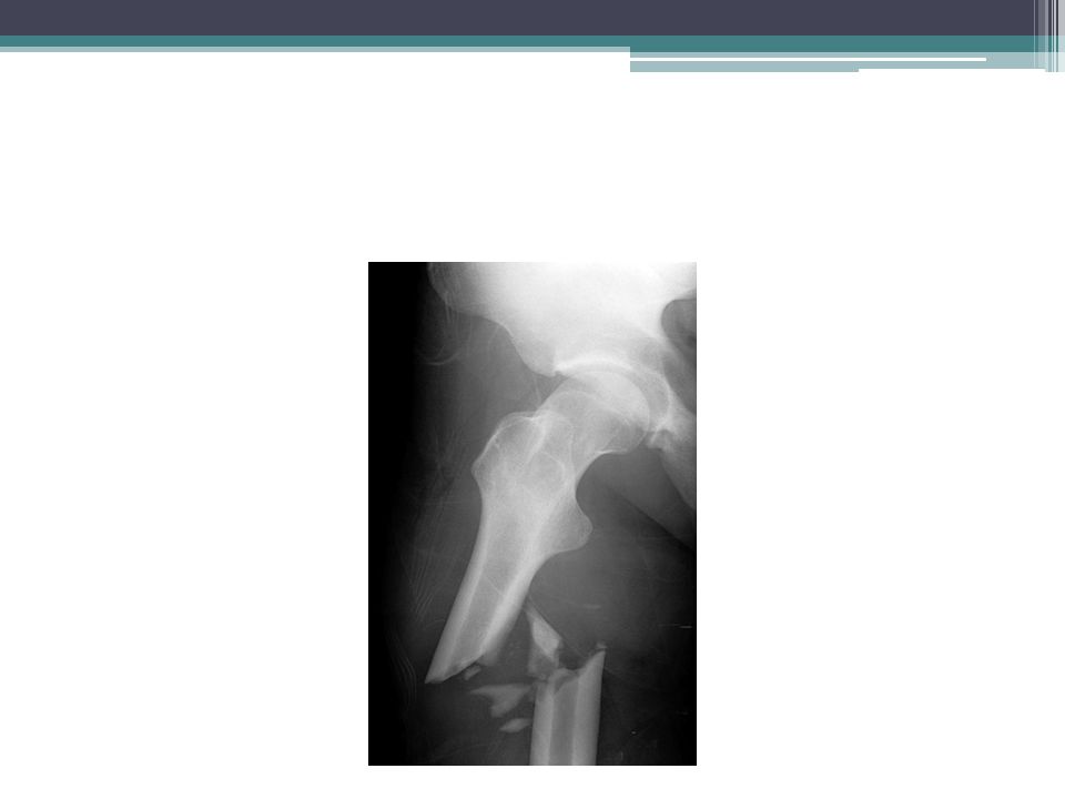
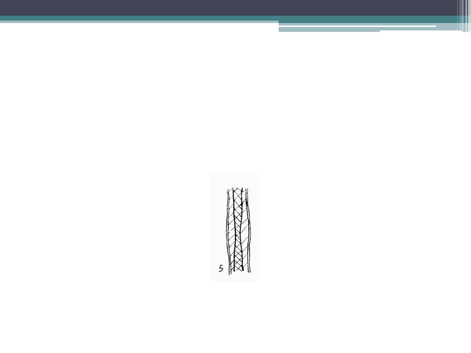
Complete fractures The bone is completely broken into two or more fragments. If the fracture is transverse, the fragment usually remains in place after reduction; if it is oblique or spiral, they tend to slip and redisplace even if the bone is splinted. In an impacted fracture the fragments are jammed tightly together and the fracture line is indistinct. A comminuted fracture is one in which there are more than two fragments: because there is poor interlocking of the fractured surfaces these lesions are often unstable.

25
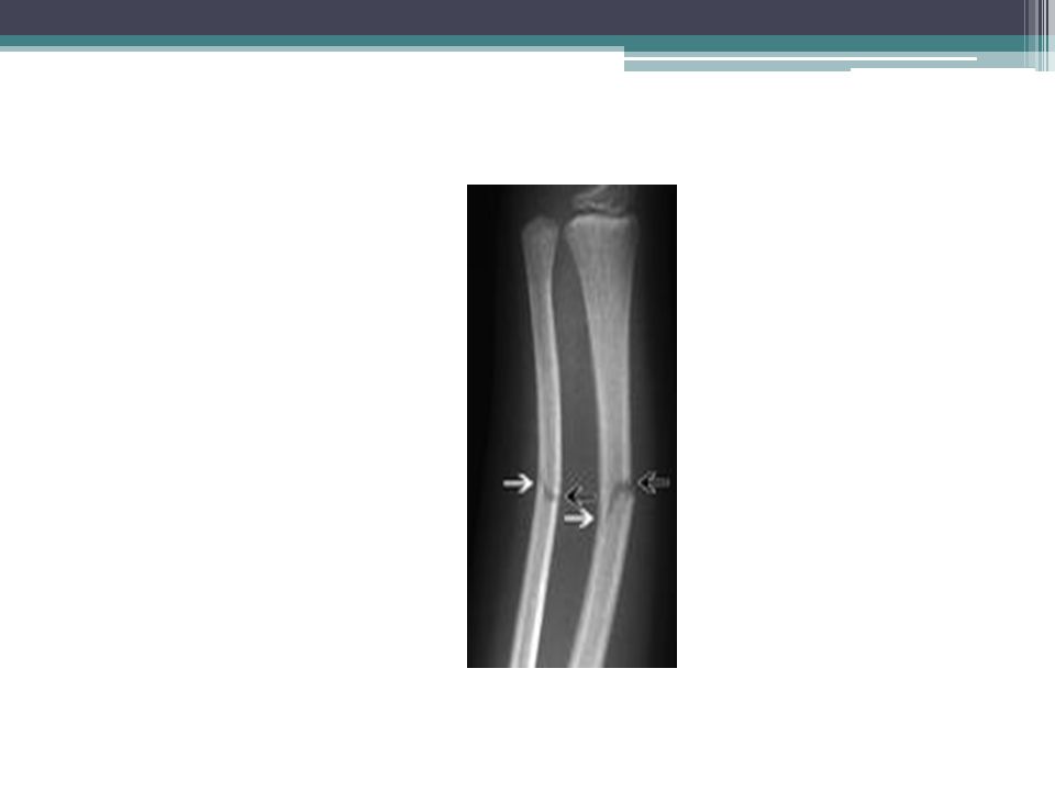
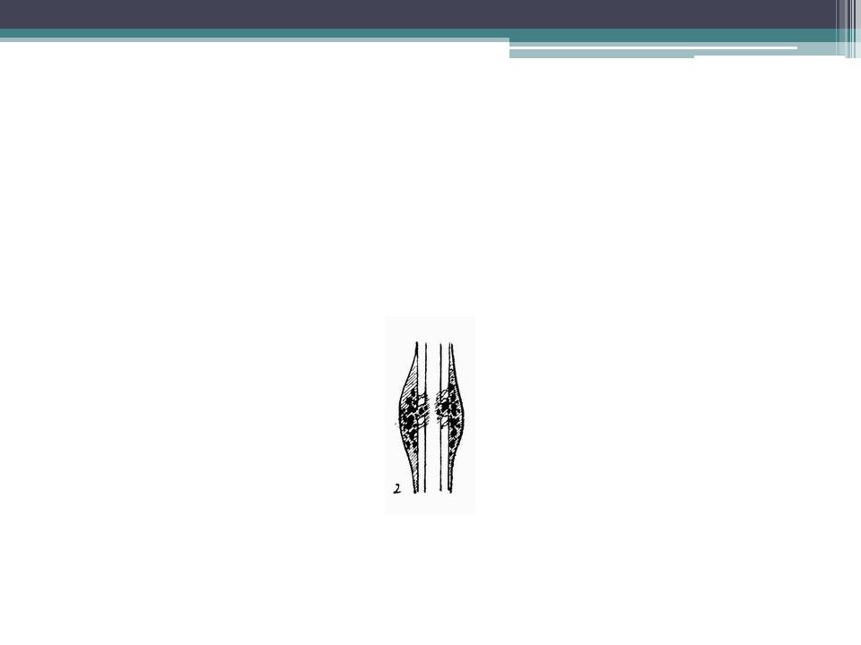
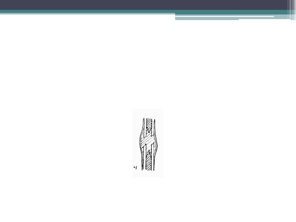
Incomplete fractures Here the bone is incompletely divided and the periosteum remains in continuity. In a greenstick fracture the bone is buckled or bent (like snapping a green twig); this is seen in children. Those bones are more springy than those of adults. Reduction is usually easy and healing is quick. Compression fractures occur when cancellous bone is crumpled. This happens in adults especially in the vertebral bodies. Unless operated upon, reduction is impossible and some residual deformity is inevitable.
; this is seen in children. Those bones are more springy than those of adults. Reduction is usually easy and healing is quick. Compression fractures occur when cancellous bone is crumpled. This happens in adults especially in the vertebral bodies. Unless operated upon, reduction is impossible and some residual deformity is inevitable..")
29
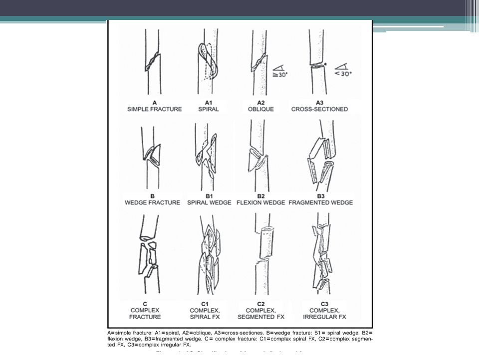
Classification of fractures An alphanumeric classification of fractures, which can be used for computer storage, has been developed (Muller et al., 1990). The first digit specifies the bone (1 = humerus, 2 = radius/ulna, 3 = femur, 4 = tibia/fibula) and the second the segment (1 =proximal, 2 = diaphyseal, 3 = distal, 4 = malleolar). A letter specifies the type of fracture (diaphysis: A = simple, B = wedge, C = complex; proximal and distal: A = extra-articular, B = partial articular, C = complete articular). Two further numbers specify the detailed morphology of the fracture.
 and the second the segment (1 =proximal, 2 = diaphyseal, 3 = distal, 4 = malleolar). A letter specifies the type of fracture (diaphysis: A = simple, B = wedge, C = complex; proximal and distal: A = extra-articular, B = partial articular, C = complete articular). Two further numbers specify the detailed morphology of the fracture..")
31
How fractures are displaced After a complete fracture the fragments usually become displaced, partly by the force of the injury, partly by gravity and partly by the pull of muscles attached to them. Displacement is usually described in terms of apposition, alignment, rotation and altered length.

32
Apposition (shift) the fragments may be shifted sideways, backwards or forwards in relation to each other, such that the fracture surfaces lose contact. The fracture will usually unite even if apposition is imperfect, or indeed even if the bone ends lies side by side with the fracture surfaces making no contact at all.

33
Alignment (tilt) the fragments may be tilted or angulated in relation to each other. Malalignment, if uncorrected, may lead to deformity of the limb.

34
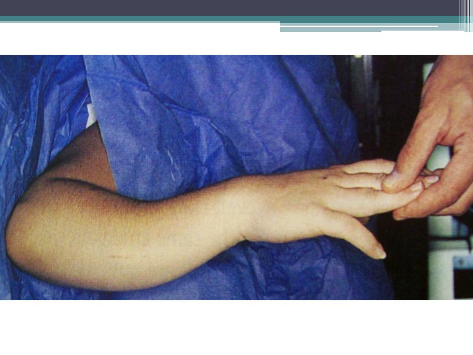
Rotation (twist) One of the fragments may be rotated on its longitudinal axis; the bone looks straight but the limb ends up with a rotational deformity.
 One of the fragments may be rotated on its longitudinal axis; the bone looks straight but the limb ends up with a rotational deformity.")
36
Length the fragments may be distracted and separated, or they may overlap, due to muscle spasm, causing shortening of the bone.

38
How fractures heal It is commonly supposed that, in order to unite, a fracture must be immobilized. This cannot be so since, with few exceptions, fractures unite whether they are splinted or not; indeed, without a built-in mechanism for union. The bone ends must at some stage be brought to rest relative to one another. But it is not mandatory for the surgeon to impose this immobility artificially.

39
Nature can do it, with callus; and callus forms in response to movement not to splintage. We splint most fractures, not to ensure union but (1) to alleviate pain, (2) to ensure that union takes place in good position and (3) to permit early movement and return of function.
 to alleviate pain, (2) to ensure that union takes place in good position and (3) to permit early movement and return of function..")
40
The process of fracture repair varies according to the type of bone involved and the amount of movement at the fracture site. In a tubular bone, and in the absence of rigid fixation, healing proceeds in five stages.

41
TISSUE DESTRUCTION AND HAEMATOMA FORMATION Vessels are torn and a haematoma forms around and within the fracture. Bone at the fracture surfaces, deprived of a blood supply, dies back for a millimeter or two.

43
INFLAMMATION AND CELLULAR PROLIFERATION Within 8 hours of the fracture there is an acute inflammatory reaction with proliferation of cells under the periosteum and within the breached medullary canal. The fragment ends are surrounded by cellular tissue, which bridges the fracture site. The clotted haematoma is slowly absorbed and fine new capillaries grow into the area.

45
CALLUS FORMATION The proliferating cells are potentially chrondrogenic and osteogenic; given the right conditions, that 'will start forming bone and in some cases also cartilage. The cell population now also includes osteoclasts (probably derived from the new blood vessels) which begin to mop up dead bone. The thick cellular mass, with its islands of immature bone and cartilage, forms the callus or splint on the periosteal and endosteal surfaces. As the immature fiber bone (or 'woven' bone) becomes more densely mineralized, movement at the fracture site decreases progressively and at 4 weeks after injury the fracture 'unites'.
 which begin to mop up dead bone. The thick cellular mass, with its islands of immature bone and cartilage, forms the callus or splint on the periosteal and endosteal surfaces. As the immature fiber bone (or woven bone) becomes more densely mineralized, movement at the fracture site decreases progressively and at 4 weeks after injury the fracture unites ..")
47
CONSOLIDATION With continuing osteoclastic and osteoblastic activity the woven bone is transformed into lamellar bone. The system is now rigid enough to allow osteoclasts to burrow through the debris at the fracture line, and close behind them osteoblasts fill in the remaining gaps between the fragments with new bone. This is a slow process and it may be several months before the bone is strong enough to carry normal loads.

49
REMODELLING The fracture has been bridged by a cuff of solid bone. Over a period of months, or even years, this crude 'Weld' is reshaped by a continuous process of alternating bone resorption and formation. Thicker lamellae are laid down where the stresses are high; unwanted buttresses are carved away the medullary cavity is reformed. Eventually and especially in children, the bone reassumes something like its normal shape.

51
Clinical studies have shown that callus is the response to movement at the fracture site. It serves to stabilize the fragments as rapidly as possible a necessary precondition for bridging by bone. If the fracture site is absolutely immobile - for example, an impacted fracture in cancellous bone, or a fracture rigidly immobilized by a metal plate - there is no need for callus. Instead, osteoblastic new bone formation occurs directly between the fragments.

52
Gaps between the fracture surfaces are invaded by new capillaries and osreoprogenitor cells growing in from the edges and new bone is laid down on the exposed surface (gap healing). Where the crevices are very narrow (less than 200 µm), osteogenesis produces lamellar bone; wider gaps are filled first by woven bone which is then remodelled to lamellar bone.
, osteogenesis produces lamellar bone; wider gaps are filled first by woven bone which is then remodelled to lamellar bone..")
53
By 3-4 weeks the fracture is solid enough to allow penetration and bridging of the area by bone remodelling units - i.e. osteoclastic 'cutting cones' followed by osteoblasts. Where the exposed fracture surfaces are in intimate contact and held rigidly from the outset, internal bridging may occasionally occur without any intermediate stages (contact healing).
..")
54
Healing by callus, though less direct (the term indirect could be used). With rigid metal fixation, on the other hand, the absence of callus means that there is a long period during which the bone depends entirely upon the metal implant for its integrity.

55
Union, consolidation and non-union Repair of a fracture is a continuous process. UNION Union is incomplete repair; the callus is calcified. Clinically the fracture site is still little tender and, though the bone moves in one piece, attempted angulation is painful. X-ray shows the fracture line still clearly visible, with fluffy callus around it. Repair is incomplete and it is not safe to subject the unprotected bone to stress.

56
COSOLIDATION Consolidation is complete repair; the calcified callus is ossified. Clinically the fracture site is not tender, no movement can be obtained and attempted angulation is painless. X-rays show the fracture line to be almost obliterated and crossed by bone trabeculae, with well- defined callus around it. Repair is complete and further protection is unnecessary.

57
TIMETABLE How long does a fracture take to unite and to consolidate? No precise answer is possible because age, constitution blood supply, type of fracture and other factors all influence the time taken.

58
Approximate prediction is possible. A spiral fracture in the upper limb unites in 3 week; for consolidation multiply by 2; for the lower limb multiply by 2 again; for transverse fractures multiply again by 2. Children's fractures, of course, join more quickly. These figures are only a rough guide; there must be clinical and radiological evidence of consolidation before full stress permitted without splintage.

59
NON-UNION Sometimes the normal process of fracture repair is thwarted and the bone fails to unite. causes of non-union are: (1) distraction and separation of the fragments; (2) interposition of soft tissues between the fragments; (3) excessive movement at the fracture line; and (4) poor local blood supply.
 distraction and separation of the fragments; (2) interposition of soft tissues between the fragments; (3) excessive movement at the fracture line; and (4) poor local blood supply..")
61
Cell proliferation is predominantly fibroblastic; the fracture gap is filled by fibrous tissue and the bone fragments remain mobile, creating a false joint or pseudarthrosis. In some cases periosteal bone formation is active so that, while new bone fails to bridge the fracture gap, the fragment ends are thickened or widened

62
this hypertrophic non-union will ultimately proceed to union provided the bone fragment are placed in contact with each other and held more or less immobile until bridging occurs. In other cases bone formation seems to peter out altogether, resulting in an atrophic non-union which will never heal unless the fragments are immobilized and grafted with cancellous bone.

Similar presentations


>")


 2.Protection: skull, vertebrae,>")

 Ortho. Consultant ( Head 0f Orthopedic Department SGH-J )>")
 fracture: involve.>")
