Download presentation
Presentation is loading. Please wait.

1
Good Morning! Happy Monday
Monday, July 22nd, 2013

2
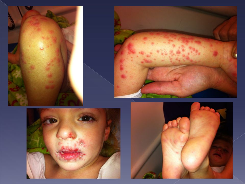
4 yo female p/w 3 days of fever (tmax 102), 2 days of progressive non-pruritic rash on face/extremities, decreased PO/UOP, emesis x 1 (non-bloody,non-bilious), diarrhea x 2 (non-bloody), increasing fatigue x 5d, refusing to eat and walk Meds: tylenol PRN Allergies: NKDA PMH: none FMH: neg Immunizations: received 4 yo shots several months ago Social: stays at home w/ mom, no travel history, older siblings with cold like symptoms, no rash

4
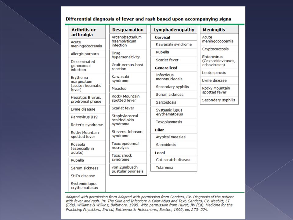
Differential Dx Arthritis/Arthralgias Desquamation Lymphadenopathy
Meningitis Enanthems (mucosal involvement) Ulcerative vesicular lesions Palm and Sole involvement Predominantly on extremities Respiratory Symptoms/Pulmonary infiltrates
 Ulcerative vesicular lesions. Palm and Sole involvement. Predominantly on extremities. Respiratory Symptoms/Pulmonary infiltrates.")
5
Problem Definition Immunized 3 yo female with acute onset of fever, progressive vesicular rash on extremities with oral mucosal involvement, mild N/V/D, non-toxic appearing

8
Enteroviruses** Single-stranded RNA viruses** “Summer viruses” **
Picornaviridae family Polioviruses Coxsackieviruses (Group A and B) Echoviruses Enteroviruses (serotypes 68-71) “Summer viruses” ** *Increased prevalence in summer months (May – October) All year round in tropical climates (NOLA)
 Echoviruses. Enteroviruses (serotypes 68-71) Summer viruses ** *Increased prevalence in summer months (May – October) All year round in tropical climates (NOLA)")
9
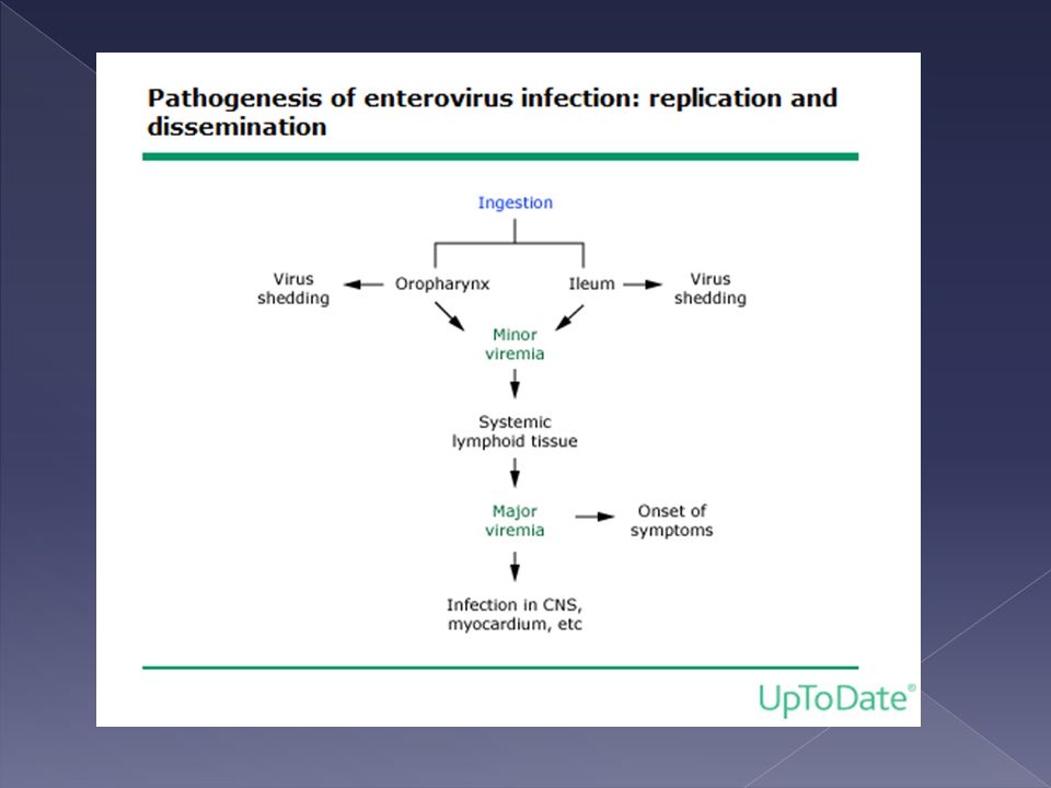
Transmission** Most cases involve children under age 5
Humans are only hosts Fecal-oral is most common route Then replicates in lymph nodes of respiratory and GI systems Initial viremia → heart, liver, skin CNS infection usually the result of second major viremia Specifically infants less than one yr of age become infected at rates that far exceed those of older children and adults Males > females by up to 50%

11
Clinical Manifestations**
Most patients are mildly ill & recover completely Most common → febrile illness, viral exanthem, vomiting, diarrhea, and malaise Others: Hemorrhagic conjunctivitis Pharyngitis Herpangina Hand-foot-and-mouth disease Paralysis Hepatitis Myocarditis Pericarditis Encephalitis Aseptic meningitis Treatment is symptomatic for most of these, no documented use for acyclovir. IVIG has been used in myocarditis and persistent meningoencephalitis

12
A 6-day-old infant is brought to the ER in August with a 1-day history of decreased feeding, decreased activity, tactile fever, and rapid breathing. He was born at term. His mother reports that she had a nonspecific febrile illness 1 week before delivery for which she received no treatment. Her GBS screen was positive at 36 weeks' gestation, and she received two doses of ampicillin (>4 hours apart) during labor. The baby received no antibiotics and was discharged at 48 hours of age. Physical examination today reveals a toxic, lethargic infant who is grunting and has a temp of 39.4°C, HR of 180, and RR of 60. His lungs are clear, with subcostal retractions. He has a regular heart rhythm with gallop, his pulses are thready, his capillary refill is 4 seconds, and his extremities are cool. Of the following, the MOST likely cause of this baby's illness is A. early-onset group B Streptococcus infection B. echovirus 11 infection C. herpes simplex virus infection D. hypoplastic left heart syndrome E. respiratory syncytial virus infection

13
Neonates** High risk for developing disseminated infection
Severe manifestations: Fulminant Hepatitis Myocarditis Pneumonitis Meningitis Encephalitis DIC Multiorgan failure Most common are Fulminant hepatitis and myocarditis – signs of hypotension, bleeding, jaundice, organ failure

14
Neonates** acquired from nurseries, or from symptomatic mothers (fever 1 week prior to delivery) Symptoms develop at 3-7 days of life Signs include mild listlessness, anorexia, transient respiratory distress, jaundice, Can be acquired from nurseries, or from symptomatic mothers (who are febrile the last week of pregnancy) – but may be exposed to virus positive cervical secretions or viremic maternal blood, not believed to be passed via the placenta Outcomes related to exposure to passive immunization – related to timing of maternal infection, and development and passage of IgG Most affected develop symptoms between 3-7 days of life Can be mild listlessness, anorexia, transient respiratory distress, biphasic – apparent well being between initial symptoms and more serious manifestations Most common are Fulminant hepatitis and myocarditis – signs of hypotension, bleeding, jaundice, organ failure
 – but may be exposed to virus positive cervical secretions or viremic maternal blood, not believed to be passed via the placenta. Outcomes related to exposure to passive immunization – related to timing of maternal infection, and development and passage of IgG. Most affected develop symptoms between 3-7 days of life. Can be mild listlessness, anorexia, transient respiratory distress, biphasic – apparent well being between initial symptoms and more serious manifestations. Most common are Fulminant hepatitis and myocarditis – signs of hypotension, bleeding, jaundice, organ failure.")
15
Diagnostic Tests** Viral culture** PCR** Serology
Stool, throat, blood, CSF, or tissue 8 to 10 days PCR** Only small sample needed Results in 24 hours Serology Based on increase in antibody titers Too many enterovirus serotypes to be practical Culture – is labor intensive and expensive, but it allows typing of the isolate for clinical/epidemiological/research purposes

16
Diagnostic Tests (cont’d)
Testing by PCR has been associated with decreased IV abx use, ancillary testing, and hospital length of stay Allows for patient isolation if necessary (ie, NICU)
")
17
Treatment Supportive care Antivirals under investigation
IVIG may benefit immunodeficient patients Also used in some with myocarditis or persistant meningoencephalitis IVIG in myocarditis= better LV function IVIG injected directly into ventricles Also used to use an experimental drug that is no longer available (pleconaril)
")
18
Prevention Contact precautions HAND WASHING!!!

19
Hand-Foot-and-Mouth Disease
1-4 yo Incubation period 3 to 7 days Prodromal phase of malaise, sore throat, mouth pain, anorexia and low grade fever Coxsackie A16 virus

20
Hand-Foot-and-Mouth Disease (cont’d)
Oral lesions

21
Hand-Foot-and-Mouth Disease (cont’d)
Painful vesicles in mouth and on hands and feet Surrounded by an erythematous margin Nonvesicular lesions on buttocks, GU and extremities less commonly

22
Hand Foot Mouth Disease
Onychomadesis – proximal separation of the nail plate from the nail bed

23
Hand-Foot-and-Mouth Disease (cont’d)
Most resolve spontaneously w/in 3d-1wk Treatment is supportive Hydration and analgesics Magic Mouthwash Maalox Benadryl Viscous lidocaine

24
Hand-Foot-and-Mouth Disease (cont’d)
Moderately contagious Spread by direct contact with nasal discharge, saliva, blister fluid, or stool Most contagious during the first week of the illness Can shed virus in stool for up to 8 weeks No day care/school during the first few days of illness and in setting of open lesions Can take any form – urticaria (coxsackie), petechiae/purpura (echovirus 9) – confusing if someones meningitic appearing, maculopapular (echo) or vesicular (coxsackie) Recent severe cases of HFM – enterovirus 71 cns disease, pulm edema and hemorrhage and heart failure, temp paralysis that resolved
, petechiae/purpura (echovirus 9) – confusing if someones meningitic appearing, maculopapular (echo) or vesicular (coxsackie) Recent severe cases of HFM – enterovirus 71 cns disease, pulm edema and hemorrhage and heart failure, temp paralysis that resolved.")
25
HFM: Parental Guidance
Analgesia: Avoid aspirin (acetaminophen and ibuprofen are ok) Diet: cold, soft foods, dairy, nothing spicy Prevent spread: wash hands often, especially after using the bathroom Avoid others during the first week of illness to prevent spread, avoid pregnant women Enteroviruses don’t cross placenta, but there are rare cases of infant death associated with echovirus, coxsackie virus – not proven cause, Advise pregnant women to avoid contact with patients suspected of having an enteroviral illness
 Diet: cold, soft foods, dairy, nothing spicy. Prevent spread: wash hands often, especially after using the bathroom. Avoid others during the first week of illness to prevent spread, avoid pregnant women. Enteroviruses don’t cross placenta, but there are rare cases of infant death associated with echovirus, coxsackie virus – not proven cause, Advise pregnant women to avoid contact with patients suspected of having an enteroviral illness.")
26
Herpangina Coxsackie group A Ages 3 -10 years
Incubation period 4-14 days Prodromal phase Malaise, HA, N/V, myalgias, anorexia sore throat and mouth pain 1-2 days prior to lesions Fever (low grade > high) Can be caused by Group B coxsackie viruses and echoviruses occasionally Can be elevated up to 105.8
 Can be caused by Group B coxsackie viruses and echoviruses occasionally. Can be elevated up to")
27
Herpangina Erythematous ring surrounds
Puntate macules vesiclulate, ulcerate Anterior tonsillar pillars, soft palate, posterior pharynx Typically 2-6 lesions, but as many as can be present Posterior oropharynx is minimally erythematous

28
Herpangina Self-limited Resolve spontaneously within 1 week
Supportive care Young children are at risk of dehydration

29
Herpetic Gingivostomatitis
Ages 6 mo – 5 yo (peaks at 2yo) Incubation 2 days – 2 weeks Prodrome: fever, irritability, malaise, HA, PO, lymphadenopathy (cervical, submandibular) Low to high grade fever
 Incubation 2 days – 2 weeks. Prodrome: fever, irritability, malaise, HA, PO, lymphadenopathy (cervical, submandibular) Low to high grade fever.")
30
Herpetic Gingivostomatitis
Red, edematous gingivae bleed easily Small vesicles ulcerate and coalesce Large ulcerations with erythema surrounding Buckle mucosa, tongue, gingiva, hard palate, pharynx, lips, perioral skin

31
Herpetic Gingivostomatitis

32
Herpetic gingivostomatitis
Diagnose with culture, PCR, or antigen testing Resolve in 10 to 14 days Treatment is supportive Hydration and analgesics Acyclovir If patients present in the first hrs of disease, unable to drink or have significant pain After resolution, reside in trigeminal ganglia Previously not recommended, few studies now that show decreased length of oral lesions, less time with mouth pain, less time with difficulty eating/drinking, decreased incidence of new lesions Acyclovir - 15mg/kg 5x per day for 5-7 days Topical acyclovir therapy not recommended

33
Aphthous stomatitis Typically found in older children and adults
Not associated with infection Can be associated with autoimmune disease (SLE, IBD) Exquisitely painful ulcers Large, yellow, pseudomembranous slough with erythematous border
 Exquisitely painful ulcers. Large, yellow, pseudomembranous slough with erythematous border.")
34
Apthous stomatitis Topical creams may help

35
Topical Analgesia Usually not recommended Benzocaine (orajel)
associated with methemoglobinemia viscous lidocaine may cause problems if absorbed systemically may choke on secretions may chew their buccal mucosa

36
Hand, Foot, Mouth Disease Herpangina Herpetic Gingivostomatitis
Aphthous Stomatitis ages 1-4 yo 3-10 yo 6mos – 5 yo Older children , adults Incubation 3-7 days 4-14 days 2 days – 2 weeks N/A prodrome Malaise, sore throat, mouth pain, anorexia Malaise, HA, N/V, sore throat, mouth pain, anorexia irritability, malaise, HA, anorexia, submandibular and cervical lymphadenitis Usually none fever Usually low grade Low-High grade fever Description of lesions Mildly painful Vesicles surrounding erythema (may ulcerate) Painful Vesicles/ulcers with surrounding erythema Vesicles that ulcerate and coalesce Beefy red gingiva Exquisitely painful Large Ulcers , yellow pseudomembranous with erythematous border Location of lesions Hands, feet, mouth (buccal mucosa and tongue), occasionally nonvesicular lesions on buttocks, genitals and extremities Anterior tonsillar pillars, soft palate, posterior pharynx Buccal mucosa, tongue, gingival, hard palate, pharynx, lips, perioral skin lips, tongue, buccal mucosa Most common virus, season Coxsackie A16 summer Group A Coxsackie HSV 1 Year round none Duration and treatment 1 week Symptomatic tx symptomatic tx 10-14 days Acyclovir, symptomatic tx Variable, can recur, symptomatic tx
 Painful Vesicles/ulcers with surrounding erythema. Vesicles that ulcerate and coalesce. Beefy red gingiva. Exquisitely painful. Large Ulcers , yellow pseudomembranous with erythematous border. Location of lesions. Hands, feet, mouth (buccal mucosa and tongue), occasionally nonvesicular lesions on buttocks, genitals and extremities. Anterior tonsillar pillars, soft palate, posterior pharynx. Buccal mucosa, tongue, gingival, hard palate, pharynx, lips, perioral skin. lips, tongue, buccal mucosa. Most common virus, season. Coxsackie A16. summer. Group A Coxsackie. HSV 1. Year round. none. Duration and treatment. 1 week. Symptomatic tx. symptomatic tx days. Acyclovir, symptomatic tx. Variable, can recur, symptomatic tx.")
37
Picture Quiz Infectious Exanthems

38
Exanthem #1 MEASLES Koplik spots

39
Exanthem #2 Coxsackie A - HFM

40
Exanthem #3 Rubella Forscheimer spots

41
Parvovirus B19- Fifth’s Disease- Erythema Infectiosum
Exanthem #4 Parvovirus B19- Fifth’s Disease- Erythema Infectiosum

42
Exanthem #5 Varicella

43
Exanthem #6 RMSF

44
Exanthem #7 HHV6- Roseola
Clue: This patient had a h/o 3 days of fever (that has since defervesced) before the appearance of the rash
 before the appearance of the rash.")
45
Scarlet Fever- Group A Strep
Exanthem #8 Scarlet Fever- Group A Strep

46
Exanthem #9 Toxic Shock Syndrome
Clue: You might be more suspicious of this illness if this picture was a hypotensive woman

47
Exanthem #10 Staph Scalded Skin

48
Steven-Johnson-Syndrome
Exanthem #11 Steven-Johnson-Syndrome

49
Exanthem #12 Kawasaki Disease

50
Exanthem #13 Meningococcemia

51
Exanthem #14 EBV- mono Clue: This patient was recently treated with Ampicillin

52
BONUS ROUND Who can name the original 6 childhood exanthems? (1st disease, etc)
")
53
Answer 1st disease: Rubeola, Measles
2nd disease: Scarlet Fever (s. pyogenes) 3rd disease: Rubella, German Measles 4th disease: Staph Scalded Skin Syndrome, Filatow-Duke’s Disease, Ritter’s Disease 5th disease: Erythema Infectiousum (parvo) 6th disease: exanthem subitum, roseola (HHV 6 or HHV 7)
 3rd disease: Rubella, German Measles. 4th disease: Staph Scalded Skin Syndrome, Filatow-Duke’s Disease, Ritter’s Disease. 5th disease: Erythema Infectiousum (parvo) 6th disease: exanthem subitum, roseola (HHV 6 or HHV 7)")
54
Have a Great Day! Noon Conference

Similar presentations


.>")


>")
 DNA virus HSV 1 and HSV 2.>")

