Download presentation
Presentation is loading. Please wait.

1
CELL STRUCTURE

2
A Liver cell

3
Liver cell TEM x9400

4
Lily Parenchyma cell x.s. (TEM x7210)
")
5
Cell and Organelle All living organisms are made of cells. A cell is made of living material called protoplasm. Different kinds of cells vary in size, shape, colours and internal structures.

9
Prokaryotic Cell

11
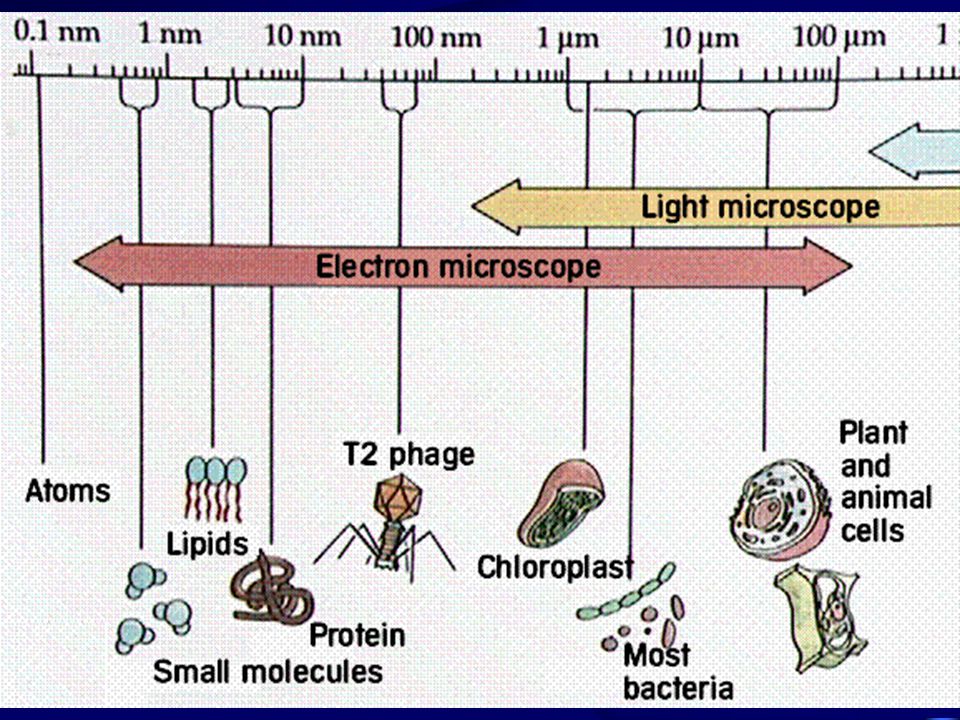
Organization of a cell Subatomic particle atomic molecule molecular complex organelle Virus Cell Tissue Organ System Organism

12
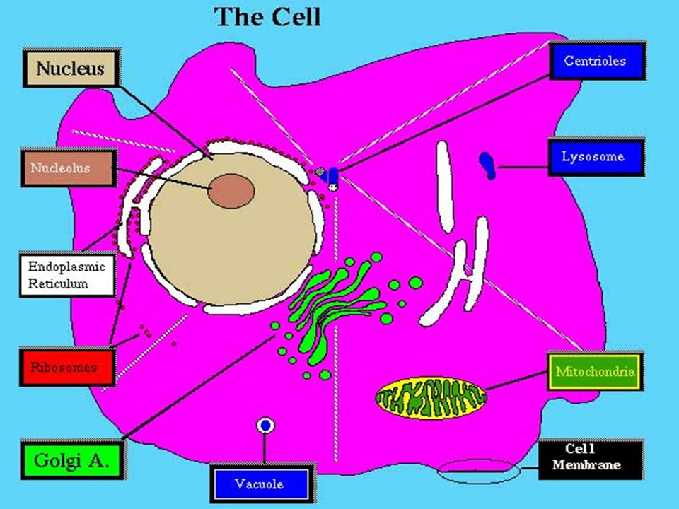
Cell Structure Cell wall Cell membrane protoplasm - the living contents within the cell: nucleus and cytoplasm The cytoplasm contains many subcellular organelles. Sub cellular organelles revealed are: Golgi apparatus, mitochondrion, food granules, centriole, secretary granules and chromatin in nucleus.

15
Cell membrane Chemical components :consists of protein and lipid Structure: Two layers of phospholipid sandwiched between two protein layers

16
Davson and Danielli model

17
Unit membrane hypothesis ( proposed by Robertson ) Under electron microscopy, membrane shows a characteristic trilaminar appearance. The 3 layers are arranged as that of Davson Danielli’s model.

18
trilaminar appearance

19
Fluid mosaic model of cell membrane

20
A mosaic of protein molecules floats in a fluid lipid layer. In this model, lipid bilayer remains as the main backbone of the unit membrane. The lipids spontaneously form a bilayer owing to their polar heads and non-polar tails. Proteins act as islands floating in the lipids. Some proteins move about freely while others are fixed in position.

21
Some proteins penetrate only part of the way into the membrane while others penetrate all the way through. Proteins are variable in function. Sugars are incorporated with protein of lipid to form glycoprotein or glycolipid respectively. These sugars are important in recognition mechanism. Both lipids and proteins show rapid lateral diffusion in the plane of the membrane. Fluidity and permeability of the cell membrane are determined by the degree of saturation and the hydrocarbon chain of the lipids.

25
Function of cell membrane Protein components: Act as a partially permeable barrier controlling the movement of substances between the cell and the surrounding Channels or pores are formed within a protein or between adjacent protein molecules Act as support to give strength Act as enzyme which catalyzes the chemical reaction within the cell membrane Act as carriers in transporting substance across the membrane Act as recognition center As a pump moving things across it

26
Lipid component: The presence of unsaturated lipids can prevent close packing of the molecules so as to make membrane fluid. Fluidity is also increased with decreasing length of the fatty acid tails. Some membranes are folded to increase surface area to facilitate exchange with environment. e.g. microvilli of intestinal epithelial cells.

27
Cell membrane can also separate the contents of cells from their external environments, i.e. form compartments for individual metabolic reaction to take place within the cell. Cell membrane is differentially permeable because cell membrane allows glucose, amino acid, fatty acid, glycerol to diffuse slowly and actively control what substances to pass through. The ability of infolding and extension to form vesicles allows endocytosis and exocytosis to take place.

28
Nucleus Contains chromatin which is involved in nuclear division Contains a nucleolus

29
Structure of Nucleus Enclosed by and envelope of two membranes that is perforated by nuclear pores

30
Function of Nucleus Necessary for survival of a cell Controls all the activities of the cell, cellular function, cell division and heredity

31
Nuclear membrane Doubled layer Similar structure as cell membrane Continuous with E.R. With microscopic pores for exchange of materials between nucleus and cytoplasm

32
Nucleus with nuclear pores

33
Nucleoplasm Nuclear sap Gel-like Denser than cytoplasm Contains proteins, nucleotides and ions

34
Chromatin Consists of DNA and protein ( histone ) Condense to rod-shape chromosome just prior to nuclear division Carry genetic materials which determine organisms’ characteristics and transmit these characteristics to next generations
 Condense to rod-shape chromosome just prior to nuclear division Carry genetic materials which determine organisms’ characteristics and transmit these characteristics to next generations")
35
Nucleolus Composed of DNA mainly Act as the manufacturing site of ribosomal DNA (rDNA) and ribosomes
 and ribosomes")
36
Endoplasm Reticulum ( E.R.) A system of parallel flattened membrane- bounded sacs called cisternae Continuous with the outer membrane of the nuclear envelope Act as an intracellular transport system There are two types of E.R - Rough ER and smooth ER
 A system of parallel flattened membrane- bounded sacs called cisternae Continuous with the outer membrane of the nuclear envelope Act as an intracellular transport system There are two types of E.R - Rough ER and smooth ER")
38
Rough E.R. Ribosomes are attached to its surface Transports proteins made by the ribosomes through the cisternae to smooth ER and then to Golgi appartus for futher modification

39
Smooth E.R. Without ribosomes attached to its surface Transport lipids Synthesis of lipids and steriod

40
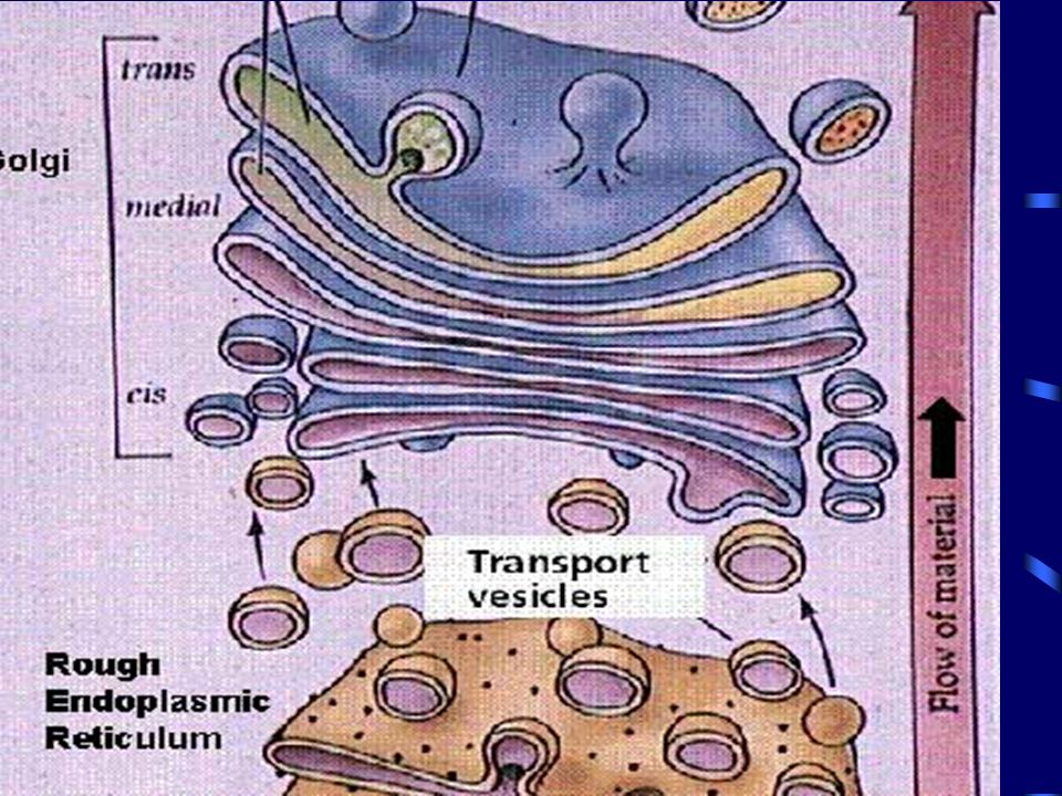
Diagram of Golgi

45
Golgi Apparatus Usually located near the nucleus consists of stacks of flattened membrane bounded sacs called cisternae and many vesicles At one end of the stacks new cisternae are constantly formed by fusion of vesicles pinched from smooth ER; at the other end, small Golgi vesicles are pinched off constantly Transport in vesicles of many cell materials, such as enzymes form ER Involved in secretion and lysosome formation

46
Ribosomes Particles synthesis in nucleolus and then pass through the nuclear pores to the cytoplasm Made of protein and rDNA The site for protein synthesis

48
Ribosomes and Polysomes

51
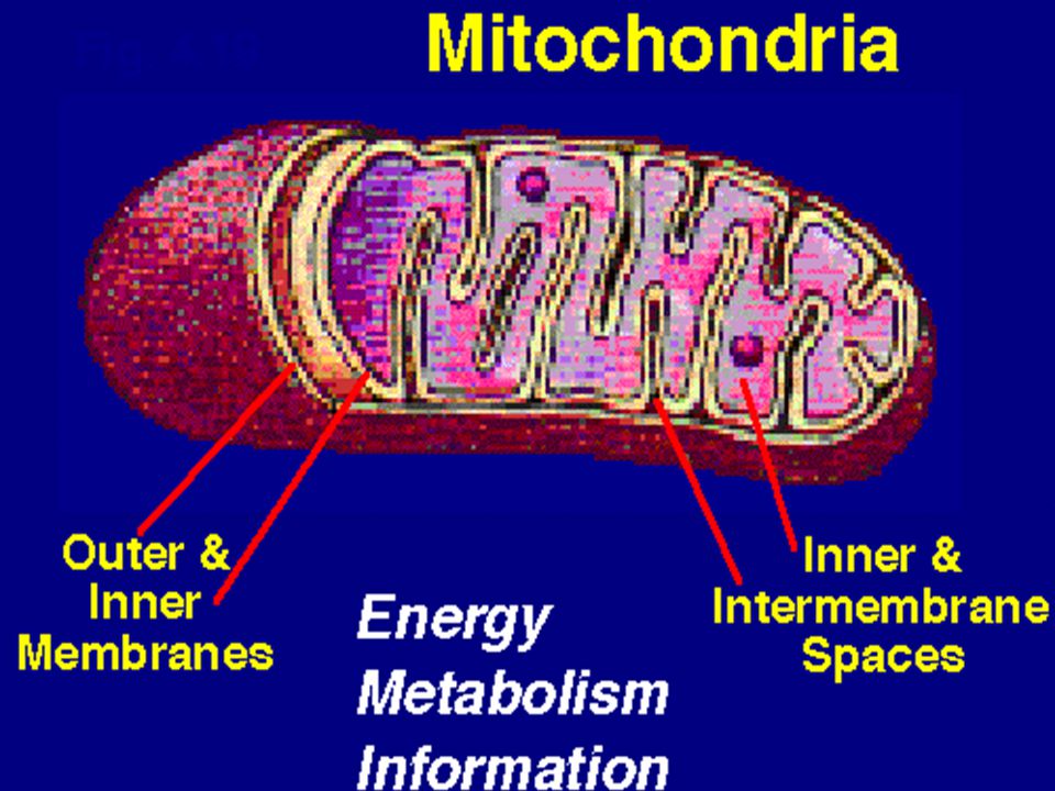
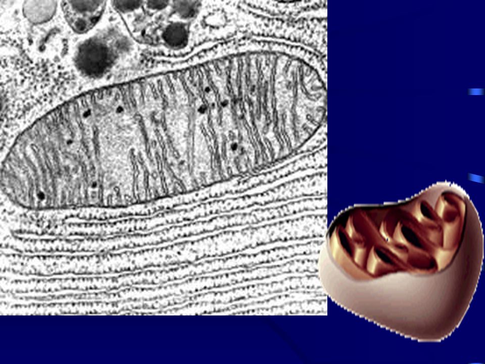
Mitochondrion I Surrounded by an envelope of two membranes, the inner being folded to form cristae Contains a matrix with respiratory enzymes for the Kreb’s cycle Rich in cell which require large amont of energy such as sperm tail, muscle cell cristae matrix outer membrane inner membrane

52
Mitochondion II The cristae increase the surface area for attachment of respiratory enzymes for the electron transfer reactions In aerobic respiration, cristae are the sites of oxidative phosporylation and electron transport

53
Function of Mitochondrion Act as power house of a cell The energy releasing reactions of respiration occur in matrix and on the cristae

54
Lysosomes A spherical sac bounded by a single membrane Contain digestive (hydrolytic) enzymes Intracellular digestion of food materials eg. Amoeba Destroy the worn-out organelles inside cell For self-destruction of cells in developmental process

56
Cell wall Only found in plant cells Rigid and rather permeable Made of cellulose Usually modified by lignin with pores which are penetrated by plasmmodesmata

57
Function of Cell wall Provides mechanical support and protection of the cell Allows a pressure potential to be developed which aids in support Prevent osmotic bursting of the cell

58
Microtubule Act as cytoskeleton which support the cell Involves in the movement of substances inside the cell Forming the spindle fibres which involve in the separation of chromatids and chromosome

59
Centrioles Adjacent to nucleus Internal structure of a centriole is similar to that of basal body of a cilium, with 9 micotubules Forming the spindle fibres and microtubules during nuclear division to control the separation of chromatids and chromosome

62
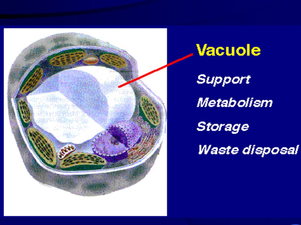
Vacuole Absence or small in animal cells Common and large in plant cells The enclosing membrane is called tonoplast Contain the internal cell sap which is a concentrated solution consists of water, sugar, salts, fat, oils, proteins and pigment

63
Function of Vacuole Store various substances eg. Food and wastes Maintenance of turgor for support Contain hydrolytic enzymes to acts as lysosomes during life and cause autolysis after death

65
Chloroplast Large plastid containing chlorophyll which absorb light for photosynthesis Bounded by two membrane Consists of chloroplast envelope, stroma, lamella and granum

67
Plant Histology Parenchyma Collenchyma Sclerenchyma Xylem Tissue Phloem Tissue

68
Parenchyma Plant cells with thin cell wall and living protoplasm Roughly isodiametric with intercellular spaces Found in cortex and pith of stems and root, mesophyll of leaves and packing tissues in xylem and phloem

69
Function of Parenchyma Act as packing tissues between more specialized tissues Turgidity of these cells can provide support in herbaceous plant Store food Intercellular air spaces allow gaseous exchange Metabolically active Their cell walls are important pathway for the water and mineral salts through the plant

70
Collenchyma Characterized by the deposition of extra cellulose at the corners of the cells so have thickening cell wall of their corners They are living cells Found in regions beneath the epidermis of stem (hypodermis) and near the vascular tissues, eg, midrib of leaves
 and near the vascular tissues, eg, midrib of leaves")
71
Sclerenchyma Plant cells with uniformly thickened cell wall which is usually lignified They are dead cells Support the cells There are two types: fibres and Sclerids

72
Fibre Long narrow cell shape with tapering ends, wall with few piths Found in cortex, pericycle, vascular tissues, surrounding vascular bundles

73
Sclereids Shorter, vary much in shape, may be spherical, polyhedral, elongated or branched with numerous pits Found in almost everywhere in plant body, especially in cortex, phloem of stems and roots, in fruit wall and seed coat Act as main cell type for mechanical support

74
Xylem Tissue Tracheary elements (tracheids, vessels) which are dead and empty cells for conducting water and support
 which are dead and empty cells for conducting water and support")
75
Tracheids Narrow elongated cell with finely tapering ends, without protoplasm at maturity, with heavily lignified and pitted secondary cell wall Passage of water from cell to cell is facilitated through pit-pairs which allow lateral transport of water Act as the only water conducting elements in gymnosperms and primitive vascular plant; small amount in angiosperms

76
Vessel I Long, pipe-like cell shape, with complete or incomplete perforation at the end wall Without protoplasm at maturity Join each other at perforated end walls to form longitudinal conducting tubes Shorter, greater in diameter than tracheids

77
Vessel II Water moves from cell to cell through perforations and pit pairs Cell wall lignified and strengthened to prevent collapse More specialized for water conducting than tracheids Only present in angiosperms

78
Diagram of Phloem

79
Phloem Tissue Sieve elements (sieve cells, sieve tube elements) for conduction of food materials Sieve elements are elongated cells, cell wall with sieve areas Sieve elements is absence of nucleus, tonoplast, decrease in number of ER and ribosomes, with thin layer cytoplasm to facilitate the translocation of food
 for conduction of food materials Sieve elements are elongated cells, cell wall with sieve areas Sieve elements is absence of nucleus, tonoplast, decrease in number of ER and ribosomes, with thin layer cytoplasm to facilitate the translocation of food")
80
Sieve Cell With sieve areas evenly distributed Present in gymnosperms and lower vascular plant

81
Sieve Tube Element Located on the end walls called sieve plates Connected by sieve plates with each other to form sieve tube Present in angiosperms With companion cell to help translocation of food

82
Blood cell Is a specialized connective tissue for transportation of materials and body defence include fluid medium (plasma 55%) and cellular constituents (erythrocytes, leukocytes and thrombocytes 45%)
 and cellular constituents (erythrocytes, leukocytes and thrombocytes 45%)")
83
Skeletal muscle Innervated by the voluntary part of nervous system contraction is neurogenic required nervous stimulation contract and fatigue rapidly attached to the skeleton long, cylindrical with tapering or rounded ends

84
Functions of skeleton muscle for the maintenance of posture for locomotion and movements of body parts

85
Neurones Including cell body and nerve fibres located in C.N.S cell body is stellate in shape nerve fibres are protoplasmic extensions of cell body, including dendrons and axon

86
Nerve a group of nerve fibres bound together by connective tissues lying outside C.N.S including sensory nerve, motor nerve and mixed nerve

Similar presentations