Download presentation
Presentation is loading. Please wait.

1
Biological Molecules (II)
")
2
There are four classes of biological molecules
3.3 Cells make a huge number of large molecules from a small set of small molecules There are four classes of biological molecules 1. Carbohydrates 2. Proteins 3. Lipids 4. Nucleic acids

3
Short polymer Unlinked monomer
Figure 3.3A Dehydration reactions build a polymer chain.

4
Short polymer Unlinked monomer Dehydration reaction Longer polymer
Figure 3.3A Dehydration reactions build a polymer chain. Longer polymer

5
Figure 3.3B Hydrolysis breaks a polymer chain.

6
Hydrolysis Figure 3.3B Hydrolysis breaks a polymer chain.

7
CARBOHYDRATES

8
3.4 Monosaccharides are the simplest carbohydrates
Carbohydrates range from small sugar molecules (monomers) to large polysaccharides Sugar monomers are monosaccharides, such as glucose and fructose These are linked together to form the polysaccharides Monosaccharides have molecular formulae that are multiples of CH2O.
 to large polysaccharides. Sugar monomers are monosaccharides, such as glucose and fructose. These are linked together to form the polysaccharides. Monosaccharides have molecular formulae that are multiples of CH2O.")
9
3.5 Cells link two single sugars to form disaccharides
Two monosaccharides (monomers) can bond to form a disaccharide in a dehydration reaction An example is a glucose monomer bonding to a fructose monomer to form sucrose, a common disaccharide Di- = two Sucrose is the sugar (disaccharide) we keep around the kitchen to sweeten coffee or use for dozens of other things. Animation: Disaccharides
 can bond to form a disaccharide in a dehydration reaction. An example is a glucose monomer bonding to a fructose monomer to form sucrose, a common disaccharide. Di- = two. Sucrose is the sugar (disaccharide) we keep around the kitchen to sweeten coffee or use for dozens of other things. Animation: Disaccharides.")
10
Glucose Glucose Figure 3.5 Disaccharide formation by a dehydration reaction.

11
Glucose Glucose Maltose
Figure 3.5 Disaccharide formation by a dehydration reaction. Maltose

12
3.7 Polysaccharides are long chains of sugar units
Polysaccharides are polymers of monosaccharides Poly- = many Animals and plants store sugars for later use. Plants store starch while animals store glycogen.

13
Starch is a storage polysaccharide composed of glucose monomers and found in plants
Glycogen is a storage polysaccharide composed of glucose, which is hydrolyzed by animals when glucose is needed Cellulose is a polymer of glucose that forms plant cell walls Chitin is a polysaccharide used by insects and crustaceans to build an exoskeleton

14
Starch granules in potato tuber cells STARCH Glucose monomer Glycogen
in muscle tissue GLYCOGEN CELLULOSE Cellulose fibrils in a plant cell wall Figure 3.7 Polysaccharides Hydrogen bonds Cellulose molecules

15
LIPIDS

16
3.8 Fats are lipids that are mostly energy-storage molecules
Lipids are water insoluble (hydrophobic, or water fearing) compounds that are important in energy storage They contain twice as much energy as a polysaccharide Fats are lipids made from glycerol and fatty acids Hydro- = of water Phob- = fear (i.e. phobia) Lipids are generally not big enough to be macromolecules. They are grouped together because they mix poorly, if at all, with water.
 compounds that are important in energy storage. They contain twice as much energy as a polysaccharide. Fats are lipids made from glycerol and fatty acids. Hydro- = of water. Phob- = fear (i.e. phobia) Lipids are generally not big enough to be macromolecules. They are grouped together because they mix poorly, if at all, with water.")
17
Figure 3.8A Water beading on the only coating of feathers.

18
Fatty acids link to glycerol by a dehydration reaction
A fat contains one glycerol linked to three fatty acids Fats are often called triglycerides because of their structure Animation: Fats

19
Glycerol Fatty acid Figure 3.8B A dehydration reaction linking a fatty acid to glycerol.

20
Figure 3.8C A fat molecule made from glycerol and three fatty acids.

21
Phospholipids The polar, hydrophilic heads are in contact with the watery environment on both sides of the membrane The hydrophobic fatty acid ‘tails’ band together in the center

22
Some fatty acids contain double bonds
This causes kinks or bends in the carbon chain because the maximum number of hydrogen atoms cannot bond to the carbons at the double bond These compounds are called unsaturated fats because they have fewer than the maximum number of hydrogens Fats with the maximum number of hydrogens are called saturated fats Most animal fat is saturated fat. Saturated fats, such as butter and lard, will pack tightly together and will be solid at room temperature. Plant and fish fats are usually unsaturated fats. They are usually liquid at room temperature. Olive oil and cod liver oil are examples. Peanut butter, margarine, and many other products are hydrogenated to prevent lipids from separating out in liquid (oil) form.
 form.")
23
Saturated fats tend to be solid at room temperature
Unsaturated fats are liquid at room temperature

25
One gram of fat stores twice as much energy as a gram of most carbohydrates

26
Fats Triglycerides (fats and oils): Store energy
Insulate (blubber, etc) Provide cushioning Prevent dehydration Help to maintain internal temperature
 Provide cushioning. Prevent dehydration. Help to maintain internal temperature.")
28
3.9 Phospholipids and steroids are important lipids with a variety of functions
Phospholipids are structurally similar to fats and are an important component of all cells They are a major part of cell membranes, in which they cluster into a bilayer of phospholipids The hydrophilic heads are in contact with the water of the environment and the internal part of the cell The hydrophobic tails band in the center of the bilayer The phospholipid bilayer provides the cell with a structure that separates the outside from the inside of the cell. The integrity of the membrane is necessary for life functions. Because of the nature of the phospholipid, many molecules cannot move across the membrane without help.

29
Phospholipids of a cell membrane
The phospholipid bilayer provides the cell with a structure that separates the outside from the inside of the cell; many molecules cannot move across the membrane without help Water (outside cell) Hydrophilic heads Hydrophobic tails interior of cell membrane Hydrophilic heads Water (inside cell)
 Hydrophilic. heads. Hydrophobic. tails. interior of cell membrane. Hydrophilic. heads. Water (inside cell)")
30
Steroids are lipids composed of fused ring structures
3.9 Phospholipids and steroids are important lipids with a variety of functions Steroids are lipids composed of fused ring structures Cholesterol is an example of a steroid that plays a significant role in the structure of the cell membrane Unfortunately, a high level of cholesterol in the blood can lead to atherosclerosis. This is a heart disease that results when deposits form in the arteries that supply the heart muscle with oxygen. The deposits block blood flow, and a heart attack results. Both saturated fats and trans fats promote higher levels of cholesterol.

31
In addition, cholesterol is the compound from which we synthesize sex hormones
Figure 3.9B Cholesterol, a steroid.

32
3.10 CONNECTION: Anabolic steroids pose health risks
Anabolic steroids are synthetic variants of testosterone that can cause a buildup of muscle and bone mass They can be sold as prescription drugs and used to treat certain diseases They may also be abused with serious consequences, such as liver damage that can lead to cancer There are several important adverse consequences to steroid use to gain an athletic edge. Sports organizations and the public have come out against their use as a means to enhance performance. Athletic governing bodies prohibit their use. The consequences of steroid abuse will likely be of great interest to your students. However, the reasons for the damaging consequences might not be immediately clear. As time permits, consider noting the diverse homeostatic mechanisms that normally regulate the traits affected by steroid abuse.

33
Anabolic Steroids Testosterone causes a build-up of muscle and bone mass in males and maintains masculine traits Anabolic steroids mimic testosterone because of their similar structure Anabolic steroids are prescribed to treat anemia and diseases that destroy muscle mass; birth control pills usually contain synthetic variants of estrogen and progesterone

34
The good, the bad and the ugly..
Anabolic steroids increase protein synthesis within cells resulting in a massive buildup of cellular tissue (“anabolism”) especially in muscle cells Anabolic steroid use has adverse side effects: Violent mood swings (“roid rage”) and deep depression, elevated blood pressure, increases in cholesterol levels, increased risk of heart attack, liver damage, reduced sexual function, infertility, breast development (in men), and more….
 especially in muscle cells. Anabolic steroid use has adverse side effects: Violent mood swings ( roid rage ) and deep depression, elevated blood pressure, increases in cholesterol levels, increased risk of heart attack, liver damage, reduced sexual function, infertility, breast development (in men), and more….")
35
Figure 3.10UN Flexed biceps.

36
PROTEINS

37
3.11 Proteins are essential to the structures and functions of life
A protein is a polymer built from various combinations of 20 amino acid monomers Proteins have unique structures that are directly related to their functions Enzymes, proteins that serve as metabolic catalysts, regulate the chemical reactions within cells Proteins are called polypeptides Proteins account for more than 50% of the dry mass of cells.

38
Many polypeptides strung together
Polypetide A chain of amino acids A protein Many polypeptides strung together

39
Structural proteins provide associations between body parts and contractile proteins are found within muscle Defensive proteins include antibodies of the immune system, and signal proteins are best exemplified by the hormones Receptor proteins serve as antenna for outside signals, and transport proteins carry oxygen

40
Proteins Antibodies are defensive proteins
Hair and nails are made of proteins (“keratin”) Muscles are pure protein (actin and myosin) Venom is protein Hemoglobin is a protein Insulin and glucagon are regulatory proteins The human growth hormone is a protein that regulates growth
 Muscles are pure protein (actin and myosin) Venom is protein. Hemoglobin is a protein. Insulin and glucagon are regulatory proteins. The human growth hormone is a protein that regulates growth.")
41
Figure 3.11 Structural proteins of hair, tendons, and ligaments; and contractile proteins of muscles.

42
3.12 Proteins are made from amino acids linked by peptide bonds
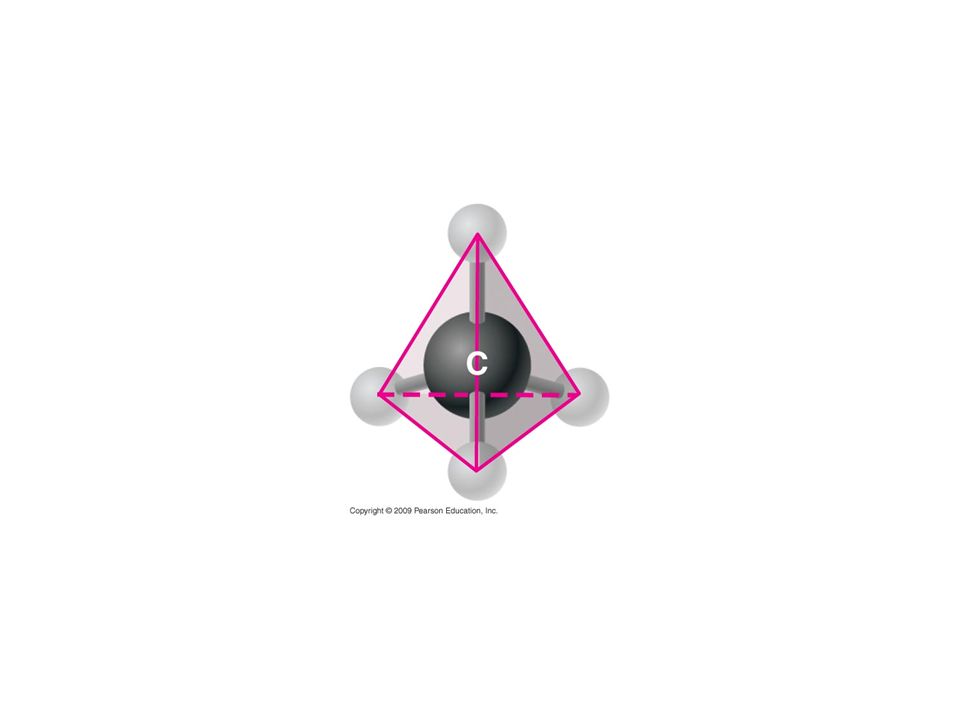
Amino acids, the building blocks of proteins, have an amino group and a carboxyl group Both of these are covalently bonded to a central carbon atom Also bonded to the central carbon is a hydrogen atom and other chemical group symbolized by “R” The “R” determines the type of amino acid and what it can build

43
3.12 Proteins are made from amino acids linked by peptide bonds
Amino acids, the building blocks of proteins I.e. The 26 letters of the alphabet are the building block for the English language

44
Amino group Carboxyl group
Figure 3.12A General structure of an amino acid.

45
Amino acids are classified as hydrophobic or hydrophilic
Some amino acids have a nonpolar R group and are hydrophobic Others have a polar R group and are hydrophilic, which means they easily dissolve in aqueous solutions

46
Leucine (Leu) Serine (Ser) Aspartic acid (Asp) Hydrophobic Hydrophilic
Figure 3.12B Examples of amino acids with hydrophobic and hydrophilic R groups. Leucine (Leu) Serine (Ser) Aspartic acid (Asp) Hydrophobic Hydrophilic
 Serine (Ser) Aspartic acid (Asp) Hydrophobic. Hydrophilic.")
47
Amino acids link together to form polymeric proteins
polymeric-consisting of many repeating structural units Linkage is accomplished by an enzyme-mediated dehydration reaction This links the carboxyl group of one amino acid to the amino group of the next amino acid The covalent linkage resulting is called a peptide bond

48
Carboxyl group Amino group Amino acid Amino acid
Figure 3.12C Peptide bond formation. As more and more amino acids are added, a chain of amino acids called a polypeptide results. The combination of amino acids is determined by expression of genes on DNA. Although there seems to be an unlimited number of combinations of 20 amino acids, the combinations are limited in an individual because of inheritance.

49
Peptide bond Carboxyl group Amino group Dehydration reaction
Amino acid Amino acid Dipeptide Figure 3.12C Peptide bond formation. As more and more amino acids are added, a chain of amino acids called a polypeptide results. The combination of amino acids is determined by expression of genes on DNA. Although there seems to be an unlimited number of combinations of 20 amino acids, the combinations are limited in an individual because of inheritance.

50
3.13 A protein’s specific shape determines its function
A polypeptide chain contains hundreds or thousands of amino acids linked by peptide bonds The amino acid sequence causes the polypeptide to assume a particular shape The shape of a protein determines its specific function Because of the molecular structure of specific proteins on brain cells, endorphins bind to them. This gives us a feeling of euphoria and pain relief. Morphine, heroin, and other opiate drugs are able to mimic endorphins and bind to the endorphin receptors in the brain. Because of the euphoria that results, we become addicted.

51
Addiction Endorphins bind to specific protein receptors on our brains, causing us to feel a sense of euphoria However, many drugs mimic these endorphins and bind to the same protein receptors.

52
Groove Figure 3.13A Ribbon model of the protein lysozyme.

53
Groove Figure 3.13B Space-filling model of lysozyme.

54
3.13 A protein’s specific shape determines its function
If for some reason a protein’s shape is altered, it can no longer function Denaturation will cause polypeptide chains to unravel and lose their shape and, thus, their function Proteins can be denatured by changes in salt concentration, temperature and pH

55
It is VERY easy to denatureate a protein , but VERY difficult to renaturate a protein

57
3.14 A protein’s shape depends on four levels of structure
A protein can have four levels of structure 1. Primary structure 2. Secondary structure 3. Tertiary structure 4. Quaternary structure For the BLAST Animation Alpha Helix, go to Animation and Video Files.

58
The primary structure of a protein is its unique amino acid sequence
The correct amino acid sequence is determined by the cell’s genetic information (genes) The slightest change in this sequence affects the protein’s ability to function, or makes an entirely new protein with a different function Sickle cell disease is manifested by an inability of hemoglobin in red blood cells to carry oxygen, the primary function of hemoglobin. This blood disorder is the result of change in a single amino acid.
 The slightest change in this sequence affects the protein’s ability to function, or makes an entirely new protein with a different function. Sickle cell disease is manifested by an inability of hemoglobin in red blood cells to carry oxygen, the primary function of hemoglobin. This blood disorder is the result of change in a single amino acid.")
59
Single amino acid changes can result in severe diseases
Single amino acid changes can result in severe diseases. Some amino acid changes result in no negative effects.

60
Protein secondary structure results from coiling or folding of the polypeptide
Coiling results in a helical structure called an alpha helix Folding may lead to a structure called a pleated sheet Coiling and folding result from hydrogen bonding between certain areas of the polypeptide chain Hydrogen bonding is an important component of the silk protein of a spider’s web. The many hydrogen bonds makes the web as strong as steel.

61
Many hydrogen bonds are responsible for the strength of spider silk.
Figure 3.14UN01 Spider in web. Many hydrogen bonds are responsible for the strength of spider silk.

62
Polypeptide chain Figure 3.14UN02 Collagen. Collagen

63
The overall three-dimensional shape of a protein is called its tertiary structure
Tertiary structure generally results from interactions between the R groups of the various amino acids Disulfide bridges are covalent bonds that further strengthen the protein’s shape

64
Two or more polypeptide chains (subunits) associate providing quaternary structure
Collagen is an example of a protein with quaternary structure Its triple helix gives great strength to connective tissue, bone, tendons, and ligaments Animation: Protein Structure Introduction Misfolding of proteins cause diseases, such as Alzheimer’s and Parkinson’s. Both are manifested by accumulations of misfolded proteins. Consider an assignment to review the organic molecules in our diets. Have students, working individually or in small groups, analyze a food label listing the components of a McDonald’s Big Mac or other fast food sandwich. Note the most abundant organic molecule class (perhaps by weight) found in each component. Animation: Primary Protein Structure Animation: Secondary Protein Structure Animation: Tertiary Protein Structure Animation: Quaternary Protein Structure
 found in each component. Animation: Primary Protein Structure. Animation: Secondary Protein Structure. Animation: Tertiary Protein Structure. Animation: Quaternary Protein Structure.")
65
Four Levels of Protein Structure
Primary structure Amino acids Figure 3.14A Primary structure.

66
Four Levels of Protein Structure
Primary structure Amino acids Hydrogen bond Secondary structure Alpha helix Pleated sheet Figure 3.14A Primary structure. Figure 3.14B Secondary structure.

67
Four Levels of Protein Structure
Primary structure Amino acids Hydrogen bond Secondary structure Alpha helix Pleated sheet Tertiary structure Figure 3.14A Primary structure. Figure 3.14B Secondary structure. Figure 3.14C Tertiary structure. Polypeptide (single subunit of transthyretin)
")
68
Four Levels of Protein Structure
Primary structure Amino acids Hydrogen bond Secondary structure Alpha helix Pleated sheet Tertiary structure Figure 3.14A Primary structure. Figure 3.14B Secondary structure. Figure 3.14C Tertiary structure. Figure 3.14D Quaternary structure. Polypeptide (single subunit of transthyretin) Transthyretin, with four identical polypeptide subunits Quaternary structure
 Transthyretin, with. four identical. polypeptide subunits. Quaternary structure.")
69
Amino acids Primary structure Figure 3.14A Primary structure.

70
Amino acids Hydrogen bond Alpha helix Pleated sheet
Figure 3.14B Secondary structure. Alpha helix Pleated sheet Secondary structure

71
Polypeptide (single subunit of transthyretin) Tertiary structure
Figure 3.14C Tertiary structure. Tertiary structure

72
Transthyretin, with four identical polypeptide subunits
Figure 3.14D Quaternary structure. Quaternary structure

73
Proteins gone bad So what happens is a protein folds incorrectly?
Many diseases, such as Alzheimer’s and Parkinson’s involve an accumulation of misfolded proteins Prions are infectious agents composed of proteins Prion diseases are currently untreatable and always fatal

74
Prions Prions infect and propogate by refolding abnormally into a structure that is able to convert normally-folded molecules into abnormally-structured form This altered form accumulates in infected tissue, causing tissue damage and cell death Prions are resistant to denaturation due to their extremely stable, tightly packed structure

75
Prions Prions are implicated in a number of diseases in a variety of mammals: Bovine Spongiform Encephalopathy (“Mad Cows Disease”) – spread by feed containing ground-up infected cattle Creutzfeldt-Jakob Disease – degenerative neurological disorder spread by skin grafts or human growth hormone products; Kuru is a similar disease spread by cannibalism among the Fore tribe of Papua New Guinea
 – spread by feed containing ground-up infected cattle. Creutzfeldt-Jakob Disease – degenerative neurological disorder spread by skin grafts or human growth hormone products; Kuru is a similar disease spread by cannibalism among the Fore tribe of Papua New Guinea.")
76
Prions Chronic Wasting Disease – found in deer, moose, elk in U.S. and Canada Fatal Familial Insomnia – very rare, inherited prion disease (50 families worldwide have the responsible gene mutation); insoluble protein causes plaques to develop in the thalamus, the region of brain responsible for the regulation of sleep; fatal within several months
; insoluble protein causes plaques to develop in the thalamus, the region of brain responsible for the regulation of sleep; fatal within several months.")
77
3.15 TALKING ABOUT SCIENCE: Linus Pauling contributed to our understanding of the chemistry of life
After winning a Nobel Prize in Chemistry, Pauling spent considerable time studying biological molecules He discovered an oxygen attachment to hemoglobin as well as the cause of sickle-cell disease Pauling also discovered the alpha helix and pleated sheet of proteins Pauling was also an advocate for halting nuclear weapons testing and won the Nobel Peace Prize for his work. He was very close to reporting the structure of DNA when Watson and Crick scooped him and correctly described its structure.

78
Figure 3.15 Linus Pauling with a model of the alpha helix in 1948.

79
NUCLEIC ACIDS

80
3.16 Nucleic acids are information-rich polymers of nucleotides
DNA (deoxyribonucleic acid) and RNA (ribonucleic acid) are composed of monomers called nucleotides Nucleotides have three parts 1. A five-carbon sugar called ribose in RNA and deoxyribose in DNA 2. A phosphate group 3. A nitrogenous base Student Misconceptions and Concerns Module 3.16 is the first time the authors present the concept of transcription and translation, discussed extensively in later chapters. The basic conceptual flow of information from DNA to RNA to proteins is essential to these later discussions.
 and RNA (ribonucleic acid) are composed of monomers called nucleotides. Nucleotides have three parts. 1. A five-carbon sugar called ribose in RNA and deoxyribose in DNA. 2. A phosphate group. 3. A nitrogenous base. Student Misconceptions and Concerns. Module 3.16 is the first time the authors present the concept of transcription and translation, discussed extensively in later chapters. The basic conceptual flow of information from DNA to RNA to proteins is essential to these later discussions.")
81
Nitrogenous base (adenine) Phosphate group Sugar
Figure 3.16A A nucleotide, consisting of a phosphate group, sugar, and a nitrogenous base. Phosphate group Sugar

82
3.16 Nucleic acids are information-rich polymers of nucleotides
There are four DNA nitrogenous bases: adenine (A), thymine (T), cytosine (C), and guanine (G) RNA also has A, C, and G, but instead of T, it has uracil (U)
, thymine (T), cytosine (C), and guanine (G) RNA also has A, C, and G, but instead of T, it has uracil (U)")
83
3.16 Nucleic acids are information-rich polymers of nucleotides
A nucleic acid polymer, a polynucleotide, forms from the nucleotide monomers when the phosphate of one nucleotide bonds to the sugar of the next nucleotide The result is a repeating sugar-phosphate backbone with protruding nitrogenous bases

84
Nucleotide Figure 3.16B Part of a nucleotide. Sugar-phosphate backbone

85
3.16 Nucleic acids are information-rich polymers of nucleotides
Two polynucleotide strands wrap around each other to form a DNA double helix The two strands are associated because particular bases always hydrogen bond to one another A pairs with T, and C pairs with G, producing base pairs RNA is usually a single polynucleotide strand

86
Base pair Figure 3.16C DNA double helix.

87
3.16 Nucleic acids are information-rich polymers of nucleotides
A particular nucleotide sequence that can instruct the formation of a polypeptide is called a gene Most DNA molecules consist of millions of base pairs and, consequently, many genes These genes, many of which are unique to the species, determine the structure of proteins and, thus, life’s structures and functions

88
Polymers The key to the great diversity of proteins and DNA is arrangement – the variation in the sequence in which monomers are strung together DNA is built of only four monomers (nucleotides) and proteins are built from only 20 kinds of amino acids; both of which are incredibly diverse: the proteins in you and a fungus are made with the same 20 amino acids
 and proteins are built from only 20 kinds of amino acids; both of which are incredibly diverse: the proteins in you and a fungus are made with the same 20 amino acids")
89
Mutations are alterations in bases or the sequence of bases in DNA
3.17 EVOLUTION CONNECTION: Lactose tolerance is a recent event in human evolution Mutations are alterations in bases or the sequence of bases in DNA Lactose tolerance is the result of mutations In many people, the gene that dictates lactose utilization is turned off in adulthood Apparently, mutations occurred over time that prevented the gene from turning off This is an excellent example of human evolution Mutations that lead to lactose tolerance are relativity recent events. The mutation was useful because it allowed people to drink milk when other foods were unavailable. In other words, it provided a survival advantage.

91
DNAs structure was ‘discovered’ in 1953 by James Watson, Francis Crick and Rosalind Franklin

92
Enzyme A Enzyme B Rate of reaction 20 40 60 80 100 Temperature (°C)
")
93
Dehydration Hydrolysis Short polymer Monomer Longer polymer

98
You should now be able to
Discuss the importance of carbon to life’s molecular diversity Describe the chemical groups that are important to life Explain how a cell can make a variety of large molecules from a small set of molecules Define monosaccharides, disaccharides, and polysaccharides and explain their functions Define lipids, phospholipids, and steroids and explain their functions Copyright © 2009 Pearson Education, Inc.

99
You should now be able to
Describe the chemical structure of proteins and their importance to cells Describe the chemical structure of nucleic acids and how they relate to inheritance Copyright © 2009 Pearson Education, Inc.

Similar presentations


>")


>")


 and Hydrogen.>")

