Download presentation
Presentation is loading. Please wait.

1
Chapter 15: Musculoskeletal System

2
Structure and Function of Joints: Important Terms
Articular Structures: include joint capsule and articular cartilege, synovium and synovial fluid, intra-articular ligaments, and juxta-articular bone Nonarticular Structures: include periarticular ligaments Ligaments: ropelike bundles of collagen fibrils that connect bone to bone Tendons: collagen fibers connecting muscle to bone Cartilege: collagen matrix overlying bony suyrfaces Bursae: pouches of synovial fluid that cushion the movement of tendons and muscles over bone or other joint structures.

3
Types of Joints: Synovial, Cartilaginous, and Fibrous
Synovial Joint Joint is freely movable Bones covered by articular cartilege Bones separated by joint capsuleoverlying synovial cavity Synovial membrane secretes synovialfluid that lubricates joint movement Examples: shoulder, knee

4
Types of Joints: Synovial, Cartilaginous, and Fibrous
Cartilaginous Joint Joint is slightly movable Bones separated by fibrocartilaginous discs Discs contain nucleus pulposis that cushion bony movement Example: vertebral bodies of the spine

5
Types of Joints: Synovial, Cartilaginous, and Fibrous
Fibrous Joint Joints have no appreciable movement Bones separated by fibrous tissue or cartilege Example: sutures of the skull

6
Types of Synovial Joints: note that anatomy determines range of movement
Type of Joint Articular Shape Movement Example Spheroidal (ball & socket) Convex surface in concave cavity Wide-ranging flexion, extension, abduction, adduction, rotation, circumduction Shoulder, hip Hinge Flat, planar Motion in one plane; flexion, extension Interphalangeal joints of hand and foot; elbow Condylar Convex or concave Movement of two articulating surfaces not dissociable Knee; temporomandibular joint
 Convex surface in concave cavity. Wide-ranging flexion, extension, abduction, adduction, rotation, circumduction. Shoulder, hip. Hinge. Flat, planar. Motion in one plane; flexion, extension. Interphalangeal joints of hand and foot; elbow. Condylar. Convex or concave. Movement of two articulating surfaces not dissociable. Knee; temporomandibular joint.")
7
Musculoskeletal System: The Health History
Common or Concerning Symptoms Low back pain Neck pain Monoarticular or polyarticular joint pain Inflammatory or infectious joint pain Joint pain with systemic features such as fever, chills, rash, anorexia, weight loss, weakness Joint pain with symptoms from other organ systems

8
Musculoskeletal System: Tips for Evaluating Joint Pain
Ask the patient to “point to the pain” This saves considerable time since patient descriptions of the location of the pain may be vague Clarify and record the mechanism of injury, particularly if the joint pain is caused by trauma Determine whether the pain is: localized or diffuse acute or chronic inflammatory or noninflammatory

9
Techniques Of Examination: Overview for Each of the Major Joints*
Inspect for joint symmetry, alignment, or any bony deformities Inspect and palpate surrounding tissues for: any skin changes, nodules, muscle atrophy, or crepitus Assess any degenerative or inflammatory changes, especially swelling, warmth, tenderness, or redness Perform range of motion; use joint-specific maneuvers to test: joint function and stability integrity of ligaments, tendons, and bursae * Includes shoulder, wrist and hands, spine, hips, knees, and ankles. For examination of the temporomandibular, elbow, and foot joints, review Bates’ 9th edition textbook, Chapter 15, The Musculoskeletal System.

10
Shoulder: Anatomy Of Bones And Muscles
Review bony anatomy Review muscle groups, Bates’ 9th edition textbook, p. 513

11
Shoulder: Identify Surface Landmarks
Acromioclavicular joint Greater tubercle Coracoid process

12
Shoulder: Examination
Inspect for swelling, deformity, or abnormal positioning Palpate over the three bony landmarks for tenderness Check range of motion: flexion, extension, abduction, adduction, and internal (hands behind small of back) and external (hands behind neck) rotation Review Bates’ 9th edition textbook, pp and perform maneuvers as needed to assess: Acromioclavicular joint Subacromial and subdeltoid bursae Overall shoulder rotation (Apley scratch test) Rotator cuff
 and external (hands behind neck) rotation. Review Bates’ 9th edition textbook, pp and perform maneuvers as needed to assess: Acromioclavicular joint. Subacromial and subdeltoid bursae. Overall shoulder rotation (Apley scratch test) Rotator cuff.")
13
Wrist and Hand: Review the Anatomy

14
WRIST and HAND: Examination
Inspect for smoothness of motion, surface contour, alignment of wrist and fingers, and any bony deformities Palpate the distal radius and ulna at the wrist, the eight carpal bones, and the MCP, PIP, and DIP joints for swelling or tenderness the “anatomic snuffbox” just distal to the radial styloid with the hand extended for tenderness

15
Wrist and Hand: Examination
Check range of motion: Wrist: flexion, extension, ulnar and radial deviation Fingers: flexion, extension, abduction (fingers spread apart), adduction (fingers back together) Thumb: flexion, extension, abduction (thumb moves away from palm), adduction (thumb moves toward palm), and opposition (thumb touches each finger) Test grip strength Test sensation on the palmar and dorsal surfaces innervated by the median, ulnar, and radial nerves (see Bates’ 9th edition textbook, p. 528)
, adduction (fingers back together) Thumb: flexion, extension, abduction (thumb moves away from palm), adduction (thumb moves toward palm), and opposition (thumb touches each finger) Test grip strength. Test sensation on the palmar and dorsal surfaces innervated by the median, ulnar, and radial nerves (see Bates’ 9th edition textbook, p. 528)")
16
Wrist and Hand: Carpal tunnel syndrome
Clinical features: Pain or numbness of the first three fingers of the hand, but not in the palm, especially at night Loss of sensation in distribution of the medial nerve: palmar surface of thumb, index, middle, and medial 4th fingers Assess for: weak abduction of the thumb – most sensitive test Tinel’s sign – tingling with tapping over the medial nerve as it enters the carpal tunnel Phalen’s sign – numbness or tingling with pressing backs of hands together in acute flexion

17
Spine: Anatomy of Representative Cervical and Lumbar Vertebrae
7 cervical, 12 thoracic, 5 lumbar vertebrae stacked on the sacrum and coccyx Review the anatomy below:

18
Spine: Muscle Groups

19
Spine: Examination — Inspection
With patient in gown, directly inspect: posture position of neck and trunk, alignment of shoulders and iliac crests, spinal curvature (concave at the neck and low back, convex over the thorax) ease of gait
 ease of gait.")
20
Spine: Examination — Palpation
Palpate all spinous processes for tenderness or “step-offs” paravertebral muscles for tenderness or spasm the sacroiliac joints the sciatic notch

21
Spine: Examination — Range of Motion
Neck: assess range of motion Flexion and extension: chin to neck, look at ceiling Rotation and lateral bending: turn head side to side, tilt each ear to shoulder Spine: assess range of motion. You may need to support the patient. Flexion and extension: bend forward toward toes, bend backward Rotation and lateral bending: rotate trunk (pull shoulder then opposite hip posteriorly), lean to the right then left
, lean to the right then left.")
22
Hip: Review Bony Anatomy and Bursae

23
Hip: Examination — Inspection and Palpation
Inspect gait, during both swing (non-weight bearing) and stance (foot on ground and bears weight) assess base – width between footsteps should be 2-4 inches rhythm should be smooth and continuous Palpate over inguinal ligament, trochanteric bursa, and ischiogluteal bursa
 and stance (foot on ground and bears weight) assess base – width between footsteps should be 2-4 inches. rhythm should be smooth and continuous. Palpate over inguinal ligament, trochanteric bursa, and ischiogluteal bursa.")
24
Hip: Examination – Range of Motion
Assess Flexion – bend and pull knee to chest. Check for flexion deformity (opposite knee goes into flexion) Extension – leg extends posteriorly while patient carefully positioned near edge of table Abduction and adduction – reach across and grasp opposite hip; grasp ankle and move leg laterally, then medially toward opposite hip External and internal rotation – flex hip and knee to 90°, grasp ankle, rotate flexed lower leg medially then laterally
 Extension – leg extends posteriorly while patient carefully positioned near edge of table. Abduction and adduction – reach across and grasp opposite hip; grasp ankle and move leg laterally, then medially toward opposite hip. External and internal rotation – flex hip and knee to 90°, grasp ankle, rotate flexed lower leg medially then laterally.")
25
Knee: Review the Anatomy
Identify bony structures, bursae, and especially the 4 ligaments and 2 menisci

26
Knee: Examination — Inspection and Palpation
contours and alignment of knees for swelling quadriceps muscle bulk for any atrophy knee action during swing and stance phases of gait Palpate, with patient sitting: Infrapatellar spaces adjacent to patella Medial and lateral femoral epicondyles and condyles Medial and lateral margins of tibial plateau Insertion of patellar tendon at the tibial tubercle

27
Knee: Examination — Palpation, cont’d
Palpate, with the knee flexed, and note any tenderness: along the joint line, including menisci and bursae along the medial and lateral collateral ligaments (MCL & LCL) over the patellar tendon. If tender, compress the patella against the femur and check knee extension Palpate: over the suprapatellar bursa above the knee the prepatellar bursa over the patella the pes anserine bursa on posteromedial knee If swelling, palpate for bulge sign or balloon sign, or balotte the patella (see Bates’ 9th edition textbook, pp , for techniques)
 over the patellar tendon. If tender, compress the patella against the femur and check knee extension. Palpate: over the suprapatellar bursa above the knee. the prepatellar bursa over the patella. the pes anserine bursa on posteromedial knee. If swelling, palpate for bulge sign or balloon sign, or balotte the patella (see Bates’ 9th edition textbook, pp , for techniques)")
28
Knee: Examination — Range of Motion and Maneuvers
Assess range of motion, with patient sitting: Flexion and extension Internal and external rotation – patient rotates foot medially and laterally If pain or swelling, use maneuvers to test stability of ligaments and integrity of menisci consult Bates’ 9th edition textbook, pp , and apply the following maneuvers as indicated: MCL – abduction or valgus stress test LCL – adduction or varus stress Anterior cruciate ligament (ACL) – anterior drawer sign, Lachman test Posterior cruciate ligament (PCL) – posterior drawer sign Medial and lateral menisci – McMurray test
 – anterior drawer sign, Lachman test. Posterior cruciate ligament (PCL) – posterior drawer sign. Medial and lateral menisci – McMurray test.")
29
Ankle and Foot: Review the Anatomy
Note: Especially the bony medial and lateral malleoli, the triangular deltoid ligament medially, and the three less stable ligaments laterally

30
Ankle and Foot: Examination — Inspection and Palpation
Inspect the surfaces of the ankles and feet for any deformities, nodules, swellings, calluses, or corns Palpate: the anterior aspect of the ankle using your thumbs the Achilles tendon for nodules or tenderness the metatarsophalangeal joints the heads of the 5 metatarsals – compress between your thumb and index finger

31
ANKLE and FOOT: Examination — Range of Motion and Maneuvers
Assess range of motion, including: tibiotalar joint (ankle) flexion and extension subtalar (talocalcaneal) joint grasp and invert and evert the heel transverse tarsal joint invert and evert the forefoot metatarsalphalangeal joints flex and extend the toes
 flexion and extension. subtalar (talocalcaneal) joint grasp and invert and evert the heel. transverse tarsal joint invert and evert the forefoot. metatarsalphalangeal joints flex and extend the toes.")
32
Diagnostic procedures Bone Marrow Aspiration
Is frequently done for evaluation of possible hematological disorders Bone marrow is usually aspirated from sternum or iliac crest in adults Preparation for procedure require explanation of the procedure and giving some tranquilizers for anxious patients. The skin area is cleansed and then a small area is anesthetized with local anesthesia. Bone marrow needle is introduced with a twisting motion and marrow is aspirated. After finishing procedure, pressure is applied to site for several minutes. The site of aspiration is observed closely for 24 hours for signs of bleeding and infection.

33
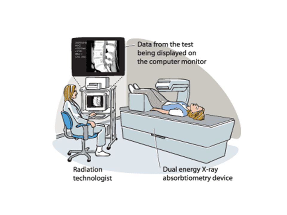
Bone Densitometry Is used to estimate bone density, through the use of x-ray or ultrasound Dual-photon absrptiometry and dual-energy x-rays determine bone mineral density at the wrist, hip, or spine to estimate the extent of osteoporosis. One peak is absorbed mainly by soft tissue and the other by bone Before the imaging study, the nurse should consider: If patient pregnant, claustrophobia, inability to tolerate position, deformity, metal implants, hair clips, hearing aid.

35
Electromyography EMG is a test that checks the activity of the muscles and the nerves that control the muscles. (electrical activity of muscles) The health care provider will insert a very thin needle electrode through the skin into the muscle. The electrode on the needle picks up the electrical activity given off by muscles. This activity appears on a nearby monitor, and may be heard through a speaker. After placement of the electrodes, patient may be asked to contract the muscle. The electrical activity seen on the monitor provides information about his muscle's ability to respond when the nerves to muscles are stimulated.

36
EMG is most often used when people have symptoms of weakness, and examination shows impaired muscle strength.( e.g. CTS) It can help to tell the difference between muscle weakness caused by injury of a nerve attached to a muscle and weakness due to neurological disorders.

Similar presentations


 Pelvic girdle.>")


