Download presentation
Presentation is loading. Please wait.

1
REPRODUCTIVE ANATOMY CHAPTER 34.1

2
REPRODUCTIVE SYSTEM This applies to all humans and can also apply to most mammals. Reproductive system – collection of specialized organs, glands, and hormones that work to make a new organism. Puberty – FSH and LH hormones are released from the brain into the body. Sexual development occurs.

3
PLANT REPRODUCTIVE ANATOMY Sepals – modified leaves that support the flower. Petal – usually brightly colored modified leaves. Used to attract pollinators to flower. Stamen – male structure of the flower. Contains filament and anther and these produce pollen. This pollen can then be carried to female part of flower. Pistol– female structure of flower. This connects down to the ovary of the plant. When pollen is absorbed it then fertilizes the plant.

4
Remember Me?

5
FEMALE ANATOMY Ovum – (Ova = plural) is an egg cell. (This is what is produced from female reproductive system) Ovaries - Pair of organs that where egg cells are produced. Uterus – (also called womb) is where fertilized cell is held. Uterine Lining – layer of cells of uterus that holds eggs.
 Ovaries - Pair of organs that where egg cells are produced. Uterus – (also called womb) is where fertilized cell is held. Uterine Lining – layer of cells of uterus that holds eggs..")
8
FEMALE ANATOMY Fallopian tube – connective tube that runs from ovaries to uterus. Transports egg and where fertilization occurs. Cervix – indicates the end of uterus and beginning of vagina. Vagina – Entrance and exit of female reproductive system.

11
FEMALE HORMONES Three hormones are released into the body during female puberty. FSH (Follicle-stimulating hormone) LH (Luteinizing hormone) Estrogen Estrogen has three main functions: Develops female characteristics Needed for egg cell development Prepares uterus for pregnancy/maintain pregnancy
 LH (Luteinizing hormone) Estrogen Estrogen has three main functions: Develops female characteristics Needed for egg cell development Prepares uterus for pregnancy/maintain pregnancy.")
12
MALE ANATOMY Main function of male reproductive system is to produce sperm. (spermatazoa) Testes – Where production of sperm takes place. Made of millions of tiny tubules called seminiferous tubules. (These actually make the sperm) Scrotum – sac that contains the testes on the exterior of the body. Epididymis – tube that sperm travels through to leave testes. Sperm wait here to be released.
 Testes – Where production of sperm takes place. Made of millions of tiny tubules called seminiferous tubules. (These actually make the sperm) Scrotum – sac that contains the testes on the exterior of the body. Epididymis – tube that sperm travels through to leave testes. Sperm wait here to be released..")
13
MALE ANATOMY Vas deferens – long tube that leads sperm from epididymis to seminal vesicle. Prostate gland / Cowlper’s gland– provide fluids to protect and help sperm move more easily. Semen – mixture of fluids and sperm that is released. Urethra – final tube of travel for semen. Contained by the penis.

15
MISCELLANEOUS Male hormone is called testosterone. This causes the main sexual characteristics of males to develop. Make the distinction here as well Males share a urinary tract as reproductive tract (urethra) Females do not. They have separated the two organ systems completely.
 Females do not. They have separated the two organ systems completely..")
16
REPRODUCTIVE PROCESS CHAPTER 34.2

17
PRODUCTION OF EGGS Eggs mature and are released according to hormonal cycles. This means hormones control pregnancy as well as the reproductive cycle Eggs (and sperm) are produced through meiosis For eggs going through meiosis not all of them mature. (Polar bodies)
 are produced through meiosis For eggs going through meiosis not all of them mature. (Polar bodies).")
19
RELEASE OF EGG Before egg cells mature they are surrounded by a follicle When the egg is ready to release the follicle ruptures When the egg is released from the ovary it is called ovulation Takes 5-7 days fro egg to travel through fallopian tube

21
MENSTRUAL CYCLE Flow Phase: This is the beginning of the menstrual cycle. Indicated by flow, or the release of blood. This is the lining of the uterus (endometrium) is being released from the body. This includes blood, tissue, and mucus. The “cramps” are when muscles in the uterus are contracting, expelling the lining.
 is being released from the body. This includes blood, tissue, and mucus. The cramps are when muscles in the uterus are contracting, expelling the lining..")
23
MENSTRUAL CYCLE Follicular Phase: From about day 6 to 14 FSH and LH is slowly added to the body and causes levels to allow ovulation to occur Around day 14 is when ovulation occurs – this is when the egg is released from the ovary Endometrium builds back up

25
MENSTRUAL CYCLE Luteal Phase: Day 15 – 28 Hormone production is allowed to decrease As this continues to thicken the uterine lining and allow it to hold a fertilized egg. (If there is one.) Remember, this length of time varies from person to person.
 Remember, this length of time varies from person to person..")
27
SPERM PRODUCTION Males do not produce any sperm until they reach maturity (female are born with eggs) They do not stop producing sperm (unless there is damage to the organs) Hormones (FSH and LH) tell the body to begin making sperm No polar bodies are produced
 They do not stop producing sperm (unless there is damage to the organs) Hormones (FSH and LH) tell the body to begin making sperm No polar bodies are produced")
29
FERTILIZATION Millions of sperm are released during sex, and only one impregnates an egg When the sperm arrives at the egg it releases an enzymes that breaks down egg cell membrane Once one reaches the inside, signal is sent from the egg to stop other sperm from entering

32
FERTILIZATION When an egg becomes fertilized it is called a zygote Sometimes each ovary will release an egg, allowing for two to become fertilized (fraternal twins) When a fertilized egg splits and two different development occur, then you have identical twins
 When a fertilized egg splits and two different development occur, then you have identical twins")
33
PROBLEMS There are some conditions that can cause infertility – the inability to reproduce Vas deferens or uterus can be too narrow Certain illness can cause sterilization (chicken pox and mumps in males) STD’s Allergies
 STD’s Allergies")
34
FETAL DEVELOPMENT CHAPTER 34.3

35
EARLY DEVELOPMENT When an egg cell becomes implanted with a sperm, we call it a zygote. After the zygote develops further (a hollow ball of cells) we call it a blastocyst. This continues to grow until we see organ cells beginning to form, called an embryo.
 we call it a blastocyst. This continues to grow until we see organ cells beginning to form, called an embryo..")
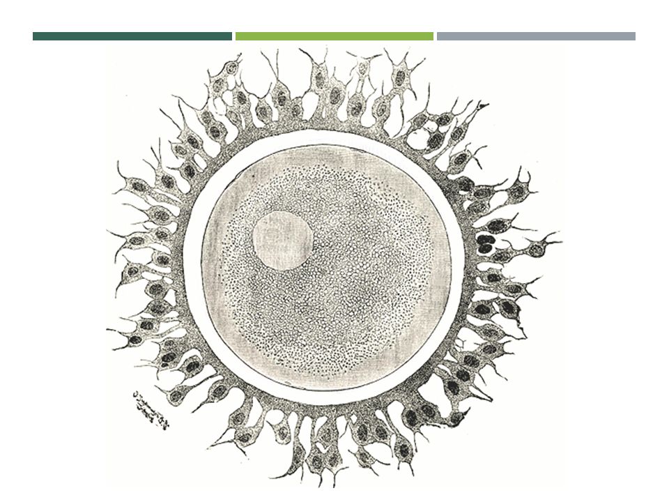
37
5 DAYS AFTER FERTILIZATION

38
CONTAINMENT As a blastocyst continues to grow, it will be grown inside of a protective fluid- filled barrier called the amniotic sac. Placenta – organ that keeps mother and child’s blood constantly exchanging oxygen. Umbilical cord – bridge between the placenta and child.

40
1 ST TRIMESTER Weeks 1 - 12 Organs start to develop separately Overall body structure is determined Heart begins beating (week 5) Early spinal cord forms Arms and legs develop Blastocyst becomes a fetus (around week 9)
 Early spinal cord forms Arms and legs develop Blastocyst becomes a fetus (around week 9)")
41
2 ND TRIMESTER Weeks 13 - 27 Physical activity Heartbeat can be heard This is when a woman begins to “show” her pregnancy Fingers and toes now form Brain rapidly develops By the end of trimester – baby is about 12 inches long

42
3 RD TRIMESTER Weeks 28 – 40 Grows to about 20 inches long and around 8 pounds Lungs begin to start breathing motion Hair develops Dreams Fetus turns down to prepare for birth Most babies born at 32 weeks can survive.

43
CAN YOU IDENTIFY THE TRIMESTER?

Similar presentations