Download presentation
Presentation is loading. Please wait.

1
HONORS ANATOMY & PHYSIOLOGY CHAPTER 19 BLOOD VESSELS

2
form a closed delivery system that begins & ends at the heart 3 types: 1. Arteries 2. Capillaries 3. Veins

3
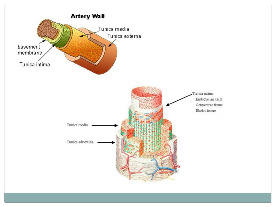
Blood Vessel Walls arteries & veins have 3 distinct layers = tunics surround a central blood-containing space = lumen 3 tunics: 1. tunica intima 2. tunica media 3. tunica externa

4
Walls of Arteries & Veins 1. tunica intima innermost layer = endothelium simple squamous epithelium: continuous with endocardium 2. tunica media middle layer : circular smooth muscle with elastin 3. tunica externa aka tunica adventitia infiltrated with nervefibers, elastic fibers, larger arteries & veins have their own smaller blood vessels = vasa vasorum (vessels of the vessels)
.")
6
Circulation of Blood left side heart aorta branches of aorta (arteries) arterioles capillaries venules veins vena cava right side of heart pulmonary circulation left side of heart
 arterioles capillaries venules veins vena cava right side of heart pulmonary circulation left side of heart")
7
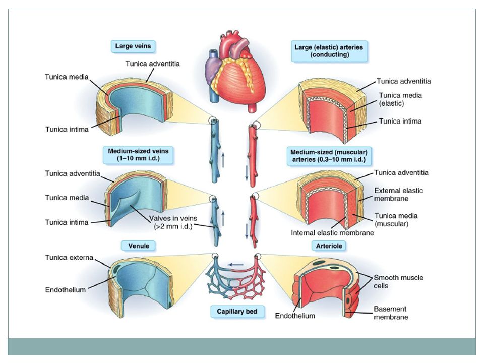
Arterial System Artery: blood vessel with blood flow AWAY from heart 3 types: 1. Elastic Arteries 2. Muscular Arteries 3. Arterioles

8
Elastic Arteries thick-walled arteries near the heart include aorta & its major branches largest of all arteries aka conducting arteries “pressure reservoirs” expand & recoil as heart ejects blood allows blood to flow continuously instead of flow & ebb

9
Muscular Arteries distal to elastic arteries deliver blood to specific organs aka distributing arteries thickest tunica media: more smooth muscle & less elastic tissue as elastic arteries more active in vasoconstriction than other types

12
Arterioles smallest of artery types lead to capillary beds small arteries (arterioles)have muscles that control their diameters (precapillary sphincters): used to control blood flow thru an organ
have muscles that control their diameters (precapillary sphincters): used to control blood flow thru an organ")
13
Capillaries where materials delivered to/from cells blood walls 1 squamous cell thick: so diffusion very fast not elastic

14
Pericytes smooth muscle-like cells surround capillaries stabilize capillaries help control capillary permeability

15
3 Types of Capillaries 1. Continuous (muscle, skin) most common endothelial cells joined by tight jcts fluids & small solutes can cross, except in brain nothing crosses (BBB) 2. Fenestrated (small intestine, endocrine organs, kidney) endothelial cells riddled with holes covered by thin membrane of basal lamina material more permeable to fluids 3. Sinusoid (liver, spleen, bone marrow, adrenal medulla) very leaky allow large molecules & blood cells to pass
 most common endothelial cells joined by tight jcts fluids & small solutes can cross, except in brain nothing crosses (BBB) 2. Fenestrated (small intestine, endocrine organs, kidney) endothelial cells riddled with holes covered by thin membrane of basal lamina material more permeable to fluids 3. Sinusoid (liver, spleen, bone marrow, adrenal medulla) very leaky allow large molecules & blood cells to pass.")
16
Types of Capillaries

17
Microcirculation capillaries don’t function solo form “beds” arteriole flow thru capillary bed venule = microcirculation

18
Veins any blood vessel with blood flowing toward the heart low pressure vessels can expand to accommodate differing volumes of blood flow contain valves to stop backflow of blood

19
Venules capillaries unite to form venules Postcapillary Venules: not much larger than capillary and almost as porous larger venules have thin layer of smooth muscle

21
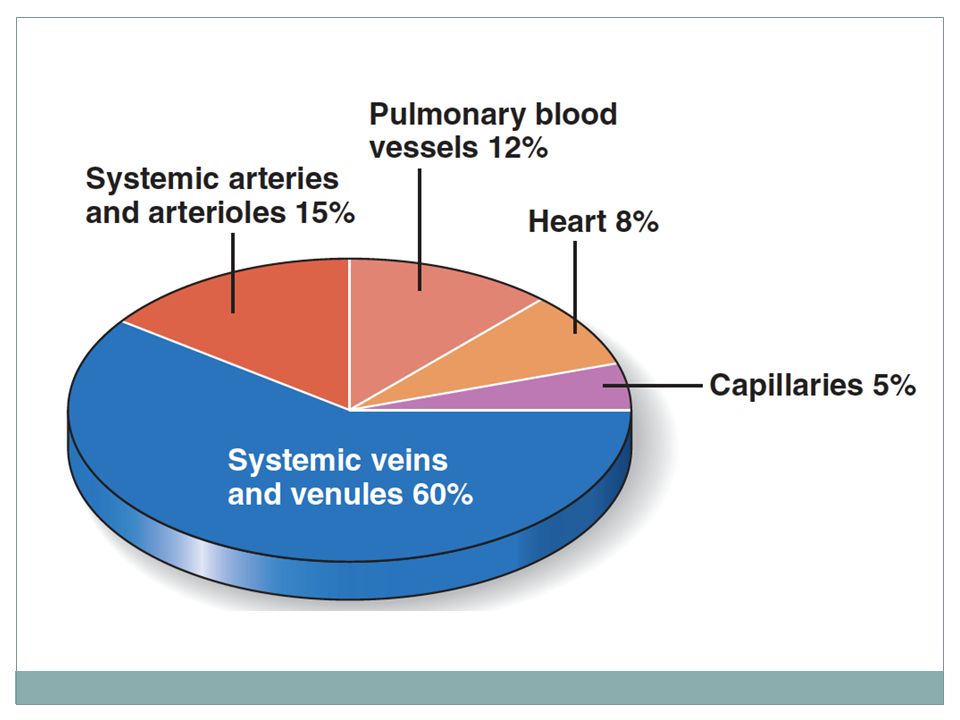
Veins venules join to form veins 3 distinct tunics larger lumens than corresponding arteries little smooth muscle or elastin in tunica media tunica externa thickest layer Veins called blood reservoirs because they can hold up to 65% of blood supply

23
Venous Valves formed from folds of tunica intima most abundant in veins of limbs where gravity opposes flow of blood Varicose Veins: tortuous, dilated veins due to incompetent valves superficial veins more susceptible because of little support, deeper veins have support from surrounding muscle

24
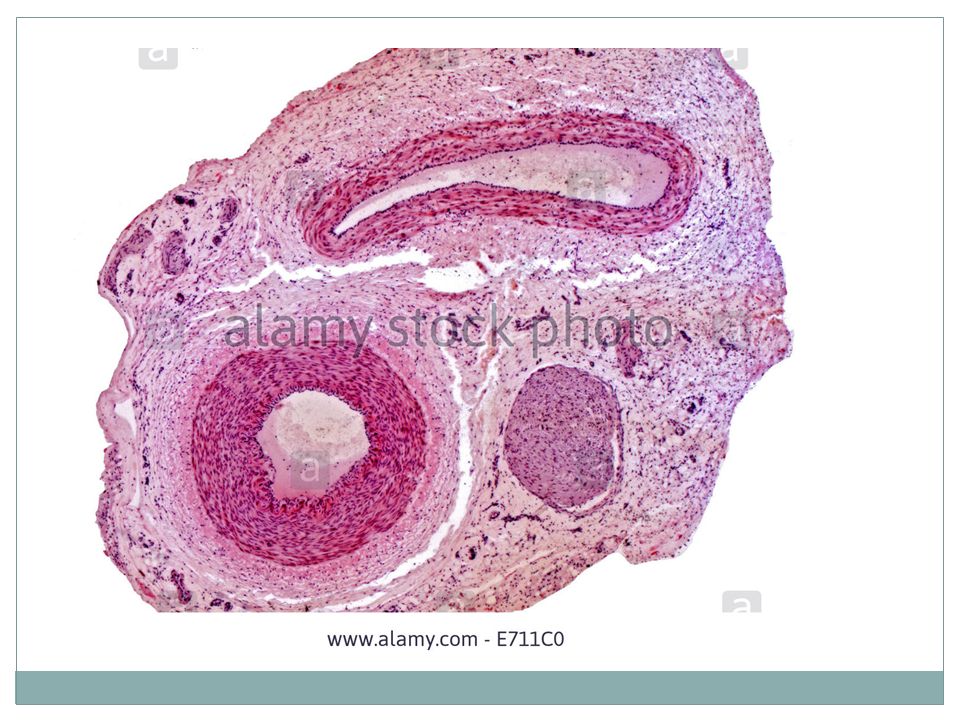
cross-section of vein with valve

25
Comparing Arteries & Veins

26
Venous Sinuses example: coronary sinus in heart dural venous sinuses of brain (CSF drains into) highly specialized, flat, veins with very thin walls only endothelial cells
 highly specialized, flat, veins with very thin walls only endothelial cells")
27
Vascular Anastomoses special interconnections between blood vessels arteries supplying same territory often merge = anastomosis provide alternate pathways = collateral channels for blood to reach a given organ or region of body common in: joints abdominal organs heart brain

29
Blood Flow volume of blood flowing through a vessel or organ, or entire circulation in a given time (mL/min) blood flow = CO at rest if considering entire vascular system is relatively constant @ any given moment blood flow through individual organs may vary widely directly proportional to BP & inversely proportional to resistance
 blood flow = CO at rest if considering entire vascular system is relatively any given moment blood flow through individual organs may vary widely directly proportional to BP & inversely proportional to resistance")
30
Blood Pressure (BP) force per unit area exerted on a vessel wall by the blood within it expressed in mm Hg BP means systemic arterial blood pressure in largest vessels *difference in BP w/in vascular system provides driving force that keeps blood flowing high low pressure
 force per unit area exerted on a vessel wall by the blood within it expressed in mm Hg BP means systemic arterial blood pressure in largest vessels *difference in BP w/in vascular system provides driving force that keeps blood flowing high low pressure")
31
Resistance opposition to flow *major determinant: small-diameter arterioles a measure of the amount of friction blood encounters flowing through vessels blood viscosity: related to thickness or stickiness of the blood fairly constant *longer the vessel the greater the resistance blood vessel diameter changes

32
Systemic BP highest in aorta/ lowest in venae cavae steepest drop in BP occurs in arterioles (resistance is the greatest) Systole: peak of BP Diasole: lowest pressure Pulse Pressure: difference between systole & diastole normal BP in adult: 120/80 mm Hg Hypotension: < 90/60 (rarely a problem) chronic HTN: 140/90 or higher indicates increased peripheral resistance
 Systole: peak of BP Diasole: lowest pressure Pulse Pressure: difference between systole & diastole normal BP in adult: 120/80 mm Hg Hypotension: < 90/60 (rarely a problem) chronic HTN: 140/90 or higher indicates increased peripheral resistance")
33
HTN Risk Factors 1. high-fat, high-salt diet 2. obesity 3. Diabetes mellitus 4. advanced age 5. smoking 6. stress 7. Black race > than others 8. family hx of HTN

34
HTN can lead to: 1. MI 2. CVA 3. Renal disease

35
Pulmonary Circulation transports O2-low, CO2-laden blood from right ventricle lungs releases CO2 & fills with O2 left atrium

36
Systemic Circulation transports O2-rich blood from left ventricle all body tissues

37
Blood Flow Through Organs regulated by nerves & chemical agents both cardiac output & blood vessel diameter controlled by hormones & nerves controlled by ANS increasing blood pressure can increase blood flow increasing blood pressure increases cardiac output constricts many arterioles more blood volume to other organs

38
Arteries & Veins all arteries deep veins: both deep & superficial superficial veins tend to have many interconnections Hepatic Portal Circulation: unique venous drainage of liver

39
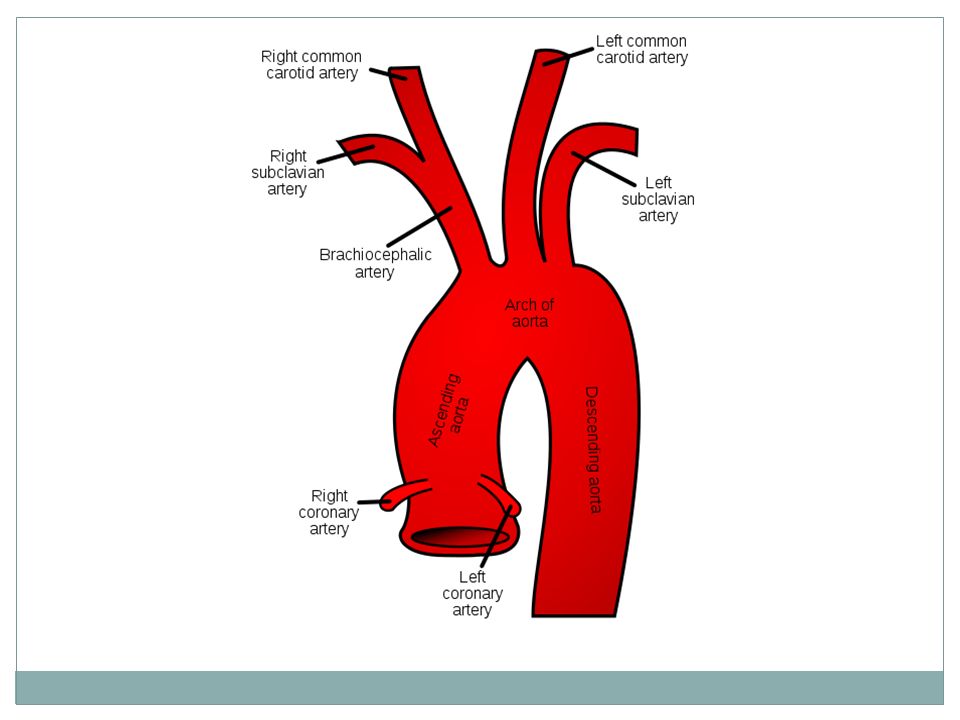
Aorta Ascending Aorta begins @ aortic semilunar valve rt & lt coronary arteries supply rt & lt sides of heart Aortic Arch 3 important branches: brachiocephalic trunk, lt common carotid, lt subclavian Descending Aorta travels posterior to heart portion in thorax called thoracic aorta portion in abdominal cavity called abdominal aorta

41
Common Carotids branch into: External Carotid arteries supply blood to neck, esophagus, pharynx, larynx, lower jaw, face Internal Carotid arteries supply blood to the brain (with the rt & lt vertebral arteries: branches of subclavian arteries)
")
42
Arteries of Upper Extremities Axillary artery: branch of subclavian artery becomes Brachial artery in the arm branches into Radial (pulse)& Ulnar arteries in lower arm
& Ulnar arteries in lower arm")
43
Branches of the Abdominal Aorta descends slightly to the left of the vertebral column retroperitoneal Branches: 1. Celiac Trunk (3 branches) Lt gastric artery: stomach Splenic artery: spleen: stomach, & pancreas Common Hepatic Artery: liver, stomach, gallbladder, & duodenum
 Lt gastric artery: stomach Splenic artery: spleen: stomach, & pancreas Common Hepatic Artery: liver, stomach, gallbladder, & duodenum.")
44
Branches of the Abdominal Aorta 2. Superior Mesenteric Artery: pancreas, duodenum, small intestines, most of large intestines 3. Inferior mesenteric Artery: terminal portion of the colon, sigmoid colon, & rectum

45
Branches of the Abdominal Aorta

46
5 Paired Arteries from Abdominal Aorta 1. Inferior phrenic arteries inferior surface of diaphragm 2. Suprarenal arteries Adrenal glands 3. Renal arteries kidneys 4. Gonadal arteries Testicular or Ovarian 5. Lumbar arteries vertebrae, spinal cord, abdominal wall

48
Iliac Arteries Abdominal Aorta branches into rt & lt Common Iliac Arteries @ L4 level each branches internal & external iliac arteries @ level of lumbosacral joint Internal Iliac Arteries: bladder, external genitalia, uterus, vagina External Iliac Arteries: blood to lower extremities

49
External Iliac Arteries when cross over to medial surface of thigh become Femoral Arteries branches to deep femoral & superficial femoral when reaches knee becomes Popliteal Artery where it branches posterior & anterior Tibial arteries Posterior Tibial Artery divides Medial & Plantar Arteries

50
Arteries of the Lower Extremities

51
Systemic Veins most veins run parallel to arteries of same name

52
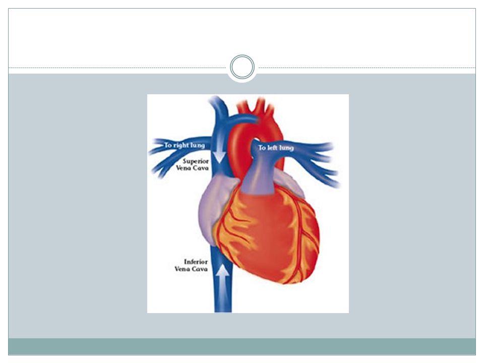
Superior & Inferior Vena Cava SVC: large vein that receives blood from upper body (head, neck, upper limbs) IVC: large vein that receives blood from the lower body (lower limbs, pelvis, abdomen) both return blood to right atrium
 IVC: large vein that receives blood from the lower body (lower limbs, pelvis, abdomen) both return blood to right atrium")
54
Systemic Veins Internal Jugular descends parallel to common carotid arteries brachiocephalic veins(just as they merge with the subclavian veins)
")
55
Veins of the Upper Extremity Radial & Ulnar veins parallel arteries of same name then merge to become Brachial vein axillary vein subclavian vein Vein draw blood from: median cubital

56
Veins of the Abdomen & Pelvis External Iliac veins receive blood from the lower extremities --> join with Internal Iliac veins to form the rt & lt Common Iliac Veins fuses with the IVC

57
Veins of Abdomen & Pelvis

58
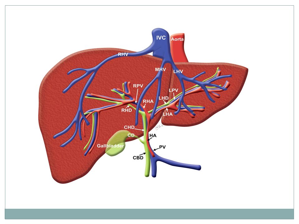
Hepatic Portal System Blood leaving the digestive organs by veins is rich in nutrients….instead of returning directly to IVC heart this blood is shunted to liver first This way liver can store, convert, detoxify, or excrete materials as necessary Hepatic Portal vein enters liver with nutrient rich blood

Similar presentations