Download presentation
Presentation is loading. Please wait.

1
Introduction to x-rays

2
What do these words mean?
Isotope Radioactivity Geiger Muller Tube Atomic mass Half life Becquerel

3
Learning objectives State qualitatively the differential absorption of X-rays by air, fat, other soft tissues and bone and the appearance of X-rays on film after passing through these media. • Explain techniques for improving quality of X-ray images; use of a grid, narrow beam, filtration. Describe how the use of image intensifying screens reduces dose rates.

4
Frequency between 1017 – 1020Hz

5
Why are they useful? Fractures Breast cancer Lung cancer Dental
Cardiac units Airports Therapeutic

6
How do they work? X-rays can be used because of the
differential absorption (attenuation) of the x-rays by different body tissues (such as bone and muscle). To obtain an image, a uniform beam of x-rays is transmitted through the area of interest. Bone absorbs most of the x-rays and as such, will appear white on an x-ray film; soft-tissue (such as muscle) only absorbs some of the x-rays and appears grey
 of the x-rays by different body tissues. (such as bone and muscle). To. obtain an image, a uniform beam. of x-rays is transmitted through. the area of interest. Bone absorbs most of the x-rays. and as such, will appear white on. an x-ray film; soft-tissue (such as. muscle) only absorbs some of. the x-rays and appears grey.")
7
How are x-rays produced
X-rays are produced when a beam of energy electrons hits a metal target. When electrons collide they lose KE some of this energy is converted to x-rays but most is converted to heat

8
X ray beam copper anode filament heavy metal target electron beam E.H.T vacuum

9
X-ray attenuation Medium Attenuation Appearance on film Air Negligible
Black Fat Small Dark grey Other soft tissues Grey Bone High White

10
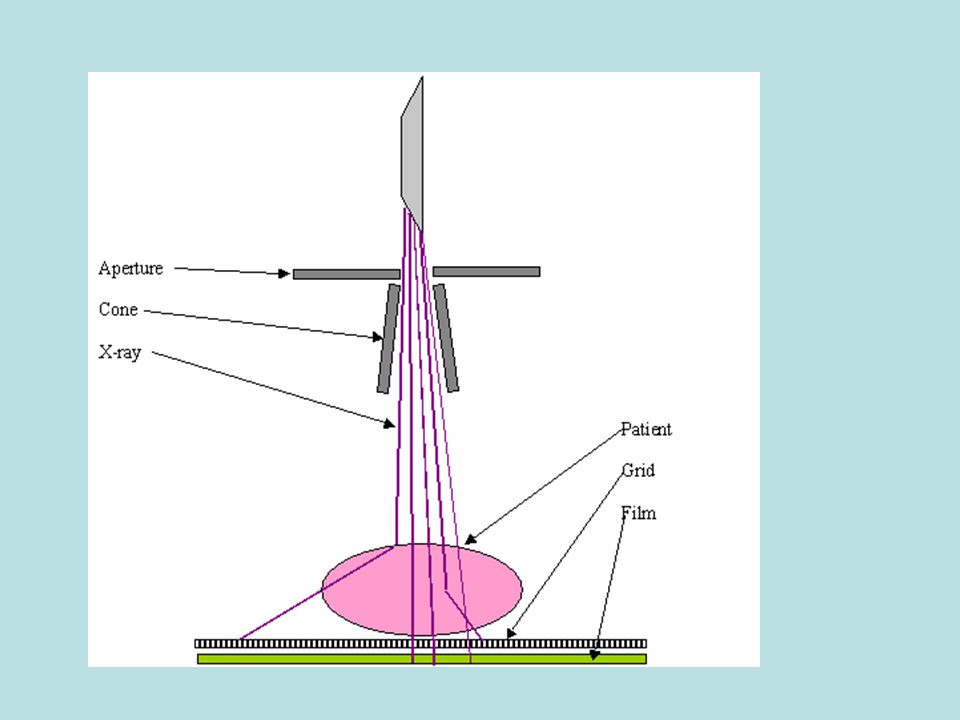
Improving image quality
The radiographer aims to produce: Sharp images by increasing resolution and reducing blur Good contrast by distinguishing clearly between different body materials. They can do this by use of a grid, narrow beam, filtration.

11
Use of a grid A grid of lead strips reduces the blurring of images caused by scattered radiation. If two crossed grids are used at right angles to each other, almost completely eliminate unwanted scatter. To prevent the grid lines appearing on the image the grid is slowly moved across the film during exposure.

13
Narrow beam The cross section of the x-ray beam can be controlled by the diaphragm, this is usually a pair of lead sheets at right angles to each other. Another type of beam definer is the cone. A narrow bean is preferred as random scatter increases with beam size and blurs the image

15
Filtration Filters reduce unwanted low-energy scatter. This increases the sharpness of the image and the contrast

16
Intensifying screens About 97% of the x-rays falling on a photographic film pass straight through without any interaction. The film alone is not a very sensitive method for detecting x-rays.

17
X-rays from patient Cassette front (plastic) Front intensifying screen Double sided film ~ 12mm Rear intensifying screen Felt padding Cassette back metal

18
X-rays from the patient
X-ray absorbed in the fluorescent screen Visible light Front fluorescent screen Double sided film Rear fluorescent screen Far fewer a-rays pass straight through Direct film blackening by x-rays Indirect film blackening by light

19
Modern screens can intensify the image by up to 250 times
Modern screens can intensify the image by up to 250 times. The increased image intensity then gives shorter exposure times and reduced radiation doses to the patient. However some detail is lost by diffusion of fluorescent light. Typical resolution is mm, whilst direct exposure film can give better than 0.1mm

Similar presentations


>")


