Download presentation
Presentation is loading. Please wait.

1
Dental Problems in the Field Setting
Roy L. Alson, PhD, MD, FACEP CO DMAT NC-1

2
Thanks Herb Johnson, DDS
Numerous Authors, etc whose photos on the web I “borrowed” NC-1 for putting up with me

3
Objectives Identify and Discuss Common Dental Problems seen in Primary Care Setting Describe management of Common Infections of the Mouth and Face Describe Management of Dental Trauma Assemble a Basic Kit for DMAT to Care for Dental Issues in the Field Setting. Will not be a discussion of complex facial fractures, etc

4
Incidence Common ED Complaint
1% of visits to DMAT NC-1 on deployments post hurricane, have been dental related Common complaint for pain medication seekers Physicians have little training in management of dental problems Data form ED and from NC-1’ s experiences shows about 1% of ED visits involve some type of dental complaint

5
Epidemiology of Mouth Pain
Infectious Trauma Post Procedure Dental Blocks Non Oral Causes Many Americans have poor dental hygiene Non oral: Sinus, other disease TMJ syndrome Systemic Diseases Too much to cover in 1 hour Dental care often delayed due to expenses, etc Appear at DMAT seeking free care

7
Assessment History Onset of Pain Location of Pain MOI
Other significant trauma Airway Status C-spine Onset of Pain Location of Pain

8
Assessment Radiation of Pain Fever, other Systemic Signs Malocclusion
Temperature Sensitivity? Recent Surgery Loss of appliance? History of Rheumatic Fever, etc? Consider non oral causes of mouth and tooth pain Sinus disease

9
Exam

10
Radiographs Limited availability in the field
Very important with deep space infections Facial trauma needs CT or a waters view

11
Water’s View From:

13
Anatomy of The Mouth 32 Adult Teeth

14
Terminology Buccal (labial): Lingual (palatal) Occlusal
: Lingual (palatal) Occlusal")
15
Anatomy of aTooth Crown Root 3 layers Enamel Dentin Pulp Gingiva: Gum
This is a first molar of the mandibular(lower jaw) with tow roots. Every tooth consists of a visual part which is the Crown and the anatomical Neck and the hidden inside the jaw bone Roots.A tooth can have 1-3 roots. In this picture you can see clearly the basic structure of a tooth.It consists the Enamel the Dentine inside of it and at the center there is the Pulp Chamber.Let's see them one by one: Enamel: This is the covering coat of the tooth's crown.It is the hardest tissue of the human body!That's the protection layer of tooth from dental caries(the acids of the bacterial plaque). It is white in color normally and it is not sensitive(no nerve endings).Once caries passes th enamel it's then that you start to feel pain. Dentine: It is the main substance of both the crown and the root of the tooth.It' yellowish-white in color and very soft and very sensitive since it is connected to the pulp through tubes(the pulp is where the nerves are).Once the caries reaches the dentine the decay is much more faster. Pulp Chamber: This is a cavity that houses the nerves and the blood vessels which carry the nutrients to the tooth.Because of the nerves it is extremely sensitive. At the roots it becomes the root canal(see below) Root Canals: These are the canals through which the blood vessels and nerves pass the roots heading from the bone to the crown of the tooth(pulp chamber) Apical Foramen The opening of the root canal to the jaw bone and place though which an infection of the pulp will pass to the periodontical tissues Cementum: Is a thin layer that covers the dentine of the roots from the outside.This is where the Periodontal Ligament(see bellow) attaches to the tooth Periodontal Ligament: The membrane that houses between the jaw bone and the tooth's roots attaching it to the bone. Bone: Part of the jaw bone that surrounds the part of the toouth that is not visual(basically the roots).This is where the tooth is anchored. Gum(Gingiva): This is the gum that we all know.Attaches to the bone underneath and all around the clinical neck of the tooth.Between the neck and the gum there is a space called Gingival Sulcus.This has a normal depth of 0.5 to 2mm and is where an infection can easily originate. Root Gingiva: Gum Periodontal Ligament Anchors tooth
 with tow roots. Every tooth consists of a visual part which is the Crown and the anatomical Neck and the hidden inside the jaw bone Roots.A tooth can have 1-3 roots. In this picture you can see clearly the basic structure of a tooth.It consists the Enamel the Dentine inside of it and at the center there is the Pulp Chamber.Let s see them one by one: Enamel: This is the covering coat of the tooth s crown.It is the hardest tissue of the human body!That s the protection layer of tooth from dental caries(the acids of the bacterial plaque). It is white in color normally and it is not sensitive(no nerve endings).Once caries passes th enamel it s then that you start to feel pain. Dentine: It is the main substance of both the crown and the root of the tooth.It yellowish-white in color and very soft and very sensitive since it is connected to the pulp through tubes(the pulp is where the nerves are).Once the caries reaches the dentine the decay is much more faster. Pulp Chamber: This is a cavity that houses the nerves and the blood vessels which carry the nutrients to the tooth.Because of the nerves it is extremely sensitive. At the roots it becomes the root canal(see below) Root Canals: These are the canals through which the blood vessels and nerves pass the roots heading from the bone to the crown of the tooth(pulp chamber) Apical Foramen The opening of the root canal to the jaw bone and place though which an infection of the pulp will pass to the periodontical tissues. Cementum: Is a thin layer that covers the dentine of the roots from the outside.This is where the Periodontal Ligament(see bellow) attaches to the tooth. Periodontal Ligament: The membrane that houses between the jaw bone and the tooth s roots attaching it to the bone. Bone: Part of the jaw bone that surrounds the part of the toouth that is not visual(basically the roots).This is where the tooth is anchored. Gum(Gingiva): This is the gum that we all know.Attaches to the bone underneath and all around the clinical neck of the tooth.Between the neck and the gum there is a space called Gingival Sulcus.This has a normal depth of 0.5 to 2mm and is where an infection can easily originate. Root. Gingiva: Gum. Periodontal Ligament. Anchors tooth.")
16
Development Primary or “Baby Teeth” Secondary or Permanent Teeth
Erupt from 6 months to 3 years “Teething” pain Treat symptomatically Secondary or Permanent Teeth Begin Erupting at 6 years Complete in Teens: “Wisdom Teeth”

18
Analgesia Dental Problems Hurt People seek care because of the pain
Blocks improve patient care May need conscious sedation Will need analgesics after visit Common complaint for “seekers” Full discussion of Sedation beyond this course Dealing with seekers is beyond this course

19
Analgesia Relief of the perception of pain
sedation not intentional sedation may be a secondary effect of medications administered for analgesia Opioids Nonopioids Local Anesthetics block pain and temperature The Patient will Feel PRESSURE!!

20
Conscious/Light Sedation
Controlled lessening of a patient’s awareness of the environment and/or pain perception. Able to maintain stable vital signs, independent airway, and adequate spontaneous respirations.

21
Conscious Sedation Who is at high risk for poor procedural analgesia and sedation? Patients at extremes of age “It’s only an LP, she won’t remember” “He’s a gome, he won’t even know he hurts” Patients with cognitive limits Ethnicity! Communication and cultural biases

22
Sedation Have a protocol in place Monitor the patient
Recover the patient Benzodiazepine and Opioids Ketamine for Pediatrics Etomidate?? Nitrous oxide!! Lack scavenger, little familiarity

23
Dental Blocks Apply topical to mucosa Introduce needle
Benzocaine Lidocaine Introduce needle ASPIRATE SYRINGE Inject slowly and use smallest effective volume Usually need only 1 to 2 cc +/- Bicarbonate Aspirate to avoid intravascular injection of agent

24
Anesthetic Agents Addition of Epinephrine prolongs anesthetic effects by decreasing absorption Rarely causes palpitations and hypertension DMAT has Marcaine (bupivicaine) 0.5% w/o Lidocaine 1% w and w/o
 0.5% w/o. Lidocaine 1% w and w/o.")
25
Duration of Analgesia Lidocaine 30-120 60-400 4.5 7.0 Bupivacaine 2.5
Anesthetic Duration without Epinephrine (min) Duration with Epinephrine (min) Maximum Dose without Epinephrine (mg/kg) Maximum Dose with Epinephrine (mg/kg) Esters Cocaine 45 - 2.8 Procaine 15-30 30-90 7.1 8.5 Chloroprocaine 30-60 11.4 14.2 Tetracaine 1.4 Amides Lidocaine 30-120 60-400 4.5 7.0 Mepivacaine Bupivacaine 2.5 3.2 Etidocaine 200 4.2 5.7 Prilocaine
 Duration with Epinephrine (min) Maximum Dose without Epinephrine (mg/kg) Maximum Dose with Epinephrine (mg/kg) Esters. Cocaine Procaine Chloroprocaine Tetracaine Amides. Lidocaine Mepivacaine. Bupivacaine Etidocaine Prilocaine.")
26
Dental Blocks Multiple Sites Useful for many facial injuries
Risk of Hitting Nerve or Vascular Injection Hematoma at Site Do Not Inject into Infected Tissue Inferior alveolar nerve block provides rapid relief of pain in all teeth on one side of the mandible and the lower lip and chin. Palpate the retromolar fossa with the index finger and identify the convexity of the mandibular ramus. Hold the syringe parallel to the occlusal surfaces of the teeth so that its barrel is in line between the first and second premolars on the opposite side of the mandible. Retract the soft tissue towards the cheek and find the pterygomandibular triangle. Puncture the triangle, making sure the needle passes through the ligaments and muscles of the medial mandibular surface. Stop advancing the needle when it reaches the bone, withdraw it a few millimeters, aspirate to be sure the tip is not in a vein, and deposit 1-2ml of local anesthetic. Supraperiosteal infiltration provides intraoral local anesthesia for pain arising from maxillary teeth. Pucture the mucobuccal fold, holding the bevel of the needle toward the bone, aspirate the area and then inject 1-2ml of anesthetic near the apex of the affected tooth. This technique usually produces full anesthesia in 5-10 minutes. For best results inject as close as possible to the tooth-bearing maxillary bone.

27
Local Infiltration Works better for maxillary teeth than mandibular teeth Thickness of bone 27 ga needle, bevel towards bome. 2 to 3 cm deep into sulcus aim for apex of tooth Palatal side similar procedure, but more uncomfortable as mucosa is tightly adherent Supraperiosteal infiltration provides intraoral local anesthesia for pain arising from maxillary teeth. Pucture the mucobuccal fold, holding the bevel of the needle toward the bone, aspirate the area and then inject 1-2ml of anesthetic near the apex of the affected tooth. This technique usually produces full anesthesia in 5-10 minutes. For best results inject as close as possible to the tooth-bearing maxillary bone.

28
Trigeminal Nerve Branches

29
Maxillary Block: Mandibular Branch

30
Inferior Alveolar Block
Palpate the retromolar fossa with the index finger and identify the convexity of the mandibular ramus. Hold the syringe parallel to the occlusal surfaces of the teeth so that its barrel is in line between the first and second premolars on the opposite side of the mandible. Retract the soft tissue towards the cheek and find the pterygomandibular triangle. Puncture the triangle, making sure the needle passes through the ligaments and muscles of the medial mandibular surface. Stop advancing the needle when it reaches the bone, withdraw it a few millimeters, aspirate to be sure the tip is not in a vein, and deposit 1-2ml of local anesthetic If you do not go from the contralateral side, you could go beyond the ramus and reach BIG RED

31
Inferior Alveolar Block
Inferior alveolar nerve block provides rapid relief of pain in all teeth on one side of the mandible and the lower lip and chin. Palpate the retromolar fossa with the index finger and identify the convexity of the mandibular ramus. Hold the syringe parallel to the occlusal surfaces of the teeth so that its barrel is in line between the first and second premolars on the opposite side of the mandible. Retract the soft tissue towards the cheek and find the pterygomandibular triangle. Puncture the triangle, making sure the needle passes through the ligaments and muscles of the medial mandibular surface. Stop advancing the needle when it reaches the bone, withdraw it a few millimeters, aspirate to be sure the tip is not in a vein, and deposit 1-2ml of local anesthetic.

32
Dental Caries Break down of Enamel by acids
Poor Dental Hygiene contributes Occlusal and between teeth Dark spot or hole Pain, tender to tooth percussion Remove Food particles Temporary Filling? Eugenol and Zinc Oxide filling

34
Lost Filling Clean area out Apply ZOE or IRM Scrape excess
Dental Referral

35
Lost Crown Clean restoration and tooth surface Dry with cotton
Apply small amount ZOE or IRM Place on tooth Bite on gauze

36
Post Extraction Hemorrhage
Prior Aspirin or Anticoagulants Coagulopathy? Sucking on Straw, etc. Clot partially retracted Have pt. apply pressure for 30 min Suction out and clean socket ? Retained fragments Gelfoam +/- Thrombin Expensive Tea bag? Presentation The patient had an extraction or other dental surgery performed earlier in the day, now has excessive bleeding at the site, and cannot reach his dentist. What to do: Ask what procedure was done. Inquire about antiplatlet drugs like aspirin, underlying coagulopathies and previous experience with unusual bleeding. Using suction and saline irrigation, clear any packing and clot from the bleeding site. Roll a 2" x 2" gauze pad, insert it over the bleeding site, and have the patient apply constant pressure upon it (biting down usually suffices) for 20 minutes. If the site is still bleeding after 20 minutes of gauze pressure, infiltrate the extraction area and inject into the socket with a local anesthetic and vasoconstrictor such as 2% lidocaine with 1:100,000 or 1:50,000 epinephrine, until the tissue blanches. Again, have the patient bite on a gauze pad for 20 minutes. The anesthetic allows the patient to bite down harder and the epinephrine helps restrict the bleeding. If this does not stop the bleeding, pack the bleeding site with Gelfoam, with gauze soaked in topical thrombin, or with bone wax (if the site is a bony socket), place the gauze pad on top, and apply pressure again. An arterial bleeder resistant to all the above may require ligation with a figure eight stitch. Assess any possible large blood loss with orthostatic vital signs. When the bleeding stops, remove the overlying gauze, have the patient leave the site alone for a day, and see his dentist in followup. What not to do: Do not routinely obtain laboratory clotting studies or hematocrits, unless there is a suspicion that they should be abnormal. Discussion Occasionally, this problem can be handled over the telephone. Some say a tea bag works even better than a gauze pad
 for 20 minutes. If the site is still bleeding after 20 minutes of gauze pressure, infiltrate the extraction area and inject into the socket with a local anesthetic and vasoconstrictor such as 2% lidocaine with 1:100,000 or 1:50,000 epinephrine, until the tissue blanches. Again, have the patient bite on a gauze pad for 20 minutes. The anesthetic allows the patient to bite down harder and the epinephrine helps restrict the bleeding. If this does not stop the bleeding, pack the bleeding site with Gelfoam, with gauze soaked in topical thrombin, or with bone wax (if the site is a bony socket), place the gauze pad on top, and apply pressure again. An arterial bleeder resistant to all the above may require ligation with a figure eight stitch. Assess any possible large blood loss with orthostatic vital signs. When the bleeding stops, remove the overlying gauze, have the patient leave the site alone for a day, and see his dentist in followup. What not to do: Do not routinely obtain laboratory clotting studies or hematocrits, unless there is a suspicion that they should be abnormal. Discussion. Occasionally, this problem can be handled over the telephone. Some say a tea bag works even better than a gauze pad.")
37
“Dry Socket” Alveolar Osteitis
Loss of clot post extraction exposes alveolar bone Local Infection Days 2-4 Painful Smoking, Use of Straws, Molars

38
Dry Socket Management Pain Relief: Local Block
Irrigate Socket and Remove Debris Fill Open Socket Commercial Products: Alvogyl, Dry Socket Paste Iodoform gauze or gauze with Eugenol may need replacement at 48 hours Eugenol on Gel foam Antibiotics may be needed: OMFS preference

39
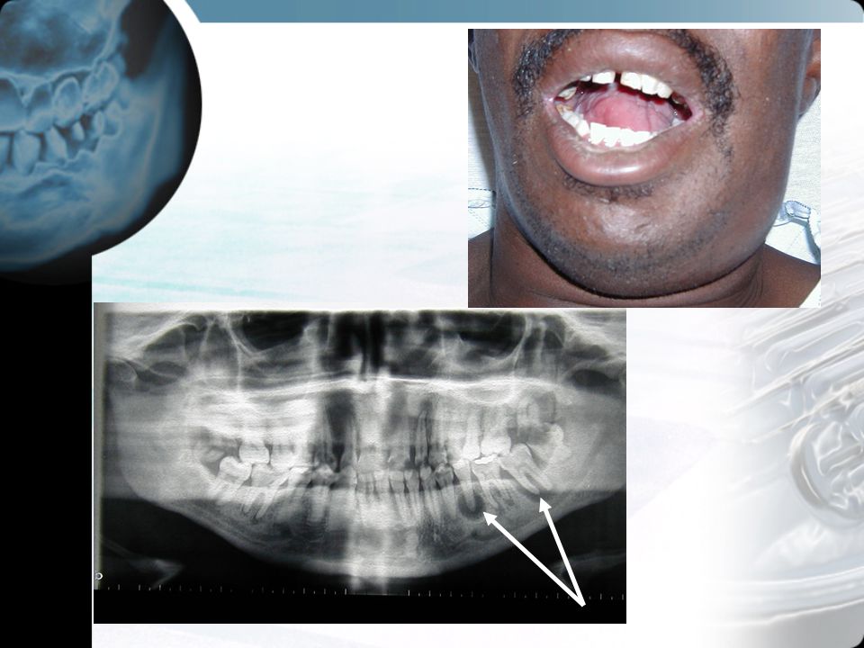
Odontogenic Infections
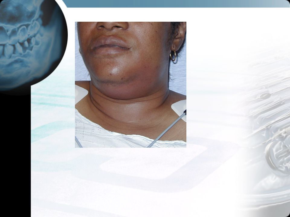
Mucosal Apical Deep Space

40
Pericoronitis Operculum of erupting wisdom teeth becomes filled with impacted food, debris Cellulitis follows Irrigate area out Oral Hygiene +/- Antibiotics Analgesics Presentation The patient is aged and seeks help because of painful swelling and infection around an erupting or impacted third molar (wisdom tooth). Occasionally, there can be trismus or pain on biting The site appears red and swollen with a flap that may reveal a partial tooth eruption and purulent drainage when pulled open. There is no pain with percussion of the tooth. What to do: Irrigate with a weak (2%) hydrogen peroxide solution. Purulent material can be released by placing the catheter tip of the irrigating syringe under the tissue flap overlying the impacted molar. Prescribe oral analgesics for comfort as well as penicillin over the next 10 days (penicillin VK 500mg qid). Instruct the patient on the importance of cleansing away any food particles that collect beneath the gingival flap. This can be accomplished by simply using a soft toothbrush or by using water jet irrigation. A dental followup should be provided to observe the resolution of the acute infection and to evaluate the need for removal of the gingival flap or molar. What not to do: Do not undertake any major blunt dissection while draining pus. This could spread a superficial infection into the deep spaces of the head and neck or follow a deep abscess posteriorly into the carotid sheath. Discussion Pericoronitis is a special type of acute periodontal abscess that occurs when ginngival tissue (operculum) overlies an erupting tooth (usually a third molar, also known as a wisdom tooth). Recurring acute symptoms are usually initiated by trauma from the opposing tooth or by impaction of food or debris under the flap of tissue that partially covers the erupting tooth. When dental referral is not readily available, one procedure for releiving the pain is surgical removal of the operculum. inject local anesthetic directly into the overlying tissue and then cut it away using the outline of the tooth as a guide for the incision. Sutures are not required.
. Occasionally, there can be trismus or pain on biting The site appears red and swollen with a flap that may reveal a partial tooth eruption and purulent drainage when pulled open. There is no pain with percussion of the tooth. What to do: Irrigate with a weak (2%) hydrogen peroxide solution. Purulent material can be released by placing the catheter tip of the irrigating syringe under the tissue flap overlying the impacted molar. Prescribe oral analgesics for comfort as well as penicillin over the next 10 days (penicillin VK 500mg qid). Instruct the patient on the importance of cleansing away any food particles that collect beneath the gingival flap. This can be accomplished by simply using a soft toothbrush or by using water jet irrigation. A dental followup should be provided to observe the resolution of the acute infection and to evaluate the need for removal of the gingival flap or molar. What not to do: Do not undertake any major blunt dissection while draining pus. This could spread a superficial infection into the deep spaces of the head and neck or follow a deep abscess posteriorly into the carotid sheath. Discussion. Pericoronitis is a special type of acute periodontal abscess that occurs when ginngival tissue (operculum) overlies an erupting tooth (usually a third molar, also known as a wisdom tooth). Recurring acute symptoms are usually initiated by trauma from the opposing tooth or by impaction of food or debris under the flap of tissue that partially covers the erupting tooth. When dental referral is not readily available, one procedure for releiving the pain is surgical removal of the operculum. inject local anesthetic directly into the overlying tissue and then cut it away using the outline of the tooth as a guide for the incision. Sutures are not required.")
41
Dentoalveolar Infection
Usually arises in Dental Pulp Periapical Abscess forms Pain and possibly swelling + / - Fever Follows path of least resistance Abscess tracks through alveolar bone into oral mucosa or skin Superficial abscess Dissects into deep spaces

43
Diagnosis Swelling of face or jaw Swelling or fluctuance in gingiva
Panorex if available, may show apical origin Consider CT for Deep Space infections

46
Treatment Antibiotics PCN or Clindamycin for the infection
May require multiple agents if deep spaces involved Surgical Drainage Dependent drainage of gingival lesions Deep space lesions should be managed by experienced surgeon. Extraction or root canal for periapical abscess Deep space infections should be admitted Airway at risk Extension into mediastinum

48
Mucosal Infections Ginigivitis Thrush Ginigivostomatitis Inflamation
Clean Debris out of sulci Mouth rinses Thrush Ginigivostomatitis Children most common Won’t eat or drink due to pain Nystatin for thrush “Magic Mouthwash” Mylanta and Benadryl +/- lidocaine

49
Gingivitis

50
ANUG Acute Necrotizing Ulcerative Gingivostomatitis Antibiotics
Trench Mouth or Vincent’s Angina Fusospirochettal infection Antibiotics Will need debridement

51
Herpangina The patient complains of a painful lesion in the mouth, and may be worried about having herpes. A pale yellow, flat, even-bordered ulcer surrounded by a red halo may be seen on the buccal or labial mucosa, lingual sulci, soft palate, pharynx, tongue, or gingiva. Lesions are usually solitary, but can be multiple and recurrent. The pain is usually greater than the size of the lesions would suggest, and major aphthae (larger than 1 cm) indicate a severe form of the disease which may last for weeks of months. What to do: Attempt to differentiate from lesions of herpes simplex and reassure the patient of the benign nature of most canker sores. Inform the patient that these lesions usually last 1-2 weeks, and that they should avoid hot, acidic or irritating food and drink. For transient pain relief, try a tablet of sucralfate crushed in a small amount of warm water, swirled in the mouth or gargled. Tetracycline elixir (or a capsule dissolved in water) not swallowed, but applied to cauterize lesions or used as a mouth wash can relieve pain after single or repeated application. Benadryl elixir mixed one-to-one with Kaopectate, Xylocaine 2% Viscous Solution, and Orabase HC applied topically can also provide symptomatic relief. For more severe cases, prescribe triamcinolone acetonide 0.1% suspension (add injectable Kenolog to sterile water without preservatives) in a 5ml oral rinse and spit out four times a day after meals and before bed, taking nothing by mouth for an hour afterward. An alternative regimen is dexamathasone elixir 1.5mg in 15ml qid rinse and swallow, tapering to three days of 0.5ml in 5ml, then three days swallowing every other dose, but discontinuing the regimen as soon as the mouth becomes comfortable. In very severe cases, try a burst dose of prednisone 40-60mg qd x5 (no tapering). Discussion Aphthous stomatitis has been studied for many years by numerous investigators. Although many exacerbating factors have been identified, the cause as yet remains unknown. Lesions can be precipitated by minor trauma, food allergy, stress, and systemic illness. Recurrent aphthous ulcers may accompany malignancy or autoimmune disease. At present, the treatment is only palliative, and may not alter the course of the syndrome. Apthous ulcers may be an immune reaction to damaged mucosa or altered oral bacteria. Herpangina and hand-foot-and-mouth disease can produce ulcers resembling aphthous ulcers, but which are instead part of coxsackie viral exanthems, usually with fever and occurring in clusters among children. Behcet's syndrome is an idiopathic condition characterized by oral ulcers clinically indistinguishable from aphthae but accompanied by genital ulcers, conjunctivitis, retinitis, iritis, leukocytosis, eosinophilia and increased erythrocyte sedimentation rate. References: Vincent SD, Lilly GE: Clinical, historic and therapeutic features of aphthous stomatitis. Oral Surg Oral Med Oral Pathol 1992;74:79-86.
 indicate a severe form of the disease which may last for weeks of months. What to do: Attempt to differentiate from lesions of herpes simplex and reassure the patient of the benign nature of most canker sores. Inform the patient that these lesions usually last 1-2 weeks, and that they should avoid hot, acidic or irritating food and drink. For transient pain relief, try a tablet of sucralfate crushed in a small amount of warm water, swirled in the mouth or gargled. Tetracycline elixir (or a capsule dissolved in water) not swallowed, but applied to cauterize lesions or used as a mouth wash can relieve pain after single or repeated application. Benadryl elixir mixed one-to-one with Kaopectate, Xylocaine 2% Viscous Solution, and Orabase HC applied topically can also provide symptomatic relief. For more severe cases, prescribe triamcinolone acetonide 0.1% suspension (add injectable Kenolog to sterile water without preservatives) in a 5ml oral rinse and spit out four times a day after meals and before bed, taking nothing by mouth for an hour afterward. An alternative regimen is dexamathasone elixir 1.5mg in 15ml qid rinse and swallow, tapering to three days of 0.5ml in 5ml, then three days swallowing every other dose, but discontinuing the regimen as soon as the mouth becomes comfortable. In very severe cases, try a burst dose of prednisone 40-60mg qd x5 (no tapering). Discussion. Aphthous stomatitis has been studied for many years by numerous investigators. Although many exacerbating factors have been identified, the cause as yet remains unknown. Lesions can be precipitated by minor trauma, food allergy, stress, and systemic illness. Recurrent aphthous ulcers may accompany malignancy or autoimmune disease. At present, the treatment is only palliative, and may not alter the course of the syndrome. Apthous ulcers may be an immune reaction to damaged mucosa or altered oral bacteria. Herpangina and hand-foot-and-mouth disease can produce ulcers resembling aphthous ulcers, but which are instead part of coxsackie viral exanthems, usually with fever and occurring in clusters among children. Behcet s syndrome is an idiopathic condition characterized by oral ulcers clinically indistinguishable from aphthae but accompanied by genital ulcers, conjunctivitis, retinitis, iritis, leukocytosis, eosinophilia and increased erythrocyte sedimentation rate. References: Vincent SD, Lilly GE: Clinical, historic and therapeutic features of aphthous stomatitis. Oral Surg Oral Med Oral Pathol 1992;74:")
52
Thrush Painful Dehydration a problem Nystatin

53
OMF Trauma Common Significant Morbidity
Assaults, MVC, Falls Significant Morbidity Potential for airway compromise Blunt versus penetrating Other associated Trauma Chest, Head, Neck

54
Oral Soft Tissue Injuries
Lacerations Cheek Tongue Gums Vascular Structures Bleed Profusely Airway at Risk Manage Airway

55
Lacerations Airway and Life Threats First
Repair of Teeth before Soft Tissue Repair Classically: Given Prophylactic Antibiotics Newer Literature suggest not necessary With large amounts of devitalized tissue, give antibiotic coverage PCN or Clindamycin Rate of oral laceration infections low

56
Buccal Lacerations Less than 2 cm: will heal on own
Close larger lacerations with absorbable sutures Through and Through Lac Check for injury to salivary ducts Stenson’s exits by upper second molar Check for Nerve Injury Close mucosa first then skin If tooth puncture, close only skin Nylon is irritating Bury knots on suture

58
Frenulum Laceration Face Plant No need to repair Anxious Parents

59
Discuss concussed tooth

60
Tongue Laceration Most small Lac heal on own
Challenge to repair, especially in child Edge Lac or gaping Lac should be repaired Prevent Bifid Tongue May need con sedation Antibiotic coverage should be considered Can close muscle and mucosa in single pass

61
Gingival Lacerations Skin is thin Hard to close
Usually heal without repair Often associated with Fractures Flaps will require closure

63
Cheek Lacerations Look for Facial Nerve Injury
Look for Parotid Duct Injury

64
Lip Lacerations Close Through and Through lacerations
Mucosa First, then skin If small, leave mucosa open Align Vermillion Border!!!

66
Dentoalveolar Trauma Blunt trauma Disrupts Dentoalveolar complex
Common Pediatric Problem Toddlers falling Sports Injuries Prevent with Mouth Guards

67
Luxation of Teeth Avulsion Intrusion Extrusion Luxation
Alveolar Ridge Fractue

68
Mandibular Anatomy

69
Assessment ABC’s Clear Airway Look for Extruded Teeth Palpate TMJ
If not Found: Get Chest Xray Palpate TMJ Assess ROM Palpate Mandible Malocclusion sensitive for Fracture

70
Assessment Tongue Blade Test Palpate oral lacerations
Step off Check for loose teeth Tooth tap for pain

71
Radiographs Panorex and CT are best Not available in Field
Plan films of mandible Should not change Field management Will require splinting Arch Bars

72
LaForte fractures Higher Force Blunt Face Injury

73
Alveolar Ridge Fracture

74
Tooth Fractures Ellis Classification 1-Enamel Only
White 2- Enamel and Dentin Yellow tint 3- Enamel, Dentin and Pulp May see blood

76
Treatment of Tooth Fracture
Pain Relief Prevent Infection of Dentin Dental Block for Analgesia Cover Exposed tooth CaOH Paste Zinc Oxide Coe-Pak Dry Area off Place Agent on Area and allow to set Patient should eat soft food till seen by dentist – 48 hr Skin adhesives not approved for oral use Dental cements similar Glass ionomers need UV system

77
Coe-Pak Tooth and surrounding gum must be dry Moisten your glove
Silly putty feel Make sure material gets into sulci between teeth Soft Diet

78
Luxation Extrusive – Partially out of socket
Lateral – displaced laterally, mesially, facially or lingually Often with associated Alveolar FX Intrusive: tooth pushed in Complete or avulsed tooth

79
Luxation with Alveolar FX
Reposition tooth Then repair Gingiva Splint

80
Intrusion Usually stable > 6mm will require surgical repair
Primary teeth Allow to grow out Permanent tooth may be damaged

81
Check socket to make sure not intruded

82
Laterally Luxated Tooth
After a direct blow to the mouth the patient may have a permanent tooth knocked from its socket. The tooth is intact, down to its root, from which hangs the delicate periodontal ligament that used to attach to alveolar bone and provide the tooth with its blood supply. What to do: In the field, avulsed teeth may be stored under the tongue or in the buccal vestibule between the gums and the teeth. If the patient is unconscious, the tooth can be stored in saline, milk or water until a better preservation solution is available. A child's tooth might be preserved, if necessary, in the parent's mouth. If the tooth has been out of its socket less than 15 minutes, take it by the crown, drop it in a tooth-preservation solution (Hank's solution, Sav-A-Tooth kit), flush the socket with the same solution, reimplant the tooth firmly, have the patient bite down firmly on a piece of gauze to help stabilize the tooth and when possible secure it to adjacent teeth with wire, arch bars, or a temporary periodontal pack (Coe- Pak). Coe-Pak is a peridontal dressing that comes in the form of a base and catalyst. Mix together and mold the resulting paste, which will eventually set semi-hard, over the gingival line and between the teeth. Put the patient on a liquid diet, prescribe penicillin VK 500mg qid x 2 weeks, and schedule a dental appointment. If the tooth was out 15 minutes to 2 hours, soak for 30 minutes to replenish nutrients. Local anesthesia will probably be needed before reimplanting as above. If the tooth was out over two hours, the periodontal ligament is dead, and should be removed, along with the pulp. The tooth sould soak 30 minutes in 5% sodium hypochlorite (Clorox), and 5 minutes each in saturated citric acid, 1% stannous fluoride and 5% doxycycline before reimplanting. The dead tooth should ankylose into the alveolar bone of the the socket like a dental implant. If the patient is between 6 and 10 years old, also soak the tooth for 5 minutes in 5% doxycycline to kill bacteria which could enter the immature apex and form an abscess. If you are not able to perform all this right away, simply keep the tooth soaking in the preservation solution until a dentist can get to it. The solution should preserve the tooth safely for up to four days. If a tooth is lost, obtain a chest x ray to rule out bronchial aspiration. Add tetanus prophylaxis if required What not to do: Do not touch a viable root with fingers, forceps, gauze or anything, or try to scrub or clean it. The periodontal ligament will be injured and unable to re-vascularize the re-implanted tooth. >Do not overlook fractures of teeth and alvolar ridges. Do not substitute the calcium hydroxide composition (Dycal) used for covering fractured teeth for the temporary periodontal pack (Coe- Pak) used to stabilized luxated teeth. They are different products. Do not replace primary deciduous teeth. Reimplanted primary teeth heal by ankylosis: they literally fuse to the bone, which can lead to cosmetic deformity since the area of ankylosis will not grow at the same rate as the rest of the dentofacial complex. Ankylosis can also interfere with the eruption of the permanent tooth. Normal developmental shedding of primary decidual teeth is preceded by absorption of the root, so that if such a tooth is brought to the ED by mistake, there is no root to reimplant in the socket, but a new permanent tooth underneath. Discussion Before commercially-available 320mOs, pH 7.2 reconstitution solutions, the best we could offer the avulsed tooth was rapid reimplantation. Without a preservation solution, the chances of successful reimplantation decline one percentage point every minute the tooth is absent from the oral cavity. In mature teeth, over age 10, the pulp will not survive avulsion even if the periodontal ligament does, and at the one-week follow-up visit with the dentist, the necrotic pulp will be removed to prevent a chronic inflammatory reaction from interfering with the healing of the periodontal ligament. References: Krasner P: Modern treatment of avulsed teeth by emergency physicians. Am J Emerg Med 1994;12:
, flush the socket with the same solution, reimplant the tooth firmly, have the patient bite down firmly on a piece of gauze to help stabilize the tooth and when possible secure it to adjacent teeth with wire, arch bars, or a temporary periodontal pack (Coe- Pak). Coe-Pak is a peridontal dressing that comes in the form of a base and catalyst. Mix together and mold the resulting paste, which will eventually set semi-hard, over the gingival line and between the teeth. Put the patient on a liquid diet, prescribe penicillin VK 500mg qid x 2 weeks, and schedule a dental appointment. If the tooth was out 15 minutes to 2 hours, soak for 30 minutes to replenish nutrients. Local anesthesia will probably be needed before reimplanting as above. If the tooth was out over two hours, the periodontal ligament is dead, and should be removed, along with the pulp. The tooth sould soak 30 minutes in 5% sodium hypochlorite (Clorox), and 5 minutes each in saturated citric acid, 1% stannous fluoride and 5% doxycycline before reimplanting. The dead tooth should ankylose into the alveolar bone of the the socket like a dental implant. If the patient is between 6 and 10 years old, also soak the tooth for 5 minutes in 5% doxycycline to kill bacteria which could enter the immature apex and form an abscess. If you are not able to perform all this right away, simply keep the tooth soaking in the preservation solution until a dentist can get to it. The solution should preserve the tooth safely for up to four days. If a tooth is lost, obtain a chest x ray to rule out bronchial aspiration. Add tetanus prophylaxis if required. What not to do: Do not touch a viable root with fingers, forceps, gauze or anything, or try to scrub or clean it. The periodontal ligament will be injured and unable to re-vascularize the re-implanted tooth. >Do not overlook fractures of teeth and alvolar ridges. Do not substitute the calcium hydroxide composition (Dycal) used for covering fractured teeth for the temporary periodontal pack (Coe- Pak) used to stabilized luxated teeth. They are different products. Do not replace primary deciduous teeth. Reimplanted primary teeth heal by ankylosis: they literally fuse to the bone, which can lead to cosmetic deformity since the area of ankylosis will not grow at the same rate as the rest of the dentofacial complex. Ankylosis can also interfere with the eruption of the permanent tooth. Normal developmental shedding of primary decidual teeth is preceded by absorption of the root, so that if such a tooth is brought to the ED by mistake, there is no root to reimplant in the socket, but a new permanent tooth underneath. Discussion. Before commercially-available 320mOs, pH 7.2 reconstitution solutions, the best we could offer the avulsed tooth was rapid reimplantation. Without a preservation solution, the chances of successful reimplantation decline one percentage point every minute the tooth is absent from the oral cavity. In mature teeth, over age 10, the pulp will not survive avulsion even if the periodontal ligament does, and at the one-week follow-up visit with the dentist, the necrotic pulp will be removed to prevent a chronic inflammatory reaction from interfering with the healing of the periodontal ligament. References: Krasner P: Modern treatment of avulsed teeth by emergency physicians. Am J Emerg Med 1994;12:")
83
Treating Avulsed Tooth
Tooth transport and storage: Socket is the best place. Save-A-Tooth: < 24 hours. Hanks Balanced buffer solution. Cold milk: < 6 hours. Saliva, saline or water: < ½ hour. Be sure patient does not swallow tooth if transported in sulcus

84
Tooth Replantation Time is tooth Analgesia Clean out clot
Gently but firmly insert tooth Splint tooth Periodontal Ligament quickly dies < 5 min Gentle suction and irrigation of socket. Preserve Peridontal ligament fibers

85
Tooth Replantation

86
Tooth Replantation

87
TMJ Syndrome Pain at TMJ Click or Pop with Chewing May have crepitus
Soft Diet Analgesics Consider occlusal problem New Filling?

89
Dental Kit Home made Commercial Cheaper Choose what you want
More expensive Easier to obtain and maintain

90
NC-1’s Ultimate Dental Kit for DMATs

91
Dental Kit Goals Control Pain Stabilize Loose Teeth Cover exposed Pulp
Secure Lost Restorations

92
Dental Kit • Dental roll gauze
Table 5. Dental equipment needed in the ED. • Packing gauze • Dental roll gauze • Calcium hydroxide paste or glass ionomer cement or zinc oxide cement • Dry Socket Paste or eugenol • Topical anesthetic gel (20% benzocaine or 5% lidocaine) • Topical bactericidal intraoral solution (Ora-5) • Periodontal paste (Coe-Pak) or self-cure composite • Bupivacaine cartridges with epinephrine • EMT ToothsaverTM Preservation System or fresh milk • Zinc oxide/eugenol temporary cement (Temrex) • Ringed injection syringe • Stainless steel spatula and mixing pads • Oral surgery tray with arch bars and ligature wires • Tongue blades and cotton-tipped applicators • Disposable electrocautery (optional) Acute Dental Emergencies In Emergency Medicine (May 2003) Emergency Medicine Practice
 • Topical bactericidal intraoral solution (Ora-5) • Periodontal paste (Coe-Pak) or self-cure composite. • Bupivacaine cartridges with epinephrine. • EMT ToothsaverTM Preservation System or fresh milk. • Zinc oxide/eugenol temporary cement (Temrex) • Ringed injection syringe. • Stainless steel spatula and mixing pads. • Oral surgery tray with arch bars and ligature wires. • Tongue blades and cotton-tipped applicators. • Disposable electrocautery (optional) Acute Dental Emergencies In Emergency Medicine (May 2003) Emergency Medicine Practice.")
93
Dental Kit www.dentalbox.net
1. 2-tray Cantilever Style Heavy Duty Plastic Utility Box (1) 2. TOPICAL ANESTHETIC 20% BENZOCAINE GEL 30GM BOTTLES (2)—used For Topical Mucosal Anesthetic 3. CALCIUM HYDROXIDE PASTE (CATALYST AND BASE) STANDARD PACKAGE (1)—used For Covering Fractured Teeth. 4. Zinc Oxide/Eugenol Temporary Cement Powder 25 Gms (1) 5. Zinc Oxide/Eugenol Temporary Cement Liquid 1 Oz. (1)--#4 & #5 Are Used In Combination To Fill Deep, Painful Caries Or To Cement Loose Fillings, Caps, Or Bridges. 6. Periodontal Dressing Standard Pkg. 90 GM BASE AND 90 GM CATALYST (1)—used For Stabilizing Loose Or Subluxed Teeth 7. Bupivocaine/Epinephrine Cartridges—canister Of 50. (1)—used As A Local Anesthetic For Odontalgia (Tooth Pain). For Use By Injection. 8. REUSABLE RINGED ASPIRATORS FOR USE WITH DISPOSABLE ANESHETIC SYRINGES (2)—for Use With #9 9. Dental Injector Disposable Syringes With 27 Gauge 1.5” NEEDLES FOR USE WITH CARTRIDGE ANESTHETIC AND RINGED ASPIRATORS (100)—used To Inject Local Anesthetic. For Use With #8. 10. Topical Oral Bactericidal Solution 1oz. MULTI-DOSE BOTTLE (1)—for Use As A Topical Antibacterial Agent In The Mouth Or Buccal Mucosa. 11. Cotton Gauze Rolls 50 Per Pkg (4) 12. Dry Socket Medicament 1 Oz Size. (1)—for Use In Sealing Dry Sockets (Alveolar Osteitis) 13. 3”x 3” Mixing Pads 100 Sheets/Pkg (2) 14. Stainless Steel Cement Spatula For Mixing Medicaments, Glues, Dressings, Etc. (1) 15. Stainless Steel Plastic Filling Instrument For Application Of Cements, Dressings, Etc. (1) 16. Laminated Quick-reference Cards With Instructional Text&photographs Depicting Use Of Each Medication And Of Each Tooth Block Type. ( The Instruction Cards Are To Be Used As A Clinical Reference Only And Are Not Designed To Replace The Individual Item’s Manufacturer’s Instructions. The Procedural Descriptions/Depictions Are Not Substitutes For Adequate Training Under A Professional Who Is Proficient In Said Procedure. ) 17. Cotton Tipped Applicators For Application Of Topical Anesthetic (50) 18. Wooden Tongue Depressors For Mixing Of Periodontal Dressing(50) 19. EMT Toothsaver, Tooth Preservation Kit (1) 20. Fax/Phone Reorder Forms (2)
 2. TOPICAL ANESTHETIC 20% BENZOCAINE GEL 30GM BOTTLES (2)—used For Topical Mucosal Anesthetic 3. CALCIUM HYDROXIDE PASTE (CATALYST AND BASE) STANDARD PACKAGE (1)—used For Covering Fractured Teeth. 4. Zinc Oxide/Eugenol Temporary Cement Powder 25 Gms (1) 5. Zinc Oxide/Eugenol Temporary Cement Liquid 1 Oz. (1)--#4 & #5 Are Used In Combination To Fill Deep, Painful Caries Or To Cement Loose Fillings, Caps, Or Bridges. 6. Periodontal Dressing Standard Pkg. 90 GM BASE AND 90 GM CATALYST (1)—used For Stabilizing Loose Or Subluxed Teeth 7. Bupivocaine/Epinephrine Cartridges—canister Of 50. (1)—used As A Local Anesthetic For Odontalgia (Tooth Pain). For Use By Injection. 8. REUSABLE RINGED ASPIRATORS FOR USE WITH DISPOSABLE ANESHETIC SYRINGES (2)—for Use With #9 9. Dental Injector Disposable Syringes With 27 Gauge 1.5 NEEDLES FOR USE WITH CARTRIDGE ANESTHETIC AND RINGED ASPIRATORS (100)—used To Inject Local Anesthetic. For Use With # Topical Oral Bactericidal Solution 1oz. MULTI-DOSE BOTTLE (1)—for Use As A Topical Antibacterial Agent In The Mouth Or Buccal Mucosa. 11. Cotton Gauze Rolls 50 Per Pkg (4) 12. Dry Socket Medicament 1 Oz Size. (1)—for Use In Sealing Dry Sockets (Alveolar Osteitis) x 3 Mixing Pads 100 Sheets/Pkg (2) 14. Stainless Steel Cement Spatula For Mixing Medicaments, Glues, Dressings, Etc. (1) 15. Stainless Steel Plastic Filling Instrument For Application Of Cements, Dressings, Etc. (1) 16. Laminated Quick-reference Cards With Instructional Text&photographs Depicting Use Of Each Medication And Of Each Tooth Block Type. ( The Instruction Cards Are To Be Used As A Clinical Reference Only And Are Not Designed To Replace The Individual Item’s Manufacturer’s Instructions. The Procedural Descriptions/Depictions Are Not Substitutes For Adequate Training Under A Professional Who Is Proficient In Said Procedure. ) 17. Cotton Tipped Applicators For Application Of Topical Anesthetic (50) 18. Wooden Tongue Depressors For Mixing Of Periodontal Dressing(50) 19. EMT Toothsaver, Tooth Preservation Kit (1) 20. Fax/Phone Reorder Forms (2)")
94
Dental Kit www.dentalbox.net
1. 2-tray Cantilever Style Heavy Duty Plastic Utility Box (1) 2. TOPICAL ANESTHETIC 20% BENZOCAINE GEL 30GM BOTTLES (2)—used For Topical Mucosal Anesthetic 3. CALCIUM HYDROXIDE PASTE (CATALYST AND BASE) STANDARD PACKAGE (1)—used For Covering Fractured Teeth. 4. Zinc Oxide/Eugenol Temporary Cement Powder 25 Gms (1) 5. Zinc Oxide/Eugenol Temporary Cement Liquid 1 Oz. (1)--#4 & #5 Are Used In Combination To Fill Deep, Painful Caries Or To Cement Loose Fillings, Caps, Or Bridges. 6. Periodontal Dressing Standard Pkg. 90 GM BASE AND 90 GM CATALYST (1)—used For Stabilizing Loose Or Subluxed Teeth 7. Bupivocaine/Epinephrine Cartridges—canister Of 50. (1)—used As A Local Anesthetic For Odontalgia (Tooth Pain). For Use By Injection. 8. REUSABLE RINGED ASPIRATORS FOR USE WITH DISPOSABLE ANESHETIC SYRINGES (2)—for Use With #9 9. Dental Injector Disposable Syringes With 27 Gauge 1.5” NEEDLES FOR USE WITH CARTRIDGE ANESTHETIC AND RINGED ASPIRATORS (100)—used To Inject Local Anesthetic. For Use With #8. 10. Topical Oral Bactericidal Solution 1oz. MULTI-DOSE BOTTLE (1)—for Use As A Topical Antibacterial Agent In The Mouth Or Buccal Mucosa. 11. Cotton Gauze Rolls 50 Per Pkg (4) 12. Dry Socket Medicament 1 Oz Size. (1)—for Use In Sealing Dry Sockets (Alveolar Osteitis) 13. 3”x 3” Mixing Pads 100 Sheets/Pkg (2) 14. Stainless Steel Cement Spatula For Mixing Medicaments, Glues, Dressings, Etc. (1) 15. Stainless Steel Plastic Filling Instrument For Application Of Cements, Dressings, Etc. (1) 16. Laminated Quick-reference Cards With Instructional Text&photographs Depicting Use Of Each Medication And Of Each Tooth Block Type. ( The Instruction Cards Are To Be Used As A Clinical Reference Only And Are Not Designed To Replace The Individual Item’s Manufacturer’s Instructions. The Procedural Descriptions/Depictions Are Not Substitutes For Adequate Training Under A Professional Who Is Proficient In Said Procedure. ) 17. Cotton Tipped Applicators For Application Of Topical Anesthetic (50) 18. Wooden Tongue Depressors For Mixing Of Periodontal Dressing(50) 19. EMT Toothsaver, Tooth Preservation Kit (1) 20. Fax/Phone Reorder Forms (2)
 2. TOPICAL ANESTHETIC 20% BENZOCAINE GEL 30GM BOTTLES (2)—used For Topical Mucosal Anesthetic 3. CALCIUM HYDROXIDE PASTE (CATALYST AND BASE) STANDARD PACKAGE (1)—used For Covering Fractured Teeth. 4. Zinc Oxide/Eugenol Temporary Cement Powder 25 Gms (1) 5. Zinc Oxide/Eugenol Temporary Cement Liquid 1 Oz. (1)--#4 & #5 Are Used In Combination To Fill Deep, Painful Caries Or To Cement Loose Fillings, Caps, Or Bridges. 6. Periodontal Dressing Standard Pkg. 90 GM BASE AND 90 GM CATALYST (1)—used For Stabilizing Loose Or Subluxed Teeth 7. Bupivocaine/Epinephrine Cartridges—canister Of 50. (1)—used As A Local Anesthetic For Odontalgia (Tooth Pain). For Use By Injection. 8. REUSABLE RINGED ASPIRATORS FOR USE WITH DISPOSABLE ANESHETIC SYRINGES (2)—for Use With #9 9. Dental Injector Disposable Syringes With 27 Gauge 1.5 NEEDLES FOR USE WITH CARTRIDGE ANESTHETIC AND RINGED ASPIRATORS (100)—used To Inject Local Anesthetic. For Use With # Topical Oral Bactericidal Solution 1oz. MULTI-DOSE BOTTLE (1)—for Use As A Topical Antibacterial Agent In The Mouth Or Buccal Mucosa. 11. Cotton Gauze Rolls 50 Per Pkg (4) 12. Dry Socket Medicament 1 Oz Size. (1)—for Use In Sealing Dry Sockets (Alveolar Osteitis) x 3 Mixing Pads 100 Sheets/Pkg (2) 14. Stainless Steel Cement Spatula For Mixing Medicaments, Glues, Dressings, Etc. (1) 15. Stainless Steel Plastic Filling Instrument For Application Of Cements, Dressings, Etc. (1) 16. Laminated Quick-reference Cards With Instructional Text&photographs Depicting Use Of Each Medication And Of Each Tooth Block Type. ( The Instruction Cards Are To Be Used As A Clinical Reference Only And Are Not Designed To Replace The Individual Item’s Manufacturer’s Instructions. The Procedural Descriptions/Depictions Are Not Substitutes For Adequate Training Under A Professional Who Is Proficient In Said Procedure. ) 17. Cotton Tipped Applicators For Application Of Topical Anesthetic (50) 18. Wooden Tongue Depressors For Mixing Of Periodontal Dressing(50) 19. EMT Toothsaver, Tooth Preservation Kit (1) 20. Fax/Phone Reorder Forms (2)")
95
Resources International association for Dental Traumatology Academy for Sports Dentistry : Local anesthesia in Dermatology dental anesthesia Guidelines for the evaluation and Management of traumatic dental injuries Authors: Flores M.T.; Andreasen J.O.; Bakland L.K. Source: Dental Traumatology, August 2001, vol. 17, no. 5, pp (4) Publisher: Blackwell Publishing
 Publisher: Blackwell Publishing.")
Similar presentations


. The first teeth which are shed and replaced by permanent teeth. There are 20 primary teeth The primary teeth are replaced starting.>")
