Download presentation
Presentation is loading. Please wait.

1
Soft Tissue Tumors

2
Reactive and Benign lesions of Fibroblastic and Histiocytic Origin
Irritation Fibroma Giant Cell fibroma Inflammatory Fibrous Hyperplasia Inflammatory Papillary Hyperplasia Fibrous Histiocytoma Fibromatosis and Myofibromatosis Oral Focal Mucinosis Pyogenic Granuloma Peripheral Giant Cell Granuloma Peripheral Ossifying Fibroma Benign Tunors of Fat tissue origin Lipoma Benign Tumors of Neural Origin Traumatic Neuroma Palisaded Encapsulated Neuroma Schwannoma Neurofibroma Granular Cell Tumor Congenital Epulis Melanotic Neuroectodermal Tumor of Infancy

3
Benign Tumors of Vascular Origin Hemangioma Lymphangioma
Benign Tumors of Muscle Origin Leiomyoma Rhabdomyoma Osseous and Cartilaginous Choristomas Malignant Tumors of Connective Tissue Fibrosarcoma Malignant Fibrous Histiocytoma Liposarcoma Neurofibrosarcoma Angiosarcoma Kaposi’s Sarcoma Leiomyosarcoma Rhabdomyosarcoma Metastases to Oral Soft Tissues

4
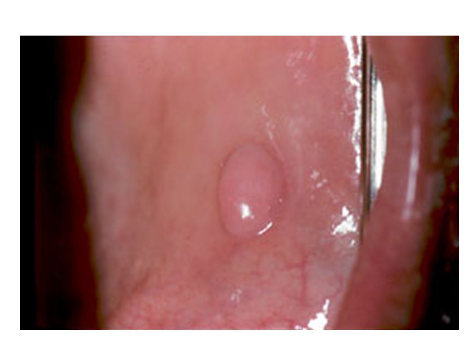
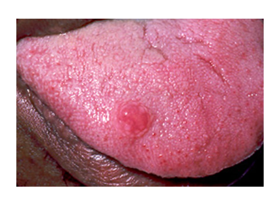
Irritation Fibroma (Traumatic Fibroma)
Clinical Features Reactive hyperplasia of fibrous connective tissue Can occur anywhere in the oral cavity that is susceptible to constant trauma – like buccal mucosa and tongue due to biting Color is similar to surrounding mucosa and is pedunculated or sessile Symptoms present only if ulcerated 4th to 6th decades of life Treatment: Conservative surgical excision

10
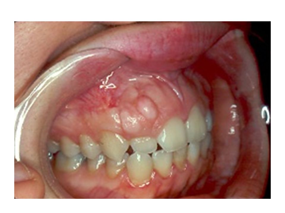
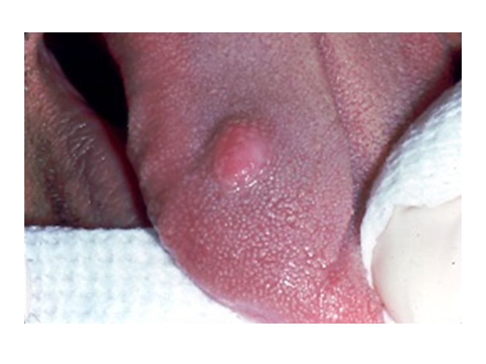
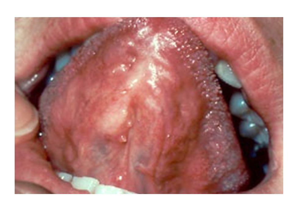
Giant Cell Fibroma Clinical Features
Occurs at a much younger age compared to fibroma and presents as asymptomatic sessile/pedunculated nodule <1cm Not associated with trauma More than half the cases occurs on the gingiva and has a papillary surface; Mandible>Maxilla Similar to retrocuspid papilla Treatment: Conservative surgical excision Recurrence is rare

13
Histology Vascular and loosely arranged fibrous connective tissue Hallmark is the presence of large, stellate shaped fibroblasts which are multinucelated Rete ridges are narrow and elongated

14
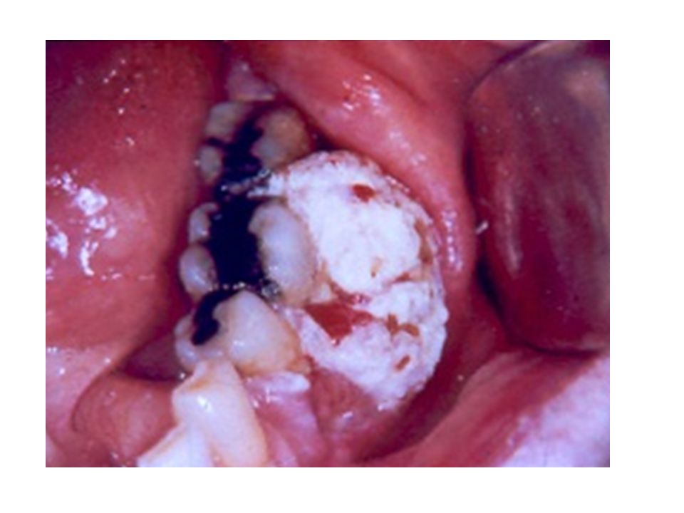
Epulis Fissuratum (Inflammatory Fibrous hyperplasia;
Denture Injury Tumor) Clinical Features Tumor-like hyperplasia of fibrous connective tissue that develops in association with the flange of an ill-fitting denture Presents as single or multiple folds of tissue in the alveolar mucosa; usually presents as two folds with denture flanges in between The size varies from < 1 cm to large lesions involving the entire length of the vestibule Appears as firm, fibrous tissue with variable ulcerations and erythema Most common location is facial aspect of alveolar ridges;anterior portions of jaws and older adults with female predilection
 Clinical Features. Tumor-like hyperplasia of fibrous connective tissue that. develops in association with the flange of an ill-fitting denture. Presents as single or multiple folds of tissue in the alveolar. mucosa; usually presents as two folds with denture flanges. in between. The size varies from < 1 cm to large lesions involving the entire. length of the vestibule. Appears as firm, fibrous tissue with variable ulcerations and. erythema. Most common location is facial aspect of alveolar ridges;anterior. portions of jaws and older adults with female predilection.")
18
Epulis Fissuratum Histology: Fibrous connective tissue hyperplasia
Overlying epithelium is hyperkeratotic and shows hyperplasia of rete ridges Pseudoepitheliomatous hyperplasia Ulceration and chronic inflammation is also seen frequently Treatment: Surgical removal and denture should be relined or remade

19
Inflammatory Papillary Hyperplasia
Reactive lesion that most commonly develops under a denture an ill-fitting denture poor oral hygiene wearing the denture 24 hours Usually occurs on the hard palate beneath a denture base Starts at the palatal vault but advanced lesions can cover the entire palate Candidiasis can also be seen associated with the lesion Mucosa is pebbly or papillary and appears erythematous Treatment: Removal of denture Surgical removal with altering the denture

22
Inflammatory Papillary Hyperplasia
Histology Papillary growths surfaced by hyperplastic startified squamous epithelium Pseudoepitheliomatous hyperplasia Chronic inflammation

23
Fibrous Histiocytoma Group of tumors which have both fibroblastic and histiocytic differentiation Most common in the skin called dermatofibroma Oral cavity – rare; buccal mucosa and vestibule Middle aged and older adults Painless nodular mass of varying size Treatment: Local surgical excision

26
Fibromatosis and Myofibromatosis
Group of fibrous proliferations that have intermediate biologic behavior Named based on clinicopathologic features: juvenile aggressive fibromatoses, extrabdominal desmoids Myofibromatosis is similar but less aggressive Painless mass occurring in children or young adults Most common site: Paramandibular soft tissues Tumor can grow to considerable size and can cause significant facial disfigurement Destruction of adjacent bone can be seen in radiographs

29
Fibromatosis and Myofibromatosis
Histology: Cellular proliferation of spindle-shaped cells arranged in fascicles Poorly circumscribed and infiltrates adjacent tissues Cells should be uniform with NO pleomorphism and hyperchromatism Treatment: Wide excision 23% recurrence rate Metastasis does not occur

31
Myofibroma (Myofibromatosis)
Rare spindle cell neoplasm that consists of myofibroblasts The multicentric disease affects infants and young children and this is called myofibromatosis Predilection to the head and neck; occurs in the first 4 decades of life with most lesions occurring in neonates and infants Most common oral site is the mandible followed by lips, cheek, and tongue Painless mass in dermis or subcutaneous tissue and intrabony cases are radiolucent Treatment: Local excision; can spontaneously regress; lesions affecting vital or visceral organs are aggressive and can be fatal

32
Oral Focal Mucinosis Uncommon tumor-like mass of unknown cause.
Maybe due to overproduction of hyaluronic acid Commonly seen in young adults with a 2:1 female-to-male ratio Most commonly seen in the gingiva followed by hard palate Sessile painless nodule of normal color Size varies from a few mm to 2 cm Histology: Well-demarcated loose myxomatous tissue surrounded by dense collagenous tissue Treatment: Surgical excision and recurrence is rare

33
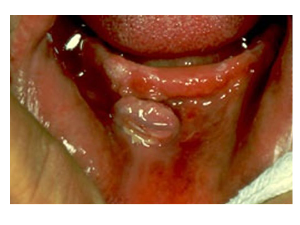
Pyogenic Granuloma Common tumor-like growth of the oral cavity
Exuberant response to irritation or trauma; periodontal irritation could be a major source Smooth or lobulated pedunculated mass which appears pink to red in color and is commonly ulcerated Range from a few mm to several cm – hormone dependent GINGIVA however other sites also affected Most common in children and young adults with females>males Develops in pregnant women during first trimester and increases through 7th months - Pregnancy tumors; Some will resolve after delivery

38
Pyogenic Granuloma Histology In spite of name, not a true granuloma
Vascular proliferation that resembles granulation tissue Surface is usually ulcerated Mixed inflammatory infiltrate Younger lesions are very vascular, but older lesions mature and are fibrous Treatment: Conservative surgical excision. Recurs if incompletely excised; Irritation also has to be removed.

40
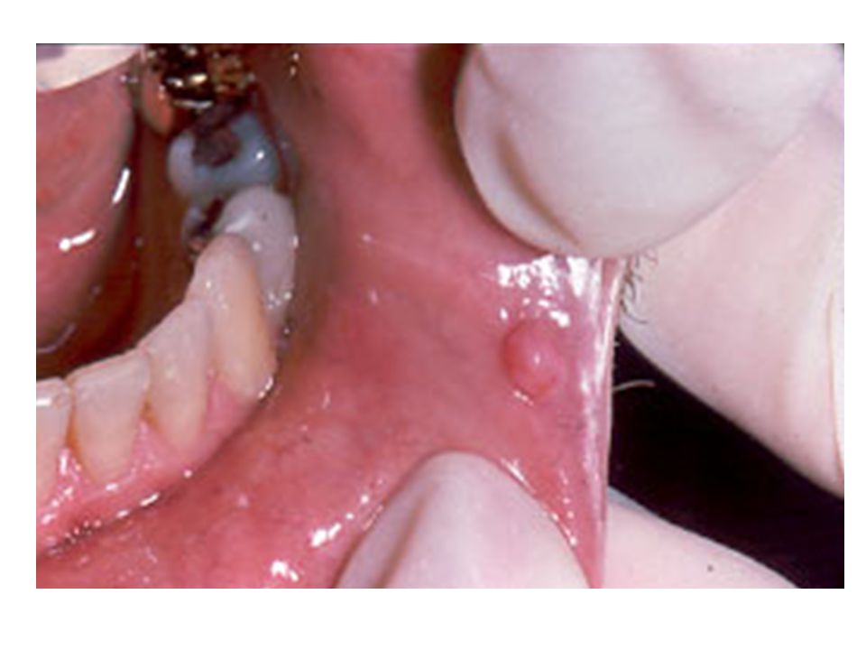
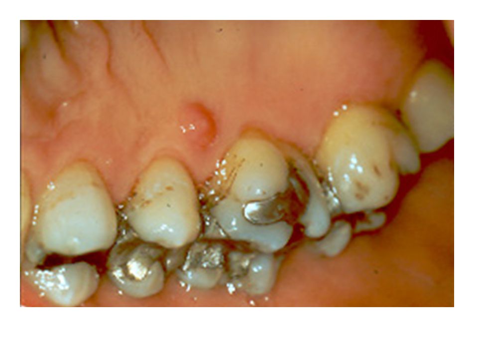
Peripheral Giant Cell Granuloma
Reactive lesion due to local irritation or trauma Resembles central giant cell granuloma GINGIVA or edentulous alveolar ridge Red or reddish-blue nodular mass which is usually < 2 cm Appearance similar to pyogenic granuloma 5th to 6th decades; F > M (60% occurs in females) Mandible > Maxilla Although occurs in soft tissues a “cupping” resorption of bone
 Mandible > Maxilla. Although occurs in soft tissues a cupping resorption of bone.")
43
Peripheral Giant Cell Granuloma
Histology: Proliferation of multinucleated giant cells in a background of plump ovoid and spindle-shaped cells Abundant hemorrhage is observed Treatment: Local surgical excision down to the underlying bone Scaling of the adjacent teeth of any source of irritation Rarely, lesions similar to this are seen in hyperparathyroidism (however these are mostly intraosseous)
")
44
Peripheral Giant Cell Granuloma

45
Peripheral Ossifying Fibroma
Reactive growth of the gingiva with uncertain histogenesis Believed to be a matured pyogenic granuloma that ultimately undergoes calcifications It does not represent central ossifying fibroma Occurs exclusively on the GINGIVA Nodular mass that is either pedunculated or sessile usually of the interdental papillae and appears red to pink and frequently ulcerated Younger adults and teens with F > M Maxilla > Mandible; >50% cases occur in the incisor/canine area

49
Peripheral Ossifying Fibroma
Histology: Fibrous proliferation associated with formation of mineralized product The surface if ulcerated, shows a fibrinopurulent membrane The mineralized component varies from bone, cementum-like material or dystrophic calcifications Treatment: Local surgical excision down to the periosteum Scaling of the adjacent teeth to remove irritants Recurrence rate of 16%

50
Peripheral Ossifying Fibroma

51
The Four “P”s Peripheral Fibroma Pyogenic Granuloma
Peripheral Giant Cell Granuloma Peripheral Ossifying Fibroma

52
Lipoma Benign tumor of fat
It represents the most mesenchymal tumor, however most of them occur in the trunk and extremities – Head and Neck are less common Oral lipomas are soft nodular masses that is sessile or pedunculated with yellow color Asymptomatic and present for several years Buccal mucosa and vestibule are the most common sites >40 years; female = male Treatment: conservative local excision

55
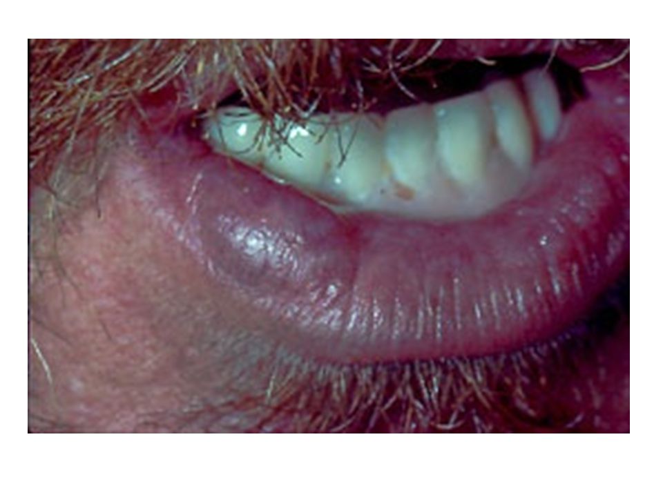
Traumatic Neuroma Reactive proliferation of neural tissue after damage to nerve bundle Smooth nodules most common in mental foramen, tongue and lower lip with a history of trauma; intraosseous lesions appear as radiolucencies Any age but mostly middle-age, with F>M Hallmark is PAIN which could be intermittent or constant and mild or severe; Mental nerve neuromas are painful especially with denture flange impingement

57
Traumatic Neuroma Histology: Haphazard proliferation of mature, myelinated nerve bundles within a fibrous connective tissue Mild chronic inflammation is also seen sometimes Treatment: Surgical excision along with a small portion of the involved nerve; low recurrence rate

58
Palisaded Encapsulated Neuroma
Benign neural tumor common in the head and neck area Trauma is considered as a major etiological factor Face: 90% of cases with majority occurring on the nose and cheek Oral cavity: hard palate and maxillary labial mucosa Smooth, PAINLESS nodules; More common in adults; F=M Histology: Well-circumscribed and encapsulated with interlacing fascicles of spindle cells (Schwann cells); wavy nuclei with no mitotic activity or pleomorphism; parallel oriented cells Treatment: Conservative surgical excision
; wavy nuclei with no. mitotic activity or pleomorphism; parallel oriented cells. Treatment: Conservative surgical excision.")
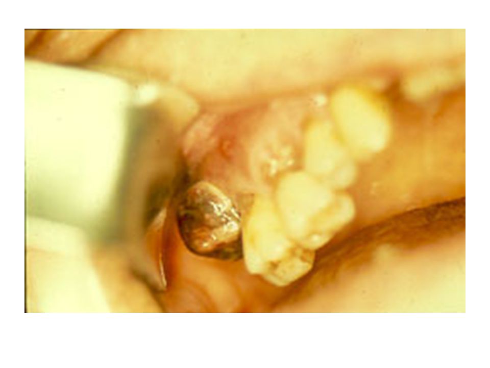
60
Palisaded Encapsulated Neuroma

61
Schwannoma (Neurilemoma)
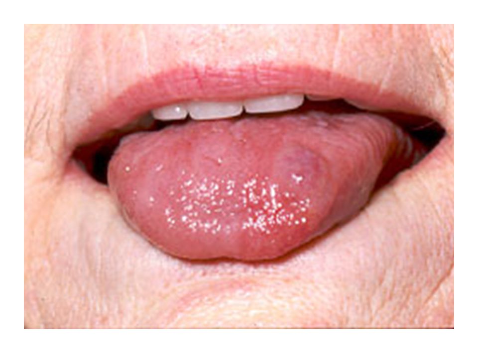
Benign neural neoplasm of Schwann cell origin Relatively uncommon, however 25-48% of all cases occur in the Head and Neck area Usually painless; slow-growing that arises in association with a nerve trunk; Asymptomatic and pushes the nerve aside Younger and middle-aged adults Tongue is the most common location Intraosseous appears as unilocular or multilocular radiolucency in posterior mandible Pain and paresthesia seen in intrabony tumors

63
Schwannoma (Neurilemoma)
Histology: Encapsulated tumor with varying amounts of Antoni A and Antoni B cells Antoni A: Streaming fascicles of spindle-shaped Schwann cells; These cells are often palisaded around acellular eosinophilic areas called Verocay bodies (which are reduplicated basement membrane and cytoplasmic processes) Antoni B: is less cellular and organized Degenerative changes are seen in older lesions Treatment: Surgical excision
 Antoni B: is less cellular and organized. Degenerative changes are seen in older lesions. Treatment: Surgical excision.")
64
Neurofibroma MOST COMMON type of peripheral nerve tumors arising from
a mixture of Schwann cells and perineural fibroblasts Can be solitary or associated with Neurofibromatosis Solitary are more common and present as slow-growing, soft, painless nodule, most common in the skin Oral cavity lesions are seem mostly in tongue and buccal mucosa Intraosseous lesions also seen as poorly defined unilocular or multilocular radiolucencies

67
Neurofibroma Histology: Not well-demarcated and consists of interlacing bundles of spindle-shaped cells that exhibit wavy nuclei Numerous mast cells are present Treatment: local surgical excision; If multiple lesions are present, patients should be evaluated for Neurofibromatosis

68
Granular Cell Tumor Benign tumor that shows predilection to oral cavity Derived from Schwann cells or neuroendocrine cells Dorsal surface of TONGUE – most common site; followed by buccal mucosa 4th to 6th decades of life and 2:1 (F:M) ratio Asymptomatic sessile nodule that is <2 cm and appears pink or yellow in color Usually solitary but multiple sometimes seen in black patients Treatment: Conservative local excision
 ratio. Asymptomatic sessile nodule that is <2 cm and appears. pink or yellow in color. Usually solitary but multiple sometimes seen in black patients. Treatment: Conservative local excision.")
71
Granular Cell Tumor Histology:
Large polygonal cells with abundant pale eosinophilic, granular cytoplasm and pale nuclei Cells arranged in sheets Lesion is not encapsulated and appears to infiltrate into surrounding tissues Overlying epithelium shows acanthosis and pseudoepitheliomatous hyperplasia Treatment: Conservative local excision

72
Granular Cell Tumor

73
Congenital Epulis Occurs exclusively in the alveolar ridge of the newborn Histologically similar to granular cell tumor, but ultrastructurally and immunohistochemical different Pink-red smooth surfaced mass on the alveolar ridge of newborns Size varies from small to over 7.5 cm with multiple tumors also occurring in 10% of cases Maxilla > Mandible in the area of lateral incisor and canine STRIKING FEMALE PREDILECTION (90% cases) Treatment: Surgical excision; spontaneous regression also seen
 Treatment: Surgical excision; spontaneous regression also seen.")
76
Melanotic Neuroectodermal Tumor of Infancy
Generally considered a benign tumor despite rapid growth of neural crest origin Rare pigmented neoplasm that occurs during the first year of life Striking predilection for the anterior maxilla (almost 2/3 of cases) Occurs as a rapidly expanding mass that is black or blue in color Destroys bone and displaces associated developing teeth Can also occur in skull, mandible, brain and testis
 Occurs as a rapidly expanding mass that is black or. blue in color. Destroys bone and displaces associated developing teeth. Can also occur in skull, mandible, brain and testis.")
79
Melanotic Neuroectodermal Tumor of Infancy

80
Melanotic Neuroectodermal Tumor of Infancy
Histology: Biphasic population of cells that form nests, tubules and alveolar structures within a dense connective tissue The 2 cell types: cuboidal epithelioid cells and neuroblastic

81
Melanotic Neuroectodermal Tumor of Infancy
Lab Tests: Vanillylmandelic acid (VMA) as in other neural crest lesions Treatment: Surgical removal Rapid growth and destruction despite being called benign 15% recurrence rate 6% behave like malignancy and metastasize
 as in other. neural crest lesions. Treatment: Surgical removal. Rapid growth and destruction despite being. called benign. 15% recurrence rate. 6% behave like malignancy and metastasize.")
82
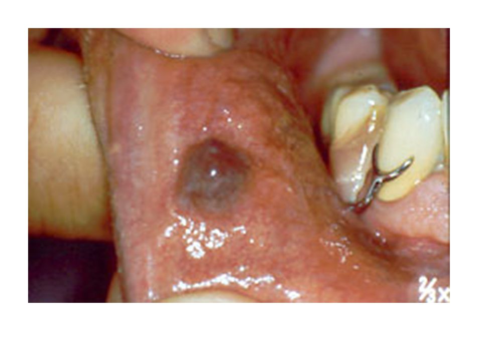
Hemangioma and Vascular Malformations
Hemangiomas are considered to be benign tumors of infancy that are characterized by a rapid growth phase with endothelial cell proliferation, followed by gradual involution Vascular malformations are structural anomalies of blood vessels without endothelial proliferation Most hemangiomas are not recognized at birth, but arise during the first 8 weeks later of life Vascular malformations are present at birth and persist throughout life

83
Hemangioma Most common tumors of infancy More common in females (3:1)
Most common in Head and Neck (60% of cases) Mostly occurs as single lesions Red/blue lesions that occur in skin, lips, tongue and buccal mucosa; The lesion blanches when compressed Intraosseous lesions also occur – Mandible > Maxilla and occurs as multilocular radiolucency
 Mostly occurs as single lesions. Red/blue lesions that occur in skin, lips, tongue and buccal. mucosa; The lesion blanches when compressed. Intraosseous lesions also occur – Mandible > Maxilla and. occurs as multilocular radiolucency.")
88
Vascular Malformations
Present at birth and persist throughout life PORT-WINE STAINS are common capillary malformation occurring most commonly on the face particularly in the area of the trigeminal nerve Port-wine stains are pink or purple macules that grows proportionally with the patient; Older patients have darker lesions and becomes nodular

89
Hemangioma Histology Cellular Hemangioma Capillary Hemangioma
Cavernous Hemangioma Treatment: Most congenital lesions will involute (“Watchful Neglect”) Surgical removal and sclerotherapy with 95% ethanol
 Surgical removal and sclerotherapy with 95% ethanol.")
91
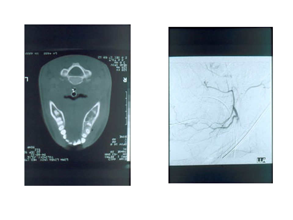
Sturge-Weber Syndrome
Hamartomatous vascular proliferation of the face and brain Dermal capillary malformation (Port wine stain) in a unilateral distribution along one or more segments of trigeminal nerve Leptomeningeal angiomas involving the ipsilateral cortex revealing “tramline” calcifications on X-rays Mental retardation and convulsions Eye involvement: glaucoma and vascular malformations Intraoral: Vascular involvement of the ipsilateral oral mucosa
 in a unilateral. distribution along one or more segments of trigeminal nerve. Leptomeningeal angiomas involving the ipsilateral cortex. revealing tramline calcifications on X-rays. Mental retardation and convulsions. Eye involvement: glaucoma and vascular malformations. Intraoral: Vascular involvement of the ipsilateral oral mucosa.")
93
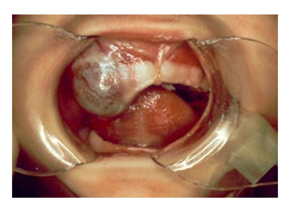
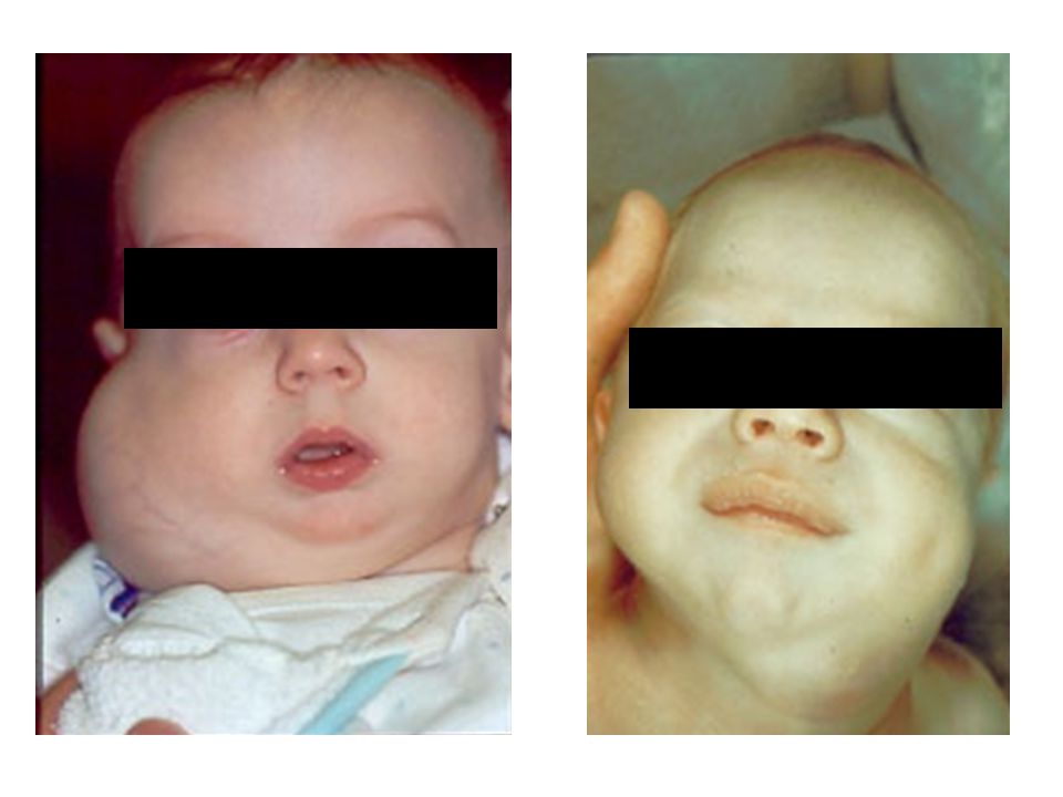
Lymphangioma Benign hamartomatous tumors of lymphatic vessels
Predilection to the head and neck with 50 – 75% occurring Three types: capillary; cavernous and cystic lymphangiomas Cavernous lymphangiomas are most common in oral cavity Most frequent site in the oral cavity - anterior 2/3 of the tongue where it causes MACROGLOSSIA Pebbly surface resembling cluster of translucent vesicles (similar to frog eggs)
")
96
Cystic Hygroma (Cystic Lymphangiomas)
Most commonly occur in the neck and axilla Cervical lymphangiomas are most common in the posterior triangle and are soft, fluctuant masses Occasionally could extend into the mediastinum or upward into oral cavity ; could also extend into the anterior triangle resulting in respiratory difficulties or dysphagia

98
Histology Treatment Intraoral: Excision and prognosis is good; recurrence does occur Cystic: Well circumscribed and have lower recurrence rate SCLEROSING AGENTS DO NOT WORK AS IN HEMANGIOMAS

99
Leiomyoma Benign neoplasms of smooth muscle
Most of these have origin in the vascular smooth muscle 3 types: SOLID, VASCULAR AND EPITHELIOID 75% of oral cases are vascular leiomyomas Can occur at any age; slow-growing mucosal nodule that occasionally can be PAINFUL Commonly seen in lips, tongue, palate and cheek Local surgical excision

100
Rhabdomyoma Benign neoplasm of skeletal muscle Adult and Fetal types
Adult: Middle-aged and older patients; M>F Intraoral lesions: FOM, soft palate and base of the tongue Nodule or mass that grows for many years Fetal: Young children with a male predilection; face and periauricular region Treatment: local surgical excision

101
Osseous and Cartilagenous Choristomas
Choristoma is a tumorlike growth of microscopically normal tissue in an abnormal location Bone, cartilage or both TONGUE (85% of cases); especially posterior tongue near the foramen cecum Gagging or dysphagia are common symptoms Histology: well-circumscribed mass of dense lamellar bone or mature cartilage Treatment: Surgical excision
; especially posterior tongue near the. foramen cecum. Gagging or dysphagia are common symptoms. Histology: well-circumscribed mass of dense lamellar bone. or mature cartilage. Treatment: Surgical excision.")
106
DENTAL CHORISTOMA: THE FIRST CASE OF ECTOPIC
DEVELOPING TOOTH IN THE TONGUE

107
Soft Tissue Sarcomas Account for less than 1% of cancers in the oral and maxillofacial area Fibrosarcoma Malignant fibrous histiocytoma Liposarcoma Malignant peripheral nerve sheath tumor Olfactory neuroblastoma Kaposi’s sarcoma Leiomyosarcoma Rhabdomyosarcoma Synovial sarcoma Alveolar soft part sarcoma

108
Rhabdomyosarcoma Malignant neoplasm of skeletal muscle origin
MOST COMMON SOFT TISSUE SARCOMA IN CHILDREN HEAD AND NECK IS THE MOST SITE (40% of cases) Primarily occurs in the first decade, teenagers and young adults 60% of cases occurs in males Painless infiltrative mass that grows rapidly Orbit > nasal cavity and nasopharynx Intraoral: PALATE
 Primarily occurs in the first decade, teenagers and young adults. 60% of cases occurs in males. Painless infiltrative mass that grows rapidly. Orbit > nasal cavity and nasopharynx. Intraoral: PALATE.")
110
3 Histologic Types Embryonal, Alveolar and Pleomorphic The head and neck cases are either embryonal or alveolar Embryonal: First 10 years of life and 60% of cases Alveolar: occurs between years and accounts for 20% - 30% of cases Treatment: Local surgical excision followed by multiagent chemotherapy (vincristine, actinomycin D and cyclophosphamide) Radiation therapy Prognosis: 5 year survival rate is 60% to 70%
 Radiation therapy. Prognosis: 5 year survival rate is 60% to 70%")
112
Metastases to Oral Soft Tissues
Uncommon representing 1% of all oral malignancies Oral metastases can occur in bone and soft tissues Lymphatic and blood-borne route Batson’s plexus: a valveless vertebral venous plexus that might allow retrograde spread of tumor cells, bypassing filtration through the lungs GINGIVA followed by the tongue Nodular masses often resembling hyperplastic or reactive growths with occasional ulcerations and loosening of adjacent teeth

115
Metastases to Oral Soft Tissues
Oral metastases is more common in males More common in middle-aged and older adults Male: Primary tumor is seen in lung cancer Female: Primary tumor is seen in breast cancer (25% of cases) In most cases, the primary tumor is known before the metastases is discovered; HOWEVER IN SOME CASES THE ORAL LESION IS THE FIRST SIGN OF MALIGNANT DISEASE Histology is similar to the primary tumor MOST CASES TO ORAL CAVITY ARE CARCINOMAS AND NOT SARCOMAS Treatment: Poor prognosis; palliative management
 In most cases, the primary tumor is known before the. metastases is discovered; HOWEVER IN SOME CASES THE. ORAL LESION IS THE FIRST SIGN OF MALIGNANT DISEASE. Histology is similar to the primary tumor. MOST CASES TO ORAL CAVITY ARE CARCINOMAS AND. NOT SARCOMAS. Treatment: Poor prognosis; palliative management.")
Similar presentations


>")

 Pathogenesis` (Mechanisms:inflammation) Clinical Features (Signs and Symptoms) Fever,>")


>")


