Download presentation
Presentation is loading. Please wait.

1
The Radiology of Benign Neoplasms
A. Ruprecht D.D.S., M.Sc.D., F.R.C.D.(C,), Dip. A.B.O.M.R. Professor and Director of Oral and Maxillofacial Radiology Professor of Radiology Professor of Anatomy and Cell Biology The University of Iowa
, Dip. A.B.O.M.R. Professor and Director of Oral and Maxillofacial Radiology. Professor of Radiology. Professor of Anatomy and Cell Biology. The University of Iowa.")
2
The Radiology of Benign Neoplasms
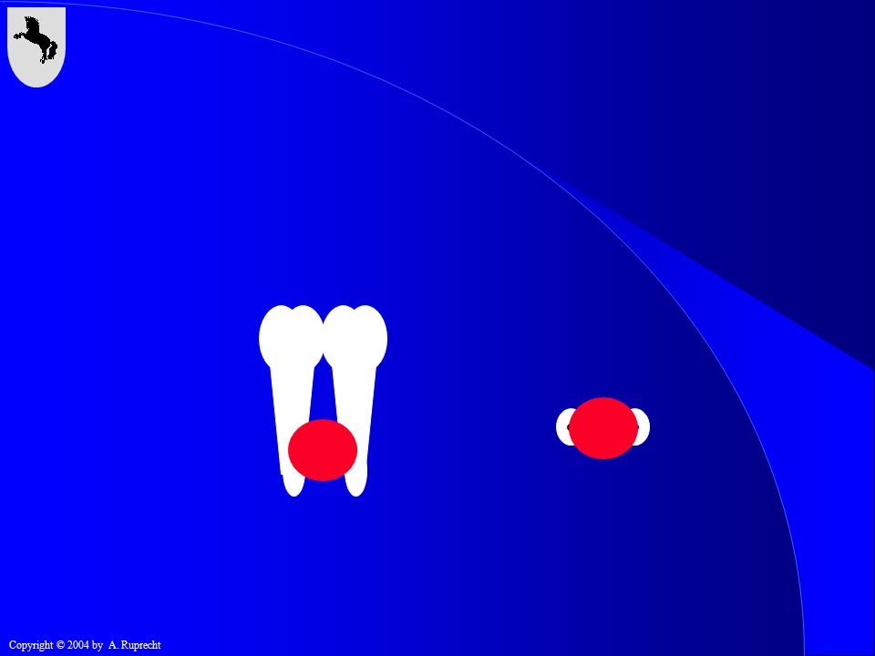
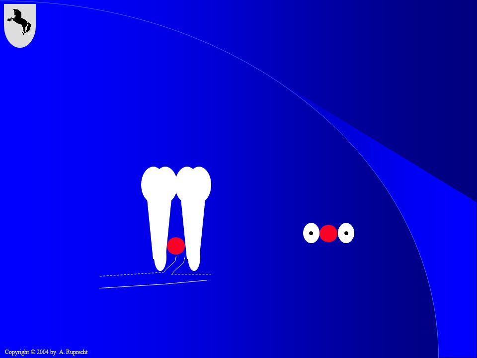
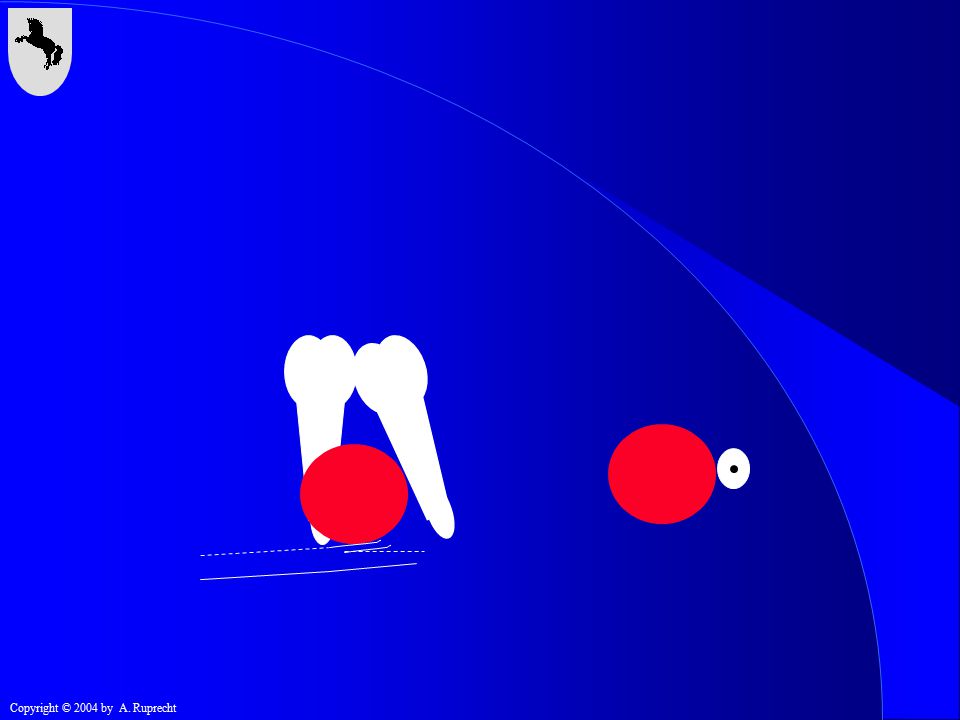
Well defined Corticated Space occupying Displacement of teeth Directional resorption of teeth Displacement of anatomical structures

3
The Radiology of Benign Neoplasms
Well defined Corticated Space occupying Displacement of teeth Directional resorption of teeth Displacement of anatomical structures

4
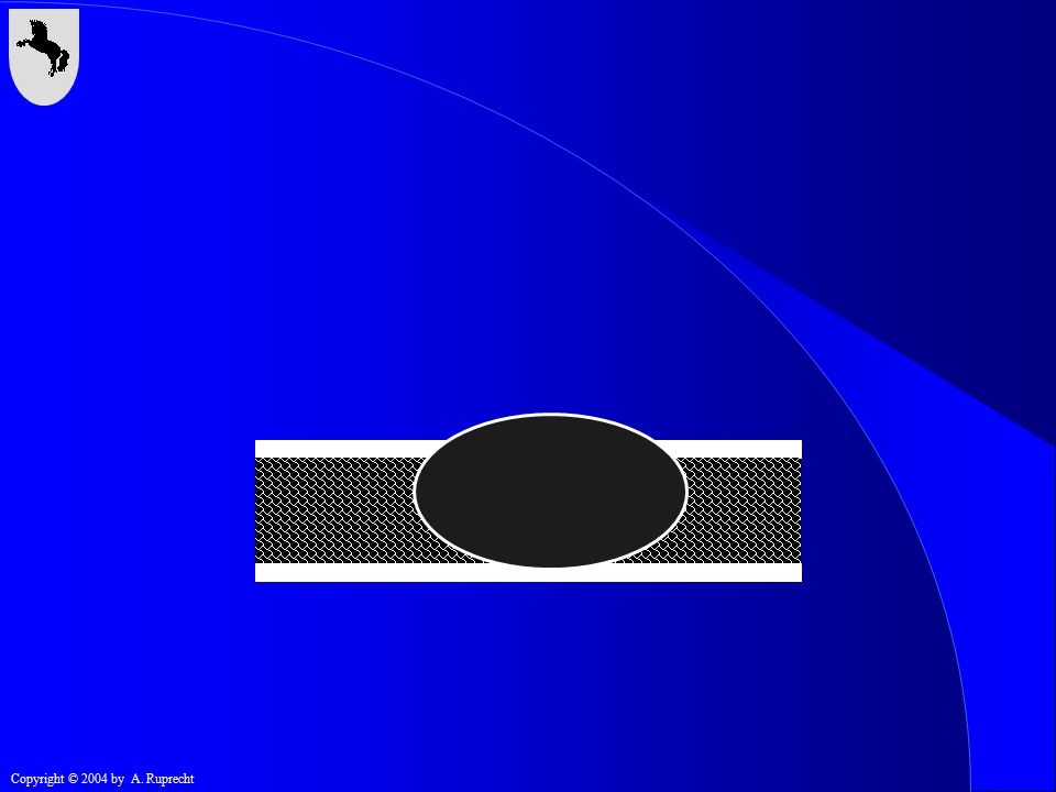
Well Defined Poorly Defined

5
The Radiology of Benign Neoplasms
Well defined Corticated Space occupying Displacement of teeth Directional resorption of teeth Displacement of anatomical structures

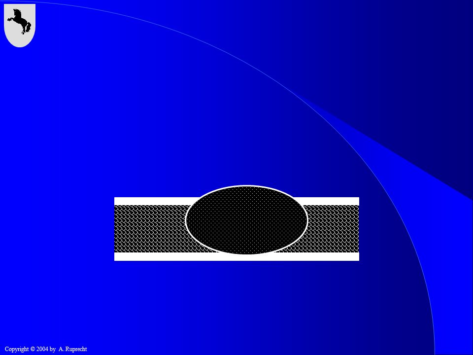
6
Corticated

7
The Radiology of Benign Neoplasms
Well defined Corticated Space occupying Displacement of teeth Directional resorption of teeth Displacement of anatomical structures

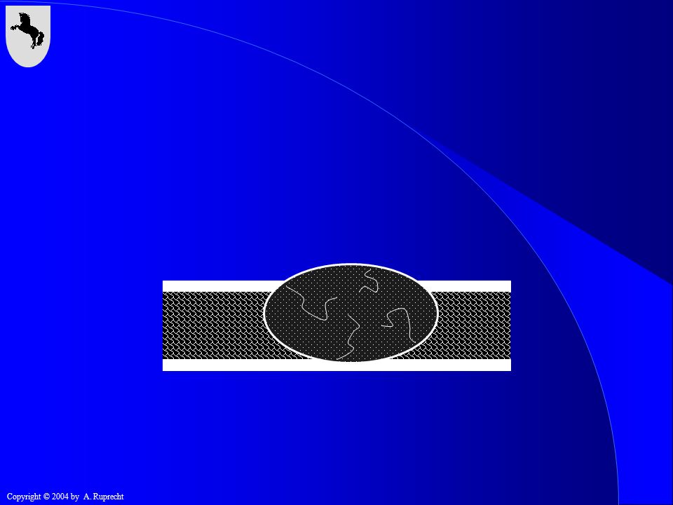
8
Space Occupying vs. Non-Space Occupying

9
Space Occupying

10
The Radiology of Benign Neoplasms
Well defined Corticated Space occupying Displacement of teeth Directional resorption of teeth Displacement of anatomical structures

16
The Radiology of Benign Neoplasms
Well defined Corticated Space occupying Displacement of teeth Directional resorption of teeth Displacement of anatomical structures

22
The Radiology of Benign Neoplasms
Well defined Corticated Space occupying Displacement of teeth Directional resorption of teeth Displacement of anatomical structures

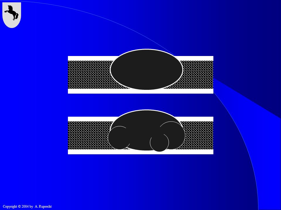
28
The Radiology of Benign Neoplasms
Displacement of teeth Directional resorption of teeth Displacement of anatomical structures

34
The Radiology of Benign Neoplasms
Displacement of periosteum Internal structure: trabeculae & calcification Unilocular / multilocular

35
The Radiology of Benign Neoplasms
Displacement of periosteum Internal structure: trabeculae & calcification Unilocular / multilocular

41
The Radiology of Benign Neoplasms
Displacement of periosteum Internal structure: trabeculae & calcification Unilocular / multilocular

45
The Radiology of Benign Neoplasms
Displacement of periosteum Internal structure: trabeculae & calcification Unilocular / multilocular

47
The Radiology of Benign Neoplasms
Cyst or Benign Neoplasm Malignant Neoplasm

48
The Radiology of Benign Neoplasms
I. Odontogenic

49
The Radiology of Benign Neoplasms
Odontogenic Epithelial Epithelial with induction/Mixed (epithelial and mesenchymal) Mesenchymal
 Mesenchymal.")
50
The Radiology of Benign Neoplasms
Odontogenic Epithelial Epithelial with induction/Mixed (epithelial and mesenchymal) Mesenchymal
 Mesenchymal.")
51
. Dental Lamina Ameloblastoma Enamel organ Adenomatoid Odontogenic
Tumor Dental Lamina Odontoma Root sheath of Hertwig Ameloblastic fibroma

52
The Radiology of Benign Neoplasms
Odontogenic Epithelial Ameloblastoma Squamous odontogenic tumor Adenomatoid odontogenic tumor Calcifying epithelial odontogenic tumor Clear cell odontogenic tumor [Melanotic neuroectodermal tumor of infancy]

53
The Radiology of Benign Neoplasms
Odontogenic Epithelial Ameloblastoma Squamous odontogenic tumor Adenomatoid odontogenic tumor Calcifying epithelial odontogenic tumor Clear cell odontogenic tumor [Melanotic neuroectodermal tumor of infancy]

54
The Radiology of Benign Neoplasms
Ameloblastoma Age: years (predominantly 30s and 40s) Mandible:Maxilla = 85:15 60% mandibular molar-ramus area
 Mandible:Maxilla = 85:15. 60% mandibular molar-ramus area.")
55
Ameloblastoma 10% 3% 2% 60% 15% 10%

56
Unilocular Multilocular (coarse septa) Soap bubble Honeycomb
The Radiology of Benign Neoplasms Ameloblastoma Unilocular Multilocular (coarse septa) Soap bubble Honeycomb
 Soap bubble. Honeycomb.")
57
The Radiology of Benign Neoplasms

58
The Radiology of Benign Neoplasms

59
The Radiology of Benign Neoplasms

60
The Radiology of Benign Neoplasms

61
The Radiology of Benign Neoplasms

62
The Radiology of Benign Neoplasms

63
The Radiology of Benign Neoplasms

64
The Radiology of Benign Neoplasms
Ameloblastoma Displacement or resorption of teeth Extends beyond radiographic limits Superior aspect often lost

65
The Radiology of Benign Neoplasms

66
The Radiology of Benign Neoplasms

67
The Radiology of Benign Neoplasms

68
The Radiology of Benign Neoplasms

69
The Radiology of Benign Neoplasms

70
The Radiology of Benign Neoplasms

71
The Radiology of Benign Neoplasms

72
Keratocyst -> Ameloblastoma
The Radiology of Benign Neoplasms Keratocyst -> Ameloblastoma

73
The Radiology of Benign Neoplasms
July 26, 1973

74
The Radiology of Benign Neoplasms
March 8, 1974

75
The Radiology of Benign Neoplasms

76
Simple Bone Cyst -> Ameloblastoma
The Radiology of Benign Neoplasms Simple Bone Cyst -> Ameloblastoma

77
The Radiology of Benign Neoplasms
February 5, 1966

78
The Radiology of Benign Neoplasms

79
The Radiology of Benign Neoplasms
December 29, 1967

80
The Radiology of Benign Neoplasms
July 19, 1968

81
The Radiology of Benign Neoplasms
January 11, 1969

82
The Radiology of Benign Neoplasms

83
The Radiology of Benign Neoplasms

84
The Radiology of Benign Neoplasms

85
The Radiology of Benign Neoplasms
July 14, 1969

86
Recurrent Ameloblastoma
The Radiology of Benign Neoplasms Recurrent Ameloblastoma

87
Recurrent Ameloblastoma
The Radiology of Benign Neoplasms Recurrent Ameloblastoma radiolucent areas circular pattern

88
The Radiology of Benign Neoplasms

89
The Radiology of Benign Neoplasms
Odontogenic Epithelial Ameloblastoma Squamous odontogenic tumor Adenomatoid odontogenic tumor Calcifying epithelial odontogenic tumor Clear cell odontogenic tumor [Melanotic neuroectodermal tumor of infancy]

90
Squamous odontogenic tumor (SOT)
The Radiology of Benign Neoplasms Squamous odontogenic tumor (SOT)
")
91
Squamous odontogenic tumor (SOT)
The Radiology of Benign Neoplasms Squamous odontogenic tumor (SOT) Age: 10s - 60s years (mean of 40) Mandible:Maxilla = 1:1 alveolar process area from rests of Malassez?
 Age: 10s - 60s years (mean of 40) Mandible:Maxilla = 1:1. alveolar process area. from rests of Malassez")
92
The Radiology of Benign Neoplasms
Squamous odontogenic tumor (SOT) radiolucent well-circumscribed semilunar associated with roots of teeth
 radiolucent. well-circumscribed. semilunar. associated with roots of teeth.")
93
The Radiology of Benign Neoplasms
Squamous odontogenic tumor (SOT) locally invasive recur with conservative treatment excision is treatment of choice
 locally invasive. recur with conservative treatment. excision is treatment of choice.")
94
The Radiology of Benign Neoplasms
Odontogenic Epithelial Ameloblastoma Squamous odontogenic tumor Adenomatoid odontogenic tumor Calcifying epithelial odontogenic tumor Clear cell odontogenic tumor [Melanotic neuroectodermal tumor of infancy]

95
Adenomatoid odontogenic tumor (AOT)
The Radiology of Benign Neoplasms Adenomatoid odontogenic tumor (AOT)
")
96
Adenomatoid odontogenic tumor (AOT)
The Radiology of Benign Neoplasms Adenomatoid odontogenic tumor (AOT) 10-20 (18 years of age) anterior maxilla or mandible radiolucent or with radiopaque foci may mimic dentigerous cyst
 (18 years of age) anterior maxilla or mandible. radiolucent or with radiopaque foci. may mimic dentigerous cyst.")
97
The Radiology of Benign Neoplasms

98
The Radiology of Benign Neoplasms

99
The Radiology of Benign Neoplasms
Odontogenic Epithelial Ameloblastoma Squamous odontogenic tumor Adenomatoid odontogenic tumor Calcifying epithelial odontogenic tumor Clear cell odontogenic tumor [Melanotic neuroectodermal tumor of infancy]

100
Calcifying epithelial odontogenic tumor (CEOT)
The Radiology of Benign Neoplasms Calcifying epithelial odontogenic tumor (CEOT)
")
101
Calcifying epithelial odontogenic tumor (CEOT, Pindborg tumor)
The Radiology of Benign Neoplasms Calcifying epithelial odontogenic tumor (CEOT, Pindborg tumor) 10-90 (mean 40) maxilla:mandible = 1:2 ramus-molar region
 (mean 40) maxilla:mandible = 1:2. ramus-molar region.")
102
Calcifying epithelial odontogenic tumor (CEOT, Pindborg tumor)
The Radiology of Benign Neoplasms Calcifying epithelial odontogenic tumor (CEOT, Pindborg tumor) usually associated with impacted teeth unilocular or multilocular radiolucent or with radiopaque foci
 usually associated with impacted teeth. unilocular or multilocular. radiolucent or with radiopaque foci.")
103
The Radiology of Benign Neoplasms

104
The Radiology of Benign Neoplasms

105
The Radiology of Benign Neoplasms

106
The Radiology of Benign Neoplasms

107
The Radiology of Benign Neoplasms
Odontogenic Epithelial Ameloblastoma Squamous odontogenic tumor Adenomatoid odontogenic tumor Calcifying epithelial odontogenic tumor Clear cell odontogenic tumor [Melanotic neuroectodermal tumor of infancy]

108
Clear cell odontogenic tumor
The Radiology of Benign Neoplasms Clear cell odontogenic tumor

109
Clear cell odontogenic tumor
The Radiology of Benign Neoplasms Clear cell odontogenic tumor rare 60 years of age females maxilla or mandible

110
Clear cell odontogenic tumor
The Radiology of Benign Neoplasms Clear cell odontogenic tumor radiolucent poorly circumscribed locally aggressive

111
The Radiology of Benign Neoplasms
Odontogenic Epithelial Ameloblastoma Squamous odontogenic tumor Adenomatoid odontogenic tumor Calcifying epithelial odontogenic tumor Clear cell odontogenic tumor [Melanotic neuroectodermal tumor of infancy]

112
Melanotic neuroectodermal tumor of infancy
The Radiology of Benign Neoplasms Melanotic neuroectodermal tumor of infancy

113
Melanotic neuroectodermal tumor of infancy (MNTI)
The Radiology of Benign Neoplasms Melanotic neuroectodermal tumor of infancy (MNTI) rare young, usually first year of life anterior maxilla moderately well circumscribed, around apical area of roots
 rare. young, usually first year of life. anterior maxilla. moderately well circumscribed, around apical area of roots.")
114
The Radiology of Benign Neoplasms

115
The Radiology of Benign Neoplasms

116
The Radiology of Benign Neoplasms

117
The Radiology of Benign Neoplasms
Odontogenic Epithelial Epithelial with induction/Mixed (epithelial and mesenchymal) Mesenchymal
 Mesenchymal.")
118
The Radiology of Benign Neoplasms
Odontogenic Epithelial Epithelial with induction/Mixed (epithelial and mesenchymal) Mesenchymal
 Mesenchymal.")
119
The Radiology of Benign Neoplasms
Odontogenic Epithelial with induction Ameloblastic fibroma Ameloblastic fibro-odontoma Odonto-ameloblastoma

120
The Radiology of Benign Neoplasms
Odontogenic Epithelial with induction Ameloblastic fibroma Ameloblastic fibro-odontoma Odonto-ameloblastoma

121
The Radiology of Benign Neoplasms

122
The Radiology of Benign Neoplasms

123
The Radiology of Benign Neoplasms
Odontogenic Epithelial with induction Ameloblastic fibroma Ameloblastic fibro-odontoma Odonto-ameloblastoma

124
The Radiology of Benign Neoplasms
Odontogenic Epithelial with induction Ameloblastic fibroma Ameloblastic fibro-odontoma Odonto-ameloblastoma

125
The Radiology of Benign Neoplasms

126
The Radiology of Benign Neoplasms

127
The Radiology of Benign Neoplasms

128
The Radiology of Benign Neoplasms

129
The Radiology of Benign Neoplasms

130
The Radiology of Benign Neoplasms

131
The Radiology of Benign Neoplasms

132
The Radiology of Benign Neoplasms

133
The Radiology of Benign Neoplasms
Odontogenic Epithelial with induction Ameloblastic fibroma Ameloblastic fibro-odontoma Compound odontoma Complex odontoma

134
The Radiology of Benign Neoplasms
Ameloblastic fibroma Ameloblastic fibro-odontoma Compound odontoma Complex odontoma

135
The Radiology of Benign Neoplasms
–>Compound Odontoma Ameloblastic Fibroma–>Ameloblastic Fibro-odontoma –>Complex Odontoma

136
The Radiology of Benign Neoplasms

137
The Radiology of Benign Neoplasms
Ameloblastic Fibroma primitive myxoid connective tissue resemblance to dental pulp strands of odontogenic epithelium 2 cells wide Ameloblastic Fibro-odontoma primitive myxoid connective tissue resemblance to dental pulp strands of odontogenic epithelium 2 cells wide cells differentiate to produce enamel and dentin

138
The Radiology of Benign Neoplasms
Compound Odontoma Complex odontoma prominent enamel matrix often seen before final maturation of hard tissue enamel and dentin in the form of compound or complex odontoma Ameloblastic Fibro-odontoma prominent enamel matrix often seen before final maturation of hard tissue enamel and dentin in the form of compound or complex odontoma

139
The Radiology of Benign Neoplasms
Odontogenic Epithelial with induction Ameloblastic fibroma Ameloblastic fibro-odontoma Odonto-ameloblastoma

140
Odonto-ameloblastoma
The Radiology of Benign Neoplasms Odonto-ameloblastoma

141
Odonto-ameloblastoma
The Radiology of Benign Neoplasms Odonto-ameloblastoma rare young, first decade ameloblastoma component odontoma component

142
The Radiology of Benign Neoplasms

143
The Radiology of Benign Neoplasms
Odontogenic Epithelial Epithelial with induction/Mixed (epithelial and mesenchymal) Mesenchymal
 Mesenchymal.")
144
The Radiology of Benign Neoplasms
Odontogenic Mesenchymal Odontogenic fibroma Odontogenic myxoma Benign cementoblastoma Cementifying fibroma

145
The Radiology of Benign Neoplasms
Odontogenic Mesenchymal Odontogenic fibroma Odontogenic myxoma Benign cementoblastoma Cementifying fibroma

146
The Radiology of Benign Neoplasms
Odontogenic fibroma Central odontogenic fibroma Peripheral odontogenic fibroma

147
Central odontogenic fibroma
The Radiology of Benign Neoplasms Central odontogenic fibroma

148
Central odontogenic fibroma
The Radiology of Benign Neoplasms Central odontogenic fibroma rare all age groups radiolucent usually unilocular

149
The Radiology of Benign Neoplasms

150
Peripheral odontogenic fibroma
The Radiology of Benign Neoplasms Peripheral odontogenic fibroma

151
The Radiology of Benign Neoplasms

152
The Radiology of Benign Neoplasms

153
The Radiology of Benign Neoplasms

154
The Radiology of Benign Neoplasms
Odontogenic Mesenchymal Central odontogenic fibroma Odontogenic myxoma Benign cementoblastoma Cementifying fibroma

155
The Radiology of Benign Neoplasms
Odontogenic myxoma

156
The Radiology of Benign Neoplasms
Odontogenic myxoma 10-50 years (15-35, mean 30) maxilla:mandible 1:1 may be infiltrative and aggressive
 maxilla:mandible 1:1. may be infiltrative and aggressive.")
157
The Radiology of Benign Neoplasms
Odontogenic myxoma “tennis racket” appearance (neither coarse nor fine septa) cortical expansion root displacement rather than resorption
 cortical expansion. root displacement rather than resorption.")
158
The Radiology of Benign Neoplasms

159
The Radiology of Benign Neoplasms

160
The Radiology of Benign Neoplasms

161
The Radiology of Benign Neoplasms

162
The Radiology of Benign Neoplasms

163
The Radiology of Benign Neoplasms
Odontogenic Mesenchymal Central odontogenic fibroma Odontogenic myxoma Benign cementoblastoma Cementifying fibroma

164
Benign cementoblastoma
The Radiology of Benign Neoplasms Benign cementoblastoma

165
Benign cementoblastoma
The Radiology of Benign Neoplasms Benign cementoblastoma second or third decade, usually before 25 continuous with root , which is resorbed pulp vitality unrelated

166
Benign cementoblastoma
The Radiology of Benign Neoplasms Benign cementoblastoma radiopaque mass surrounded by radiolucent line surrounded by radiopaque line

167
The Radiology of Benign Neoplasms

168
The Radiology of Benign Neoplasms

169
The Radiology of Benign Neoplasms

170
The Radiology of Benign Neoplasms

171
The Radiology of Benign Neoplasms

172
The Radiology of Benign Neoplasms

173
The Radiology of Benign Neoplasms

174
The Radiology of Benign Neoplasms

175
The Radiology of Benign Neoplasms

176
The Radiology of Benign Neoplasms
Odontogenic Mesenchymal Central odontogenic fibroma Odontogenic myxoma Benign cementoblastoma Cementifying fibroma

177
Cementifying fibroma Ossifying fibroma Cemento-ossifying fibroma
The Radiology of Benign Neoplasms Cementifying fibroma Ossifying fibroma Cemento-ossifying fibroma

178
The Radiology of Benign Neoplasms
Cementifying fibroma all age groups, mainly around 40 radiolucent, mixed, radiopaque usually unilocular

179
The Radiology of Benign Neoplasms

180
The Radiology of Benign Neoplasms

181
The Radiology of Benign Neoplasms

182
The Radiology of Benign Neoplasms

183
The Radiology of Benign Neoplasms

184
The Radiology of Benign Neoplasms

185
The Radiology of Benign Neoplasms

186
The Radiology of Benign Neoplasms

187
The Radiology of Benign Neoplasms

188
The Radiology of Benign Neoplasms

Similar presentations


 Gilbert E. Lilly Professor of Diagnostic.>")

 There is a well-defined unilocular oval shaped radiolucence with a corticated margin. It extend from apex of tooth.>")


