Download presentation
Presentation is loading. Please wait.

1
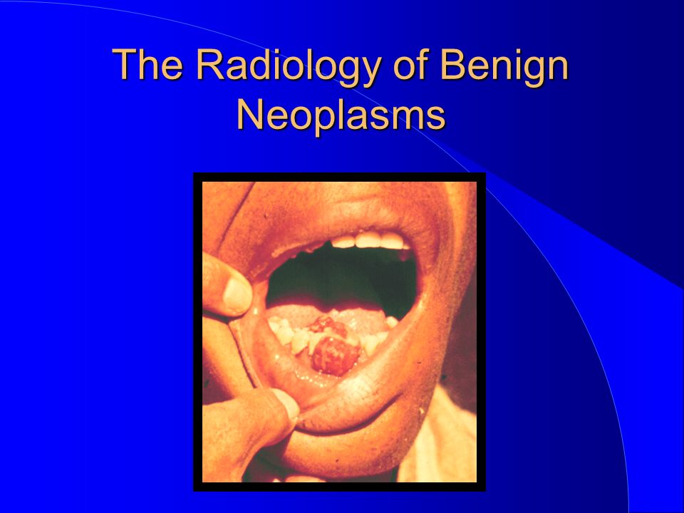
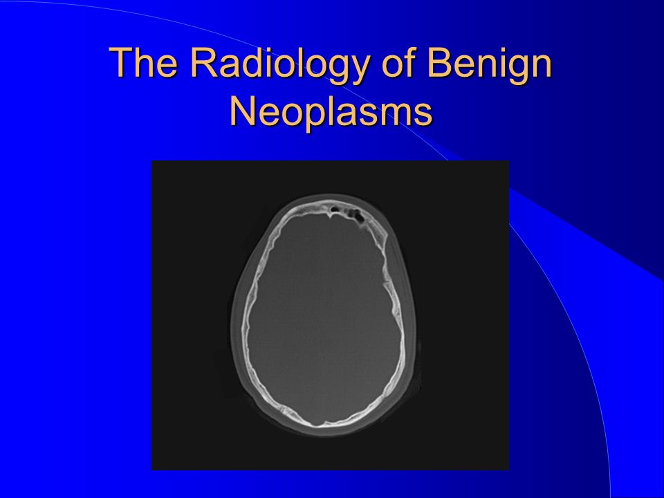
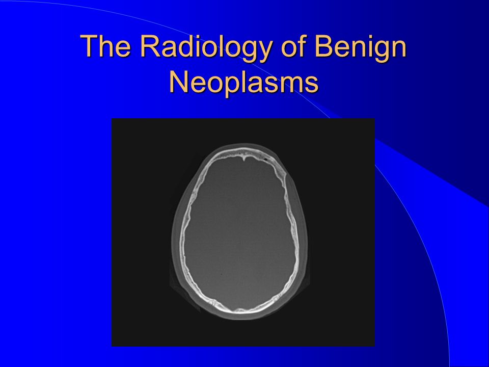
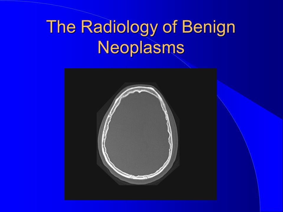
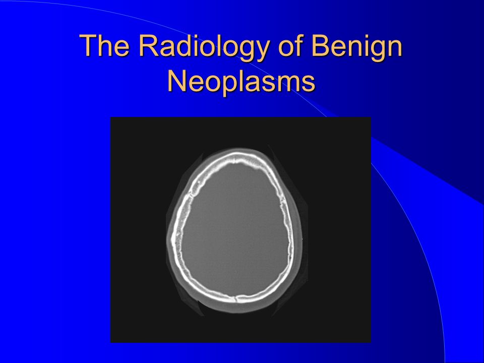
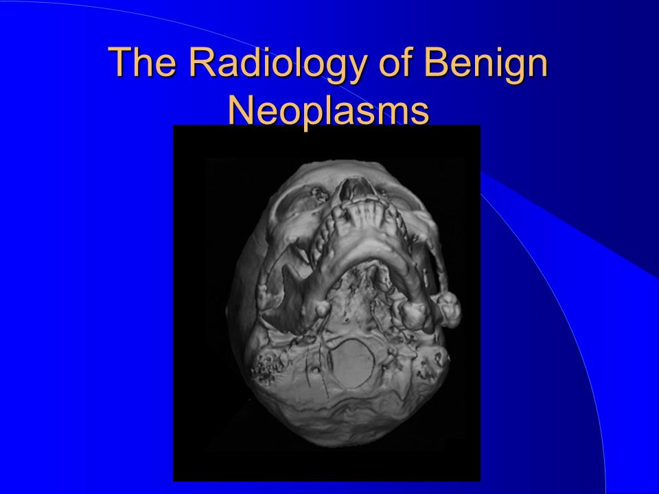
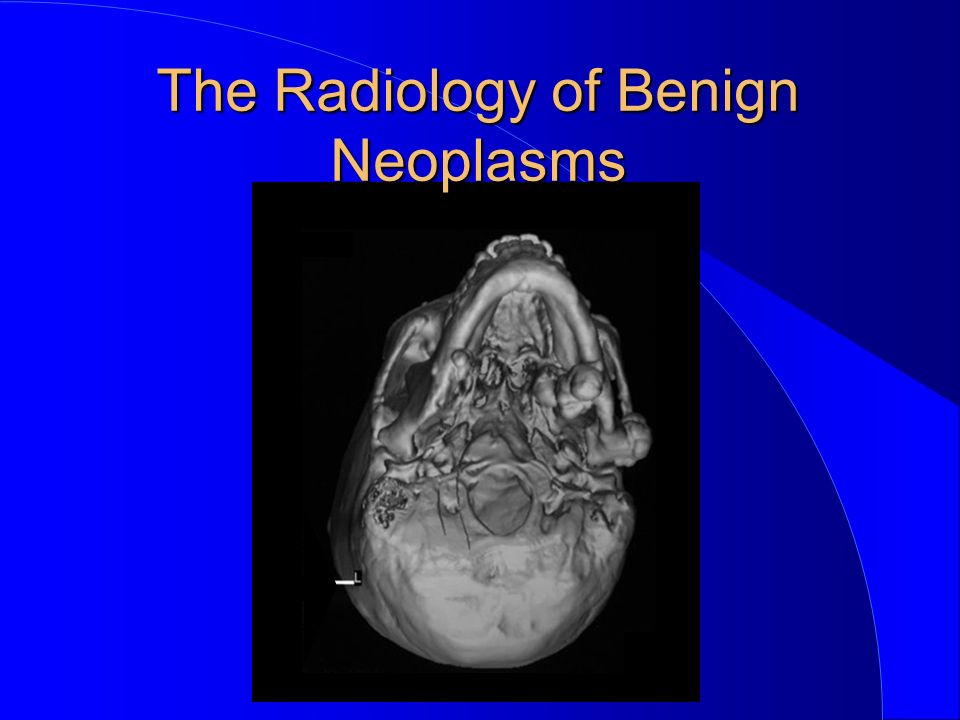
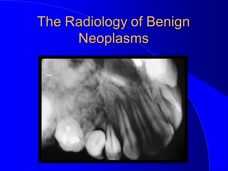
The Radiology of Benign Neoplasms

2
II. Non-Odontogenic

3
Giant cell lesion Hemangioma (Haemangioma) Neurofibroma Fibroma Osteoma Osteoblastoma Chondroma Non-Odontogenic The Radiology of Benign Neoplasms
 Neurofibroma Fibroma Osteoma Osteoblastoma Chondroma Non-Odontogenic The Radiology of Benign Neoplasms")
4
Giant cell lesion Hemangioma (Haemangioma) Neurofibroma Fibroma Osteoma Osteoblastoma Chondroma Non-Odontogenic The Radiology of Benign Neoplasms
 Neurofibroma Fibroma Osteoma Osteoblastoma Chondroma Non-Odontogenic The Radiology of Benign Neoplasms")
5
Giant cell lesion Central giant cell lesion Central giant cell granuloma Peripheral giant cell lesion Peripheral giant cell granuloma Giant cell tumor True giant cell tumor The Radiology of Benign Neoplasms

6
Giant cell lesion Central giant cell lesion Central giant cell granuloma Peripheral giant cell lesion Peripheral giant cell granuloma Giant cell tumor True giant cell tumor The Radiology of Benign Neoplasms

7
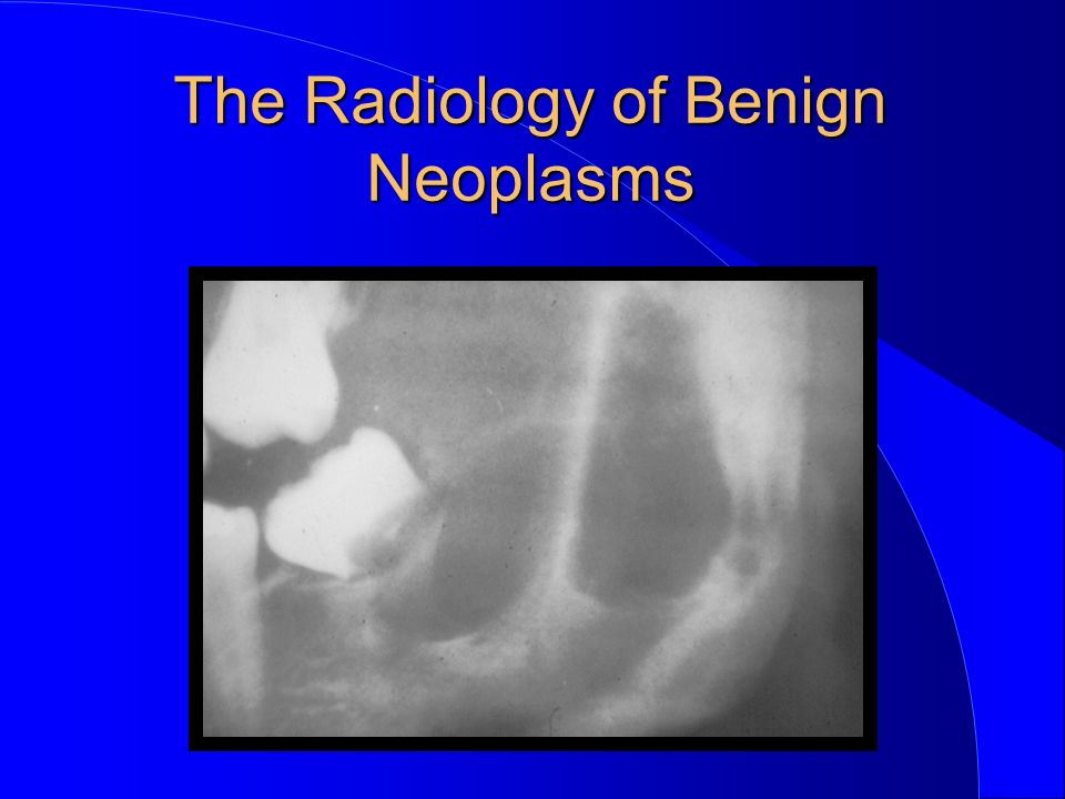
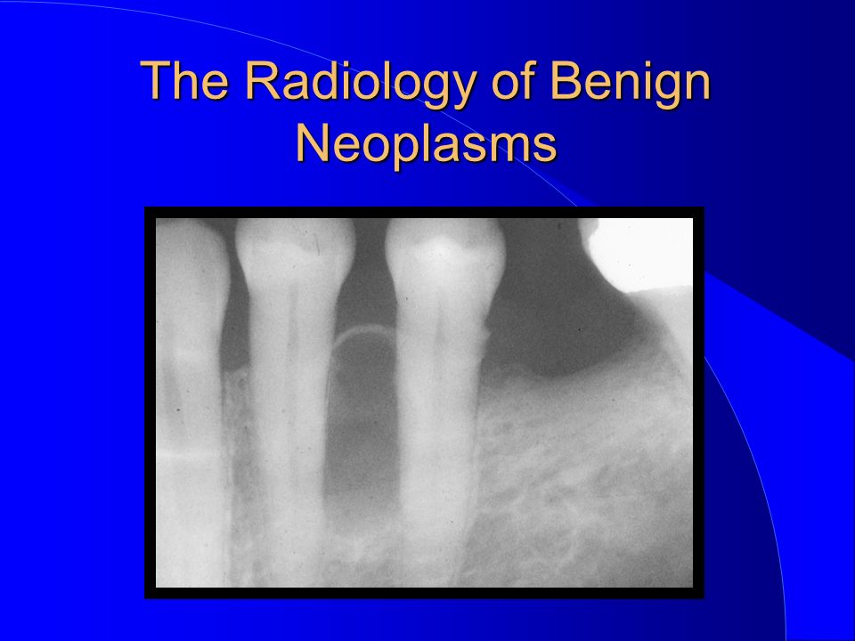
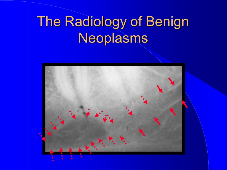
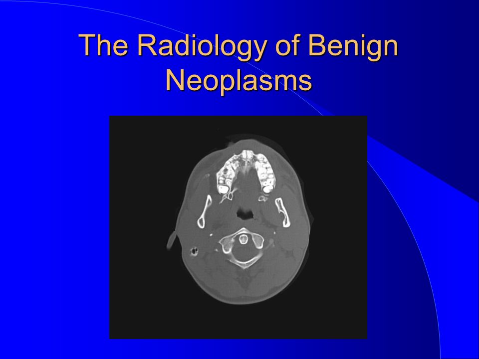
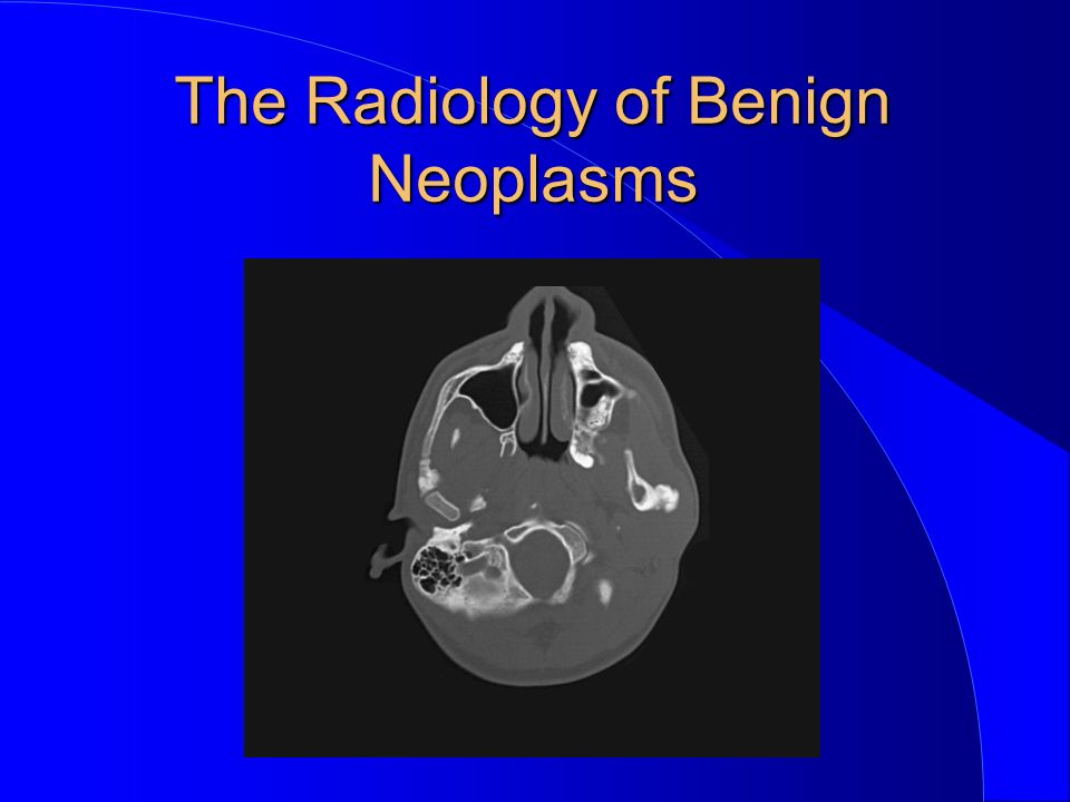
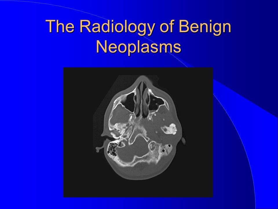
Central giant cell lesion Central giant cell granuloma before 21 years unknown nature anterior to first permanent molars painless The Radiology of Benign Neoplasms

8
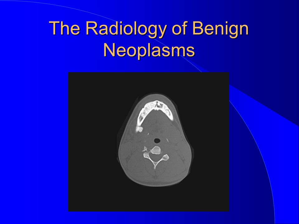
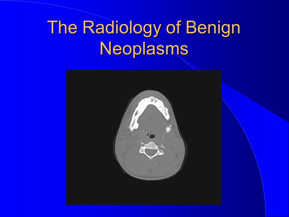
radiolucent, with “salt and pepper “ calcification thin, wispy septa resorption of teeth very common Central giant cell lesion Central giant cell granuloma The Radiology of Benign Neoplasms

23
Giant cell lesion Central giant cell lesion Central giant cell granuloma Peripheral giant cell lesion Peripheral giant cell granuloma Giant cell tumor True giant cell tumor The Radiology of Benign Neoplasms

24
Central giant cell lesion Central giant cell granuloma The Radiology of Benign Neoplasms

28
Giant cell lesion Central giant cell lesion Central giant cell granuloma Peripheral giant cell lesion Peripheral giant cell granuloma Giant cell tumor True giant cell tumor The Radiology of Benign Neoplasms

29
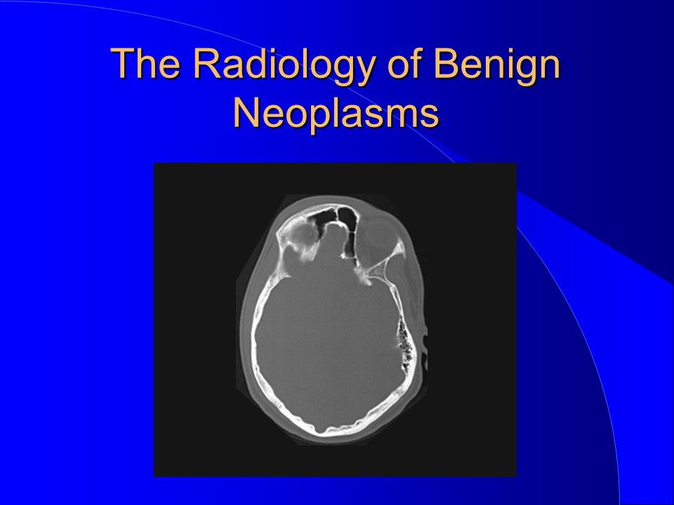
after 21 years rare in jaws posterior to first permanent molars painful Giant cell tumor True giant cell tumor The Radiology of Benign Neoplasms

30
considered by some to be variant of GCG radiographic appearance similar to GCG Giant cell tumor True giant cell tumor The Radiology of Benign Neoplasms

31
Giant cell lesion Hemangioma (Haemangioma) Neurofibroma Fibroma Osteoma Osteoblastoma Chondroma Non-Odontogenic The Radiology of Benign Neoplasms
 Neurofibroma Fibroma Osteoma Osteoblastoma Chondroma Non-Odontogenic The Radiology of Benign Neoplasms")
32
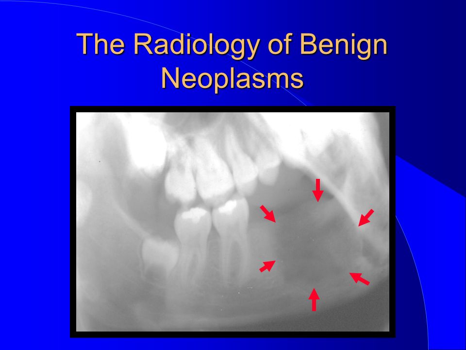
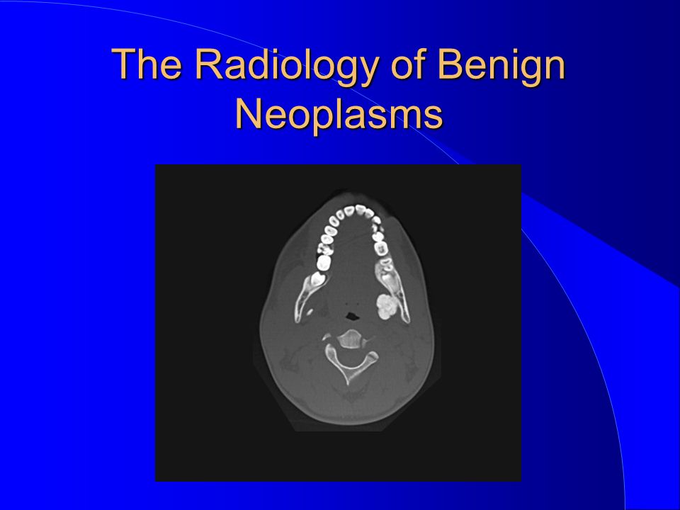
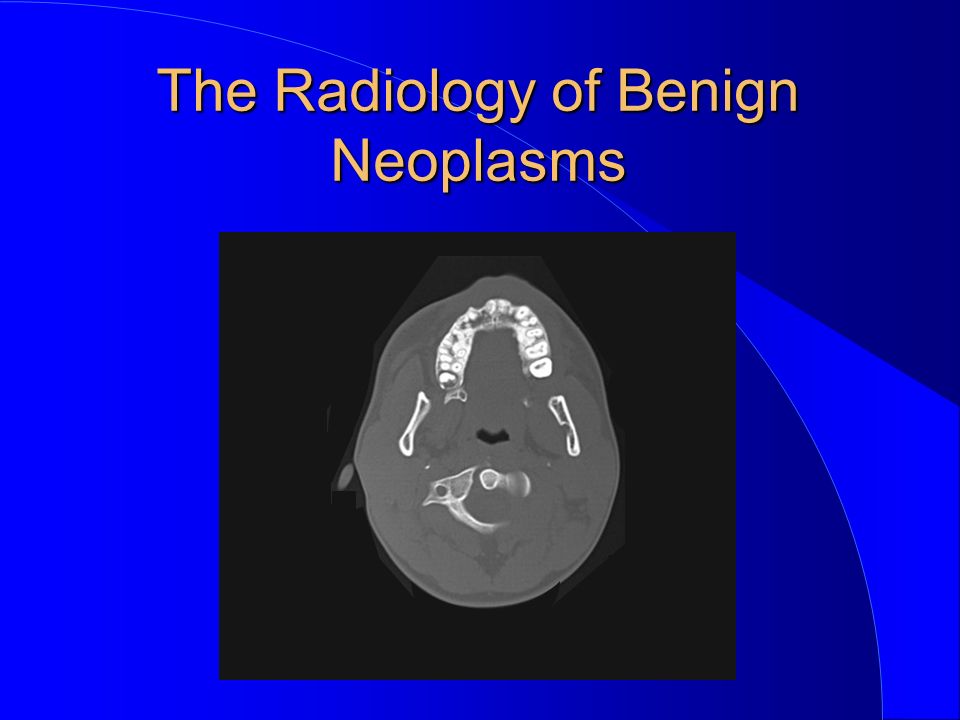
Intraosseous hemangioma Extraosseous hemangioma Hemangioma Haemangioma The Radiology of Benign Neoplasms

33
Intraosseous hemangioma Extraosseous hemangioma The Radiology of Benign Neoplasms Hemangioma Haemangioma

34
some may be true neoplasms, most are probably developmental uncommon no phleboliths Intraosseous Hemangioma The Radiology of Benign Neoplasms

35
altered bone pattern “moth-eaten” bone pattern hypoplastic teeth The Radiology of Benign Neoplasms Intraosseous Hemangioma

36
The Radiology of Benign Neoplasms

39
Intraosseous hemangioma Extraosseous hemangioma Hemangioma Haemangioma The Radiology of Benign Neoplasms

40
may be some true neoplasms, most are probably developmental common in head and neck area phleboliths common The Radiology of Benign Neoplasms Extraosseous Hemangioma

41
may have hypo- or hyperplasia of neighboring bone The Radiology of Benign Neoplasms Intraosseous Hemangioma

42
The Radiology of Benign Neoplasms

54
X

57
Giant cell lesion Hemangioma (Haemangioma) Neurofibroma Fibroma Osteoma Osteoblastoma Chondroma Non-Odontogenic The Radiology of Benign Neoplasms
 Neurofibroma Fibroma Osteoma Osteoblastoma Chondroma Non-Odontogenic The Radiology of Benign Neoplasms")
58
Neurofibroma Solitary Neurofibromatosis Non-Odontogenic The Radiology of Benign Neoplasms

59
Neurofibroma Solitary Neurofibromatosis Non-Odontogenic The Radiology of Benign Neoplasms

60
Neurofibroma radiolucent well-defined, corticated unilocular or multilocular hypo- or hyperplasia of neighboring bone Non-Odontogenic The Radiology of Benign Neoplasms

73
Giant cell lesion Hemangioma (Haemangioma) Neurofibroma Fibroma Osteoma Osteoblastoma Chondroma Non-Odontogenic The Radiology of Benign Neoplasms
 Neurofibroma Fibroma Osteoma Osteoblastoma Chondroma Non-Odontogenic The Radiology of Benign Neoplasms")
74
Fibroma radiolucent unilocular corticated may look like cyst Non-Odontogenic The Radiology of Benign Neoplasms

75
Giant cell lesion Hemangioma (Haemangioma) Neurofibroma Fibroma Osteoma Osteoblastoma Chondroma Non-Odontogenic The Radiology of Benign Neoplasms
 Neurofibroma Fibroma Osteoma Osteoblastoma Chondroma Non-Odontogenic The Radiology of Benign Neoplasms")
76
Osteoma Cortical osteoma Cancellous osteoma Osteoid osteoma Osteoma cutis Non-Odontogenic The Radiology of Benign Neoplasms

77
Osteoma Cortical osteoma Cancellous osteoma Osteoid osteoma Osteoma cutis Non-Odontogenic The Radiology of Benign Neoplasms

78
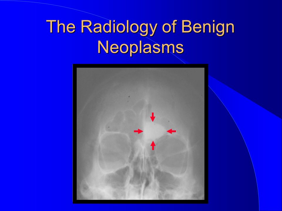
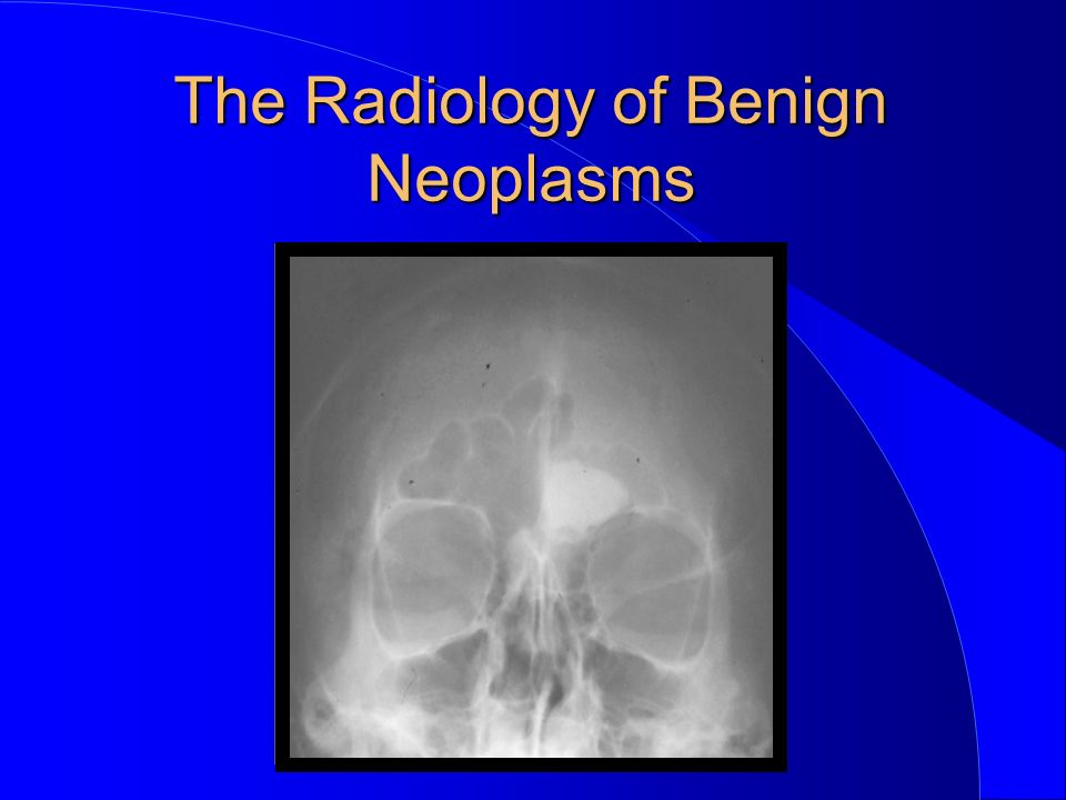
Cortical and Cancellous osteoma almost exclusively in the skull and facial bones most common in the paranasal sinuses (frontal>ethmoid>maxillary>sphenoid) Non-Odontogenic The Radiology of Benign Neoplasms
 Non-Odontogenic The Radiology of Benign Neoplasms")
79
Cortical and Cancellous osteoma uncommon in the jaws many so-called osteoma probably reactive or “burned-out fibrous dysplasia Non-Odontogenic The Radiology of Benign Neoplasms

80
Osteoma Cortical osteoma Cancellous osteoma Osteoid osteoma Osteoma cutis Non-Odontogenic The Radiology of Benign Neoplasms

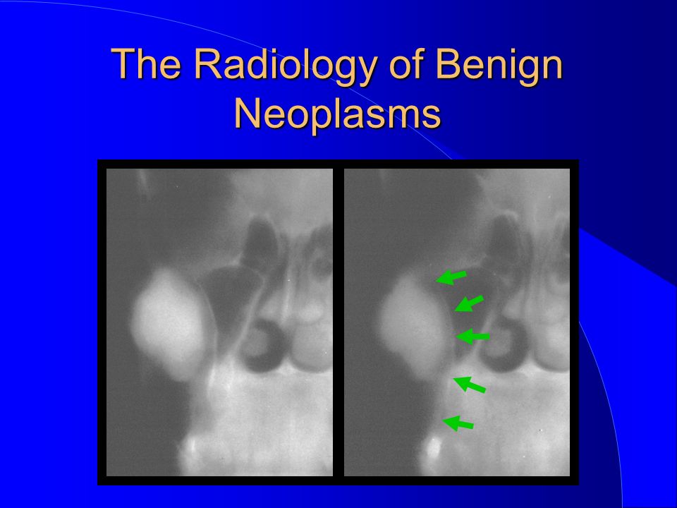
81
Cortical osteoma (Ivory osteoma) osseous neoplasm of cortical bone many so-called osteoma probably reactive or “burned-out fibrous dysplasia Non-Odontogenic The Radiology of Benign Neoplasms
 osseous neoplasm of cortical bone many so-called osteoma probably reactive or burned-out fibrous dysplasia Non-Odontogenic The Radiology of Benign Neoplasms")
90
Osteoma Cortical osteoma Cancellous osteoma Osteoid osteoma Osteoma cutis Non-Odontogenic The Radiology of Benign Neoplasms

91
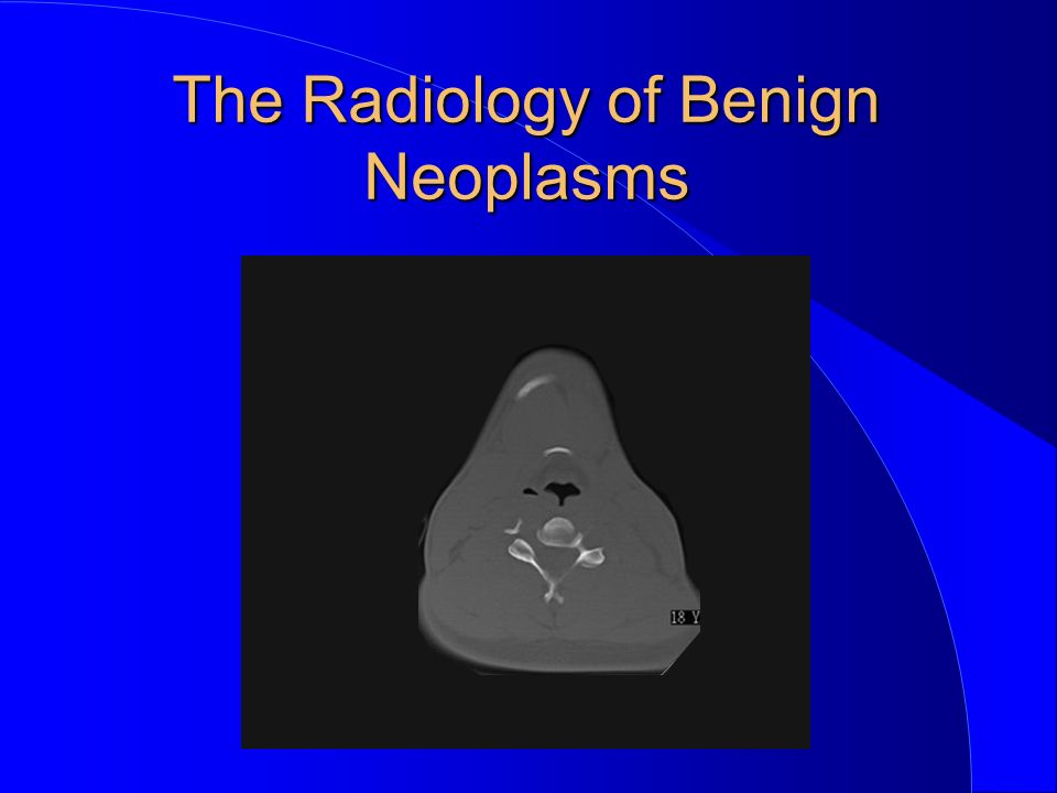
Cancellous osteoma osseous neoplasm of cancellous bone uncommon in the jaws many so-called osteoma probably reactive or “burned-out fibrous dysplasia Non-Odontogenic The Radiology of Benign Neoplasms

95
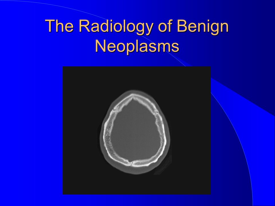
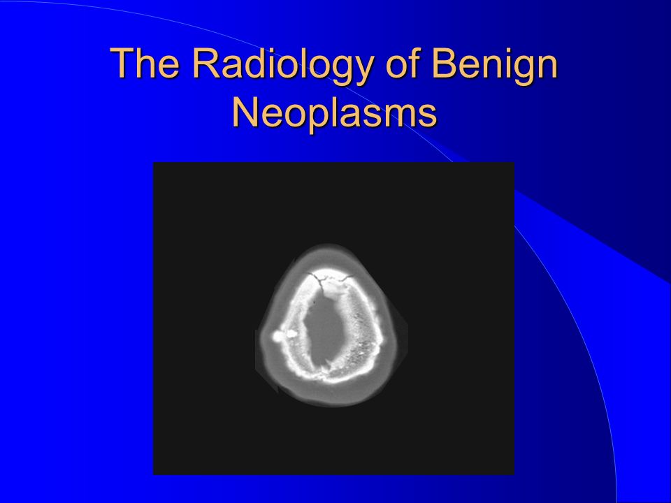
Gardner Syndrome Non-Odontogenic The Radiology of Benign Neoplasms

96
Gardner Syndrome AD multiple osteomas (one of first features) polyposis of colon (polyps become malignant) epidermoid and sebaceous cysts Non-Odontogenic The Radiology of Benign Neoplasms
 polyposis of colon (polyps become malignant) epidermoid and sebaceous cysts Non-Odontogenic The Radiology of Benign Neoplasms")
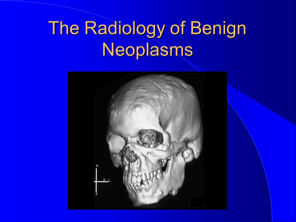
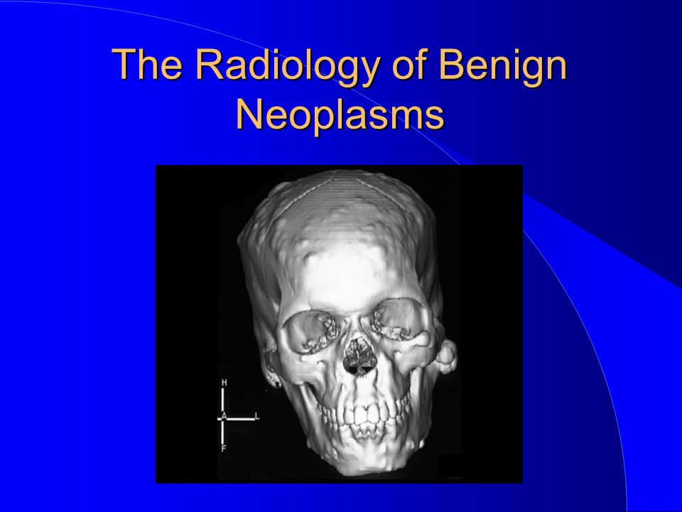
97
Gardner Syndrome desmoid skin tumors impacted supernumerary and permanent teeth odontomas Non-Odontogenic The Radiology of Benign Neoplasms

184
Osteoma Cortical osteoma Cancellous osteoma Osteoid osteoma Osteoma cutis Non-Odontogenic The Radiology of Benign Neoplasms

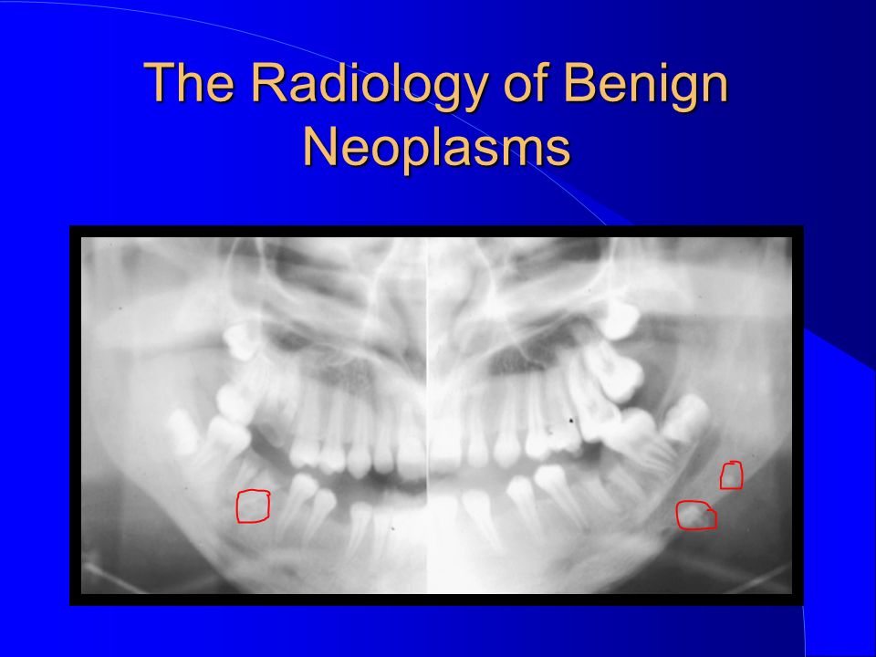
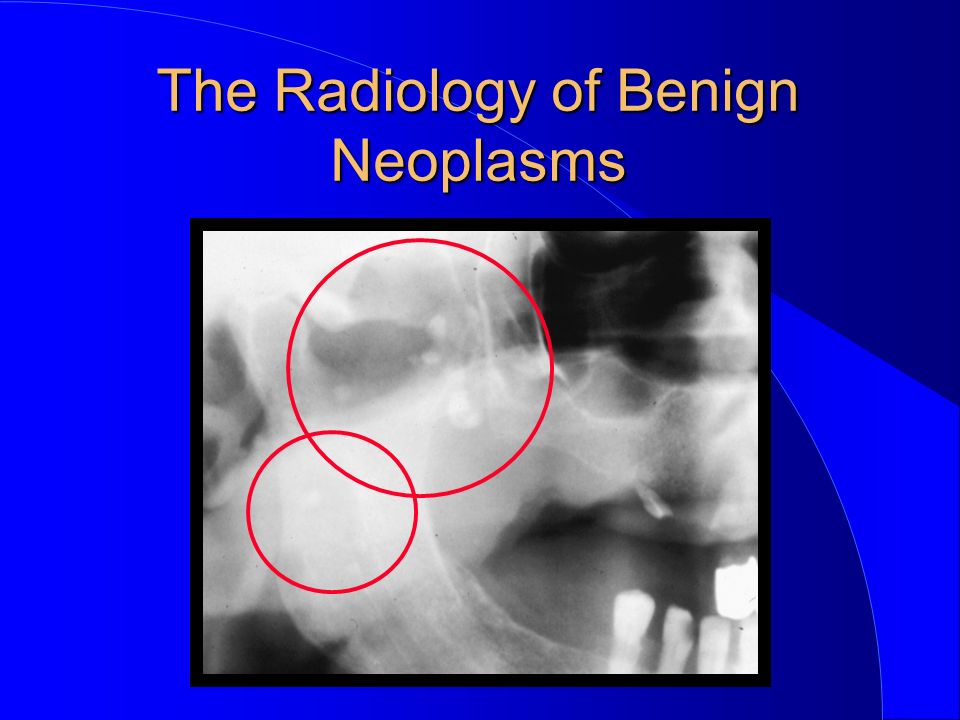
185
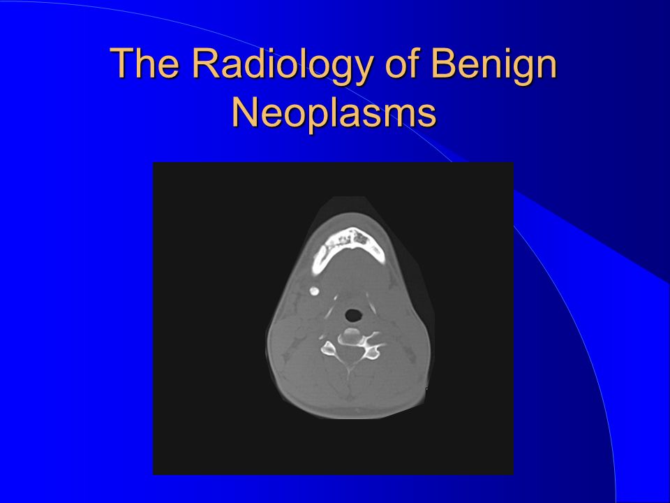
Osteoid osteoma osseous neoplasm of bone uncommon in the jaws may be painful Non-Odontogenic The Radiology of Benign Neoplasms

186
Osteoid osteoma bull’s-eye appearance subperiosteal radiopaque nidus, radiolucent band, sclerotic border Non-Odontogenic The Radiology of Benign Neoplasms

187
Osteoma Cortical osteoma Cancellous osteoma Osteoid osteoma Osteoma cutis Non-Odontogenic The Radiology of Benign Neoplasms

188
Osteoma cutis non-neoplasm of bone found in facial region calcification in scar tissue Non-Odontogenic The Radiology of Benign Neoplasms

194
Giant cell lesion Hemangioma (Haemangioma) Neurofibroma Fibroma Osteoma Osteoblastoma Chondroma Non-Odontogenic The Radiology of Benign Neoplasms
 Neurofibroma Fibroma Osteoma Osteoblastoma Chondroma Non-Odontogenic The Radiology of Benign Neoplasms")
195
Osteoblastoma uncommon primary lesion of bone occasionally in maxilla or mandible mandible more than maxilla Non-Odontogenic The Radiology of Benign Neoplasms

196
Osteoblastoma may be rapid and cause pain tender to palpation “radiolucent” Non-Odontogenic The Radiology of Benign Neoplasms

203
Giant cell lesion Hemangioma (Haemangioma) Neurofibroma Fibroma Osteoma Osteoblastoma Chondroma Non-Odontogenic The Radiology of Benign Neoplasms
 Neurofibroma Fibroma Osteoma Osteoblastoma Chondroma Non-Odontogenic The Radiology of Benign Neoplasms")
Similar presentations


 normal cell of origin Most are classified.>")


. General considerations Primary bone tumors are much less than secondary tumors. All age groups affected,>")


