Download presentation
Presentation is loading. Please wait.

1
Ref: Maxillofacial Imaging ,T A Larheim , P L Westesson 2006
Malignant Jaw tumors By: Nour-Eldin Mohammed Ref: Maxillofacial Imaging ,T A Larheim , P L Westesson 2006

2
Squamous Cell Carcinoma
Synonyms: Epidermoid carcinoma Clinical Features Most common malignancy of oral cavity Males more frequent than females Older age groups (50 years and older), but also younger than 30 years Most frequent in tongue,floor of mouth,mandibular gingiva; retromolar trigone, and anterior tonsillar pillar, soft palate
, but also younger than 30 years. Most frequent in tongue,floor of mouth,mandibular gingiva; retromolar trigone, and anterior tonsillar pillar, soft palate.")
3
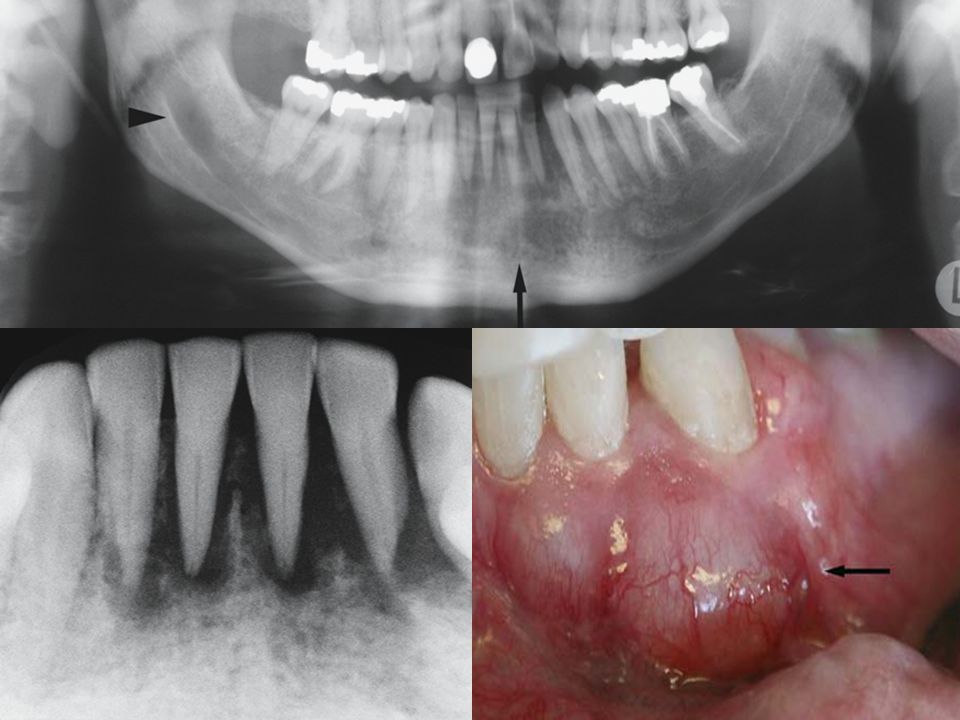
Clinical photograph shows leukoplakia that transformed to gingival cancer
Intraoral panoramic view shows diffuse bone destruction

4
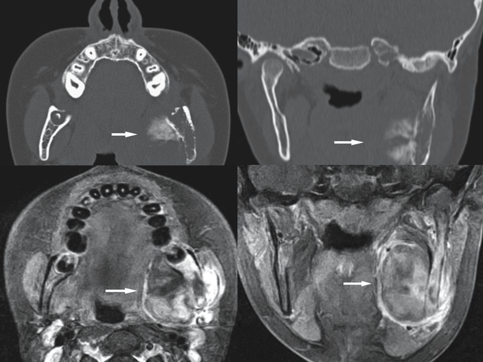
Coronal CT image Coronal T2- fat suppressed MRI Squamous cell carcinoma, maxilla; 70-year-old female with some bleeding from tender soft tissue mass of right gingival mucosa.

5
Mucoepidermoid Carcinoma
Clinical Features Swelling with or without pain Mandible, posterior regions, more frequent than maxilla Females more frequent than males (unlike most oral carcinomas) Fourth and fifth decades, but may occur in any age group Spread to regional lymph nodes in less than 10%
 Fourth and fifth decades, but may occur in any age group. Spread to regional lymph nodes in less than 10%")
6
Panoramic view Axial CT image Coronal CT image Coronal T1-weighted post-Gd MRI

7
Adenoid Cystic Carcinoma
Clinical Features Most commonly seen in minor salivary glands of head and neck, usually palate. Mostly as a painless mass, slowly growing Unlike most carcinomas, seldom metastasizes to regional lymph nodes Lung most common site of metastasis Perineural spread in more than 50%; frequent distant metastasis Slight female predominance Fourth to sixth decades

8
Axial CT image Axial T2-weighted MRI Coronal T1-weighted pre-Gd MRI Coronal T1-weighted post-Gd

10
Non-Hodgkin’s Lymphoma
Clinical Features Non-Hodgkin’s lymphoma of extranodal sites (as opposed to Hodgkin’s lymphoma which is predominantly nodal) Extranodal involvement may include maxillary sinus and maxilla or, less frequently, mandible All age groups, adults in particular Burkitt’s lymphoma affects children; shows rapid growth and may involve one or both jaws
 Extranodal involvement may include maxillary sinus and maxilla or, less frequently, mandible. All age groups, adults in particular. Burkitt’s lymphoma affects children; shows. rapid growth and may involve one or both jaws.")
11
Non-Hodgkin’s lymphoma, maxilla; 49-year-old male painless swelling

12
Multiple Myeloma Clinical Features (Myeloma)
Most common primary bone malignancy in adults Males more frequent than females Older age groups (50 years) Bone pain, malaise Plasmacytoma is a solitary form of myeloma
 Bone pain, malaise. Plasmacytoma is a solitary form of myeloma.")
14
Osteosarcoma Clinical Features
Only 5–10% in head and neck; mostly in jaws Usually painless swelling in jaws Mandible slight predominance Males slight predominance May occur in any age group; peak in fourth decade Prognosis of jaw sarcoma is poor

17
Chondrosarcoma Clinical Features
Mostly in adults in fourth to sixth decades Less aggressive, more slowly growing than osteosarcoma Better prognosis; metastasizes more seldom than osteosarcoma Mandible and maxilla, but rare

19
Ewing Sarcoma Clinical Features
Only 1–4% in head and neck area; most commonly mandible Hard swelling, pain or pain-free Males more frequent than females Usually first and second decades, but may occur at any age

21
Thank you

Similar presentations


.>")


 is the branch of medicine and surgery that specializes in the diagnosis and treatment.>")