Download presentation
Presentation is loading. Please wait.

1
G622 Monitoring the activity of the human body
DATE: 12th March 2014 Uses of physiological measurements LO: to understand why we use physiological indicators to assess health and fitness – blood analysis Lesson outcomes: All D= state normal values or ranges of vital signs Most C = describe how blood tests are used to indicate or diagnose diseases and detect chemicals Some B = explain why and how blood tests are used to diagnose and monitor diabetes Starter - get out exam questions, Q11 Jun 11, Q4 Jun 11, Q4 Jan 11 (set ) and swap papers ready to mark
 and swap papers ready to mark")
2
Starter - get out exam question
June 2011 Q3 p8-10 (a-c) (9) – resp system (HW) June 2011 Q4 p11 – respiration (HW) (10) Jan 2011 Q4 p12 – resp system (HW) (9)Set Swap papers ready to mark
 (9) – resp system (HW) June 2011 Q4 p11 – respiration (HW) (10) Jan 2011 Q4 p12 – resp system (HW) (9)Set Swap papers ready to mark.")
3
What can we learn from our blood?
There are hundreds of different blood tests used to measure the function of an individual organ or systems. Many are used to identify a specific medical condition or disease. Some are used to monitor the response of your body to types of treatment i.e.cancer treatment. Many measure particular chemical constituents in our bodies while some measure the number of cells in our blood.

4
Blood and the transport system:
Blood is the transport system for all animals and is the easiest component of our bodies to gain access to - tiny blood test. Average adult contains about 5 litres of blood and it is vital for life and the loss of even a litre is life threatening and it needs to be replaced quickly. If left to stand it will separate out into the plasma and cell layers.

5
The plasma is the main component- it contains all the dissolved substances and the protein fibrinogen which is essential for blood clotting. The fibrinogen can be removed very simply by allowing the blood to clot. This leaves serum. Serum contains all the dissolved substances but without the interfering effects of fibrinogen - sample of choice for most biochemical determinations. The rest of the blood contains the cells - majority are red blood cells which carry oxygen and much small number of white cells (buffy coat layer). White cells are responsible for fighting infection. Platelets are also found in tiny numbers and are involved in the clotting process. They are fragments of larger cells called the megakaryocytes found in the bone marrow.
. White cells are responsible for fighting infection. Platelets are also found in tiny numbers and are involved in the clotting process. They are fragments of larger cells called the megakaryocytes found in the bone marrow.")
6
We have on average 5 – 6 litres of blood
Blood – a connective tissue We have on average 5 – 6 litres of blood Blood is made of cells (45%) and fluid plasma (55%) Plasma (liquid part – 55%) – 90% water + 10% substances Transports substances around the body: CO2 from cells to lungs; urea from liver to kidneys; hormones; enzymes; antibodies; fibrinogen (protein for blood clotting); it distributes heat. White blood cells (Leucocytes) Defence Lymphocytes (produce antibodies and antitoxins) Phagocytes – engulf and destroy pathogens (microbes) Red blood cells (Erythrocytes) Transport oxygen from lungs to cells via the Haemoglobin No nuclei
 and fluid plasma (55%) Plasma (liquid part – 55%) – 90% water + 10% substances. Transports substances around the body: CO2 from cells to lungs; urea from liver to kidneys; hormones; enzymes; antibodies; fibrinogen (protein for blood clotting); it distributes heat. White blood cells (Leucocytes) Defence. Lymphocytes (produce antibodies and antitoxins) Phagocytes – engulf and destroy pathogens (microbes) Red blood cells (Erythrocytes) Transport oxygen from lungs to cells via the Haemoglobin. No nuclei.")
7
Functions of Blood Transport Defence Blood clotting red blood cells
Against pathogens - white blood cells - phagocytosis (neutrophils); immune response - production of antibodies and antitoxins (lymphocytes) Defence Blood clotting To prevent blood loss at site of damage & prevent entry of pathogens (platelets)
; immune response - production of antibodies and antitoxins (lymphocytes) Defence. Blood clotting. To prevent blood loss at site of damage & prevent entry of pathogens (platelets)")
8
Defence - phagocytosis Contain enzymes
Red blood cells (erythrocytes) Contains haemoglobin (Hb) - formation requires Fe Hb transports O2 from lungs to cells Biconcave discs – large surface area for diffusion of O2 No nucleus – more space for Hb Flexible – squeeze through capillaries in single file Phagocytes Defence - phagocytosis Contain enzymes Engulf (ingest) microbes and digest them Lymphocytes Defence – immune response Produce antibodies and antitoxins Killer cells – destroy cells infected with viruses Platelets Rupture and release enzyme at site of cut – initiates a cascade of reactions – converts insoluble blood protein fibrinogen to insoluble fibrin threads which trap blood cells and platelets to form a blood clot
 Contains haemoglobin (Hb) - formation requires Fe. Hb transports O2 from lungs to cells. Biconcave discs – large surface area for diffusion of O2. No nucleus – more space for Hb. Flexible – squeeze through capillaries in single file. Phagocytes. Defence - phagocytosis. Contain enzymes. Engulf (ingest) microbes and digest them. Lymphocytes. Defence – immune response. Produce antibodies and antitoxins. Killer cells – destroy cells infected with viruses. Platelets. Rupture and release enzyme at site of cut – initiates a cascade of reactions – converts insoluble blood protein fibrinogen to insoluble fibrin threads which trap blood cells and platelets to form a blood clot.")
10
Blood cells can be counted using a special counting slide called a haemocytometer. Nowadays automated counting machines are used which can count simultaneously all blood cells in a very short time interval. Low red blood count may indicate anaemia from for example a low iron intake. An excess of white blood cells may indicate an underlying infection or if very high may be indicative of cancer (leukaemia).
.")
11
Determination of drugs
Blood tests are sometimes used to test for the presence and amount of drugs within the body. Urine tends to be used for screen a for a large range of drugs and their metabolites. Testing for drugs may be to monitor compliance, optimum treatment levels or to determine abuse (for recreational or performance-enhancing drugs). A range of quantitative techniques are used depending on type of drug. GLC and HPLC are useful quantitative and qualitative techniques. Specific groups of drugs can now be detected with test kits (Police, Schools or Clinics) using techniques based upon antibody-enzymic techniques. Do some research and find how alcohol, a named recreational and a named performance-enhancing drug are determined.
. A range of quantitative techniques are used depending on type of drug. GLC and HPLC are useful quantitative and qualitative techniques. Specific groups of drugs can now be detected with test kits (Police, Schools or Clinics) using techniques based upon antibody-enzymic techniques. Do some research and find how alcohol, a named recreational and a named performance-enhancing drug are determined.")
12
When do we have to take blood for drug tests?
There are lots of situations listed below are some: Accident testing Health/life insurance medicals Pre-employment screening Evidence of illegality Comply with drug rehabilitation programme Screen of company work-force Sports related drug abuse

14
ELISA tests ELISA stands for Enzyme Linked Immuno Sorbent Assay. There are several variants of the assay – these can be direct or indirect detection and the competitive assay Direct is where antigen is directly detected with primary labelled antibody or indirect with a labelled secondary antibody . Are used to detect the presence of antibody indicators for certain infective diseases such as AIDs and Hepatitis. They use the principle of antigen-antibody complex. These type of assays are collectively called immunoassays. Specific diseases possess specific antigens and these are recognised by specific antibodies to form these antigen-antibody complexes. The antibodies are labelled with a marker, which can be An enzyme A radioactive isotope label A fluorescent or chemi-luminescent compound

16
TASK: Read the notes provided on ELISA and in your own words, using diagrams explain how ELISA can be used to detect the presence of an antigen or antibody for a disease. Find out how an HCG (human chorionic gonadotrophin) pregnancy test kit works. AntiHCG is used in these kits.
 pregnancy test kit works. AntiHCG is used in these kits. v=DqX7VxW3wL0.")
17
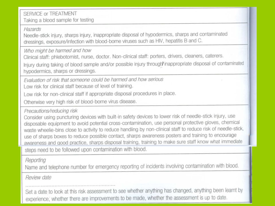
Health and safety Student activities:
find out and make notes on the health and safety requirements for the following: 1. Regulations for disposal of hazardous biological waste e.g. sharps and hypodermic needles used in obtaining blood for testing 2. Procedures for the treatment of materials that may be contaminated with microbiological hazards e.g. used petri dishes, materials from antibody testing 3. How to carry out a risk assessment for a blood test, state hazards and explain how to minimse the risks to the person carrying out blood test

18
Disposal of syringes and hypodermic needles
Sharps and hypodermic needles are disposed of in special containers to avoid contact. Specialist waste disposal companies provide a service to collect and dispose of the waste material in the sharp boxes. Similar services exist for disposal of clinical waste that might be contaminated with blood borne viruses ,such as swabs and other soiled waste. The clinical waste is finally disposed of in special incinerators. Disposal of microbiological hazards Pathology laboratories produce large volumes of clinical waste. This will include microbiological cultures, antibody testing kits, including ELISA plates, contaminated plastic tubes and even radioactive waste. All of this is potentially infected waste. High hazard waste, like microbiological cultures are autoclaved before disposing as clinical waste and then incinerated. Non disposable apparatus is sterilised by being autoclaved, washed before being reused.

19
Risk assessment for a blood test:
The volume of blood my differ depending on the type of analysis required. The details of a general blood test i.e. the method used is always designed to minimise the risks involved with that hazard. Techniques may alter in some way to take the blood but the hazards, risks and precautions will be similar. The risk assessment on the next slide might be used for a blood test but it is only one example of how the assessment might appear.

21
Diabetes types and control of blood sugar levels
Some people are unable to control their blood glucose level properly because they have insufficient insulin. This is known as diabetes mellitus. There are two types I – insulin dependent and type II- non-insulin dependent (sometimes called early and late onset diabetes). Insulin and glucagon: Insulin and glucagon are both secreted by the pancreas and regulate out blood glucose (sugar) levels. The endocrine glands of the pancreas consists of the islets of langerhans and these secrete hormones directly into our blood stream. The alpha cells are sensitive to low levels of glucose and secrete the hormone glucagon. The beta cells detect increases in blood glucose and secrete the hormone insulin.
. Insulin and glucagon: Insulin and glucagon are both secreted by the pancreas and regulate out blood glucose (sugar) levels. The endocrine glands of the pancreas consists of the islets of langerhans and these secrete hormones directly into our blood stream. The alpha cells are sensitive to low levels of glucose and secrete the hormone glucagon. The beta cells detect increases in blood glucose and secrete the hormone insulin.")
22
Islets of Langerhan in the pancreas

23
Diabetes symptoms and causes:
Diabetics are unable to control their blood glucose levels because they are unable to produce enough insulin – Clinical condition is called diabetes mellitis (sweet urine). After a meal diabetics are , because of lack of insulin, unable to increase the permeability of the cell membranes to glucose. So the cells starve of fuel!! The cells, unable to get glucose start to respire proteins and fats instead, which results in loss of weight. Another symptom is thirst due to increased water potential of the blood. Presence of glucose in the urine – kidneys being unable to reabsorb the high levels of glucose filtered by the tubules. Blood plasma glucose (fasting) is 3.5 to 7.5 mmol/l and urine appears in urine above 9.0 mmol/l and would indicate diabetes and further investigation – Glucose Tolerance Test.
. After a meal diabetics are , because of lack of insulin, unable to increase the permeability of the cell membranes to glucose. So the cells starve of fuel!! The cells, unable to get glucose start to respire proteins and fats instead, which results in loss of weight. Another symptom is thirst due to increased water potential of the blood. Presence of glucose in the urine – kidneys being unable to reabsorb the high levels of glucose filtered by the tubules. Blood plasma glucose (fasting) is 3.5 to 7.5 mmol/l and urine appears in urine above 9.0 mmol/l and would indicate diabetes and further investigation – Glucose Tolerance Test.")
24
Type 1 Diabetes: Sometimes called early onset as often occurs in children or young adults. Develops because the insulin producing cells are attacked by the body – immune disorder. May be initiated by a virus or other infections. Type 2 Diabetes: Or late onset diabetes occurs when body does not produce enough insulin. Very strong link with poor diet, obesity, lack of exercise. Increasing now this type of diabetes is seen in the young i.e. children to obesity and lack of exercise.

25
Starter questions: What is the fasting blood sugar range?
What is level of concern for blood sugar? What does insulin do? What does glucagon do? What is the relationship between these two hormones? Where are the islets of Langerhans found? Name the two types of diabetes? What is the commonest type caused by? What are the symptoms of diabetes? How can it be controlled?

26
Answers: 3.5 to 7.5 mmol/dm3 9.0 mmol/dm3
Lowers blood glucose by increasing glycogen and fat stores (allows cells to take up glucose) Increases blood glucose by stimulating glycogenolysis and gluconeogenesis Both work together to keep blood level of glucose in narrow band – homeostasis Pancreas islets of Langerhans Type 1 or early onset and type 2 late onset Obesity and lack of exercise Thirst, weight loss, excessive urination, infections – thrush, passing out. Insulin and diet control.
 Increases blood glucose by stimulating glycogenolysis and gluconeogenesis. Both work together to keep blood level of glucose in narrow band – homeostasis. Pancreas islets of Langerhans. Type 1 or early onset and type 2 late onset. Obesity and lack of exercise. Thirst, weight loss, excessive urination, infections – thrush, passing out. Insulin and diet control.")
27
Monitoring Physiological changes:
There are many ways in which doctors, nurses and other health care specialists monitor the working of the human body. This is necessary to ensure that everything is working as it should. Some examples are temperature, pulse and breathing rate, BP and ECG. Often they are concerned with a major organs function because any problem will have a major affect on the well being of the individual i.e. heart or lungs. In this lesson we will look at temperature, pulse rate and ECG. The normal values are those that the OCR will use in the exam, so learn them.

28
Temperature: We measure body temperature using a clinical thermometer - either mercury or electronic. The reason body temperature is measured is that our body is controlled within a relatively narrow temperature range and a range of diseases (notable infections) can alter that balance. First student activity: Find out how a clinical thermometer works - mercury and electronic. Take each others body temperature. (Caution-hazard) Copy the average normal values for body temperature. Answer the question on worksheet please. How do we maintain a normal body temperature? Use internet to supplement your notes and answer above question.
 can alter that balance. First student activity: Find out how a clinical thermometer works - mercury and electronic. Take each others body temperature. (Caution-hazard) Copy the average normal values for body temperature. Answer the question on worksheet please. How do we maintain a normal body temperature Use internet to supplement your notes and answer above question.")
30
Direct measurements: Heart sounds: The beat of the heart produces a sound: the heartbeat. It has two sounds - lub-dup - which is the blood hitting the heart valves. The lub sound is made when blood is forced back against the bi and tri-cuspid valves as the ventricles contract. The dup comes from the back flow of blood as it hits the semi-lunar valves of the pulmonary artery and aorta as the ventricles relax Can be heard using a stethoscope. This magnifies and isolates the heart sounds. It will pick up unusually sounds or murmurs in the heart which may indicate a valve is leaking or that the chambers are not working together.

31
Monitoring heart function:
Monitoring the heart can by direct or indirect measurement. Indirect measurement: is the monitoring the pulse. Each time the ventricles contract, blood is forced out into the aorta under high pressure, and then onto the arteries,. This pressure surge is felt as a pulse. Simple to measure on any of the arteries lieing close to the skin in the wrist or neck. Usual to count it for 15 seconds and then multiply by 4 to give number of beats per minute. Alternatively can be measured by a pulse meter. Normal adult rate is beats per minute. It will tell if heart is beat too fast or slow or if it is an irregular beat.

32
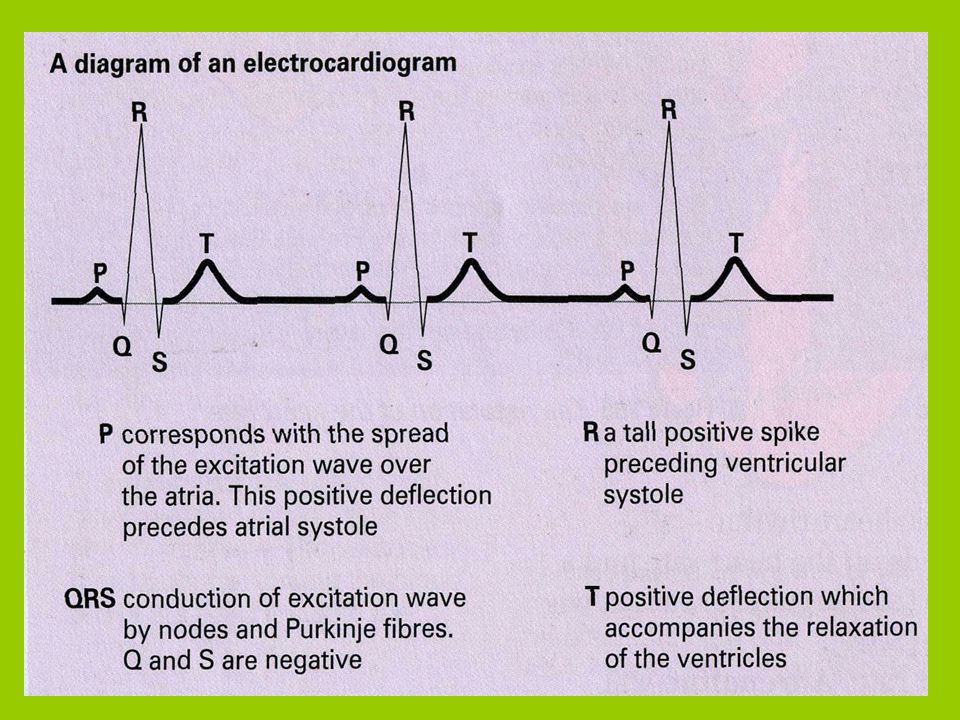
ECG or electrocardiogram;
The wave of excitation through the Purkinje tissue of the heart is measured by an electrocardiograph and this produces a graphical display called an electrocardiogram or ECG.

33
Each part of the ECG curve represents a particular electrical event in the heart:
P wave is caused by the excitation spreading from the sinoatrail node (SAN) to the aterioventricular node (AVN) causing atrail systole (contraction) QRS complex is the spread of excitation through the Purkinje tissue causing ventricular systole T wave is the recovery or depolarization of the ventricles after contraction Flat area of the heart is caused by diastole, when there is no excitation of the heart. ECG is able to identify a number of heart problems: sinus tachycardia (abnormally rapid fast heartbeat), bradycardia (abnormally slow heartbeat), sinus arrhyythmia and ventricular fibrillation.
 to the aterioventricular node (AVN) causing atrail systole (contraction) QRS complex is the spread of excitation through the Purkinje tissue causing ventricular systole. T wave is the recovery or depolarization of the ventricles after contraction. Flat area of the heart is caused by diastole, when there is no excitation of the heart. ECG is able to identify a number of heart problems: sinus tachycardia (abnormally rapid fast heartbeat), bradycardia (abnormally slow heartbeat), sinus arrhyythmia and ventricular fibrillation.")
35
Normal Atrail fibrillation Ventricular fibrillation Ventricular tachycardia

36
A Atrioventricular valves close (1st louder heart sound “LUB”)
Atrial Systole Ventricular Systole Diastole “DUB” “LUB” A Atrioventricular valves close (1st louder heart sound “LUB”) B Semilunaris valves open C Semilunaris valves close (2nd softer heart sound “DUB”) D Atrioventricular valves open
 B Semilunaris valves open. C Semilunaris valves close (2nd softer heart sound DUB ) D Atrioventricular valves open.")
37
Pressures changes in the aorta, left ventricle and left atrium during one
heartbeat © Pearson Education Ltd 2008 This document may have been altered from the original 37

38
A normal ECG trace compared with others indicating an unhealthy heart
38

39
Transport of CO2 in the blood
There are 3 ways in which carbon dioxide is transported in the blood: DISSOLVED CO2 About 5 % of carbon dioxide is transported unchanged, simply dissolved in the plasma BOUND TO HAEMOGLOBIN About 10 % of carbon dioxide is transported bound to haemoglobin. Carbon dioxide combines reversibly with haemoglobin to form carbamino-haemoglobin. BICARBONATE IONS (HCO3-) 85% of carbon dioxide is transported in this way
 85% of carbon dioxide is transported in this way.")
41
Measuring blood pressure
bp animation

Similar presentations


 55% total volume - Blood cells – 45% total volume.>")


