Download presentation
Presentation is loading. Please wait.

1
Anatomy & Physiology The Nervous System

2
Organization of the Nervous System Central Nervous System (CNS): consists of the brain and spinal cord, which occupy the dorsal body cavity. Central Nervous System (CNS): consists of the brain and spinal cord, which occupy the dorsal body cavity. Peripheral Nervous System (PNS): the part of the nervous system outside the CNS, consisting mainly of the nerves that extend from the brain and spinal cord. Peripheral Nervous System (PNS): the part of the nervous system outside the CNS, consisting mainly of the nerves that extend from the brain and spinal cord. A) Spinal nerves- carry impulses to and from the spinal cord. A) Spinal nerves- carry impulses to and from the spinal cord. B) Cranial nerves- carry impulses to and from the brain. B) Cranial nerves- carry impulses to and from the brain.
: consists of the brain and spinal cord, which occupy the dorsal body cavity. Peripheral Nervous System (PNS): the part of the nervous system outside the CNS, consisting mainly of the nerves that extend from the brain and spinal cord. Peripheral Nervous System (PNS): the part of the nervous system outside the CNS, consisting mainly of the nerves that extend from the brain and spinal cord. A) Spinal nerves- carry impulses to and from the spinal cord. A) Spinal nerves- carry impulses to and from the spinal cord. B) Cranial nerves- carry impulses to and from the brain. B) Cranial nerves- carry impulses to and from the brain..")
4
Peripheral Nervous System (PNS) The PNS has two functional subdivisions: The PNS has two functional subdivisions: 1) Sensory (Afferent) division- consists of nerve fibers that convey impulses to the central nervous system from sensory receptors located throughout the body. 1) Sensory (Afferent) division- consists of nerve fibers that convey impulses to the central nervous system from sensory receptors located throughout the body. a) Somatic afferent fibers- convey impulses from the skin, skeletal muscles, and joints a) Somatic afferent fibers- convey impulses from the skin, skeletal muscles, and joints b) Visceral afferent fibers- sensory fibers transmitting impulses from the visceral organs b) Visceral afferent fibers- sensory fibers transmitting impulses from the visceral organs
 Sensory (Afferent) division- consists of nerve fibers that convey impulses to the central nervous system from sensory receptors located throughout the body. a) Somatic afferent fibers- convey impulses from the skin, skeletal muscles, and joints a) Somatic afferent fibers- convey impulses from the skin, skeletal muscles, and joints b) Visceral afferent fibers- sensory fibers transmitting impulses from the visceral organs b) Visceral afferent fibers- sensory fibers transmitting impulses from the visceral organs.")
5
PNS, cont. 2) Motor (efferent) division- transmits impulses from the CNS to effector organs, the muscles and glands. 2) Motor (efferent) division- transmits impulses from the CNS to effector organs, the muscles and glands. The motor division has two main parts: The motor division has two main parts: a) Somatic nervous system- composed of somatic nerve fibers that conduct impulses from the CNS to skeletal muscles (voluntary) a) Somatic nervous system- composed of somatic nerve fibers that conduct impulses from the CNS to skeletal muscles (voluntary) b) Autonomic nervous system- consists of visceral motor nerve fibers that regulate the activity of smooth muscles, cardiac muscles, and glands. b) Autonomic nervous system- consists of visceral motor nerve fibers that regulate the activity of smooth muscles, cardiac muscles, and glands. 1. Sympathetic- mobilizes body systems during emergencies 1. Sympathetic- mobilizes body systems during emergencies 2. Parasympathetic- conserves energy, promotes nonemergency functions. 2. Parasympathetic- conserves energy, promotes nonemergency functions.
 Motor (efferent) division- transmits impulses from the CNS to effector organs, the muscles and glands. 2) Motor (efferent) division- transmits impulses from the CNS to effector organs, the muscles and glands. The motor division has two main parts: The motor division has two main parts: a) Somatic nervous system- composed of somatic nerve fibers that conduct impulses from the CNS to skeletal muscles (voluntary) a) Somatic nervous system- composed of somatic nerve fibers that conduct impulses from the CNS to skeletal muscles (voluntary) b) Autonomic nervous system- consists of visceral motor nerve fibers that regulate the activity of smooth muscles, cardiac muscles, and glands. b) Autonomic nervous system- consists of visceral motor nerve fibers that regulate the activity of smooth muscles, cardiac muscles, and glands. 1. Sympathetic- mobilizes body systems during emergencies 1. Sympathetic- mobilizes body systems during emergencies 2. Parasympathetic- conserves energy, promotes nonemergency functions. 2. Parasympathetic- conserves energy, promotes nonemergency functions..")
6
Central Nervous System (CNS) Consists of the brain and spinal cord Consists of the brain and spinal cord Receives messages from the body Receives messages from the body Sends out signals to the body in response Sends out signals to the body in response
 Consists of the brain and spinal cord Consists of the brain and spinal cord Receives messages from the body Receives messages from the body Sends out signals to the body in response Sends out signals to the body in response")
7
CENTRAL NERVOUS SYSTEM

8
Organization of the Brain The brain is part of the CNS The brain is part of the CNS Consists of: Consists of: Cerebral hemispheres (right and left) Cerebral hemispheres (right and left) Diencephalon (hypothalamus, thalamus) Diencephalon (hypothalamus, thalamus) Brain stem ( midbrain, pons, and medulla) Brain stem ( midbrain, pons, and medulla)
 Cerebral hemispheres (right and left) Diencephalon (hypothalamus, thalamus) Diencephalon (hypothalamus, thalamus) Brain stem ( midbrain, pons, and medulla) Brain stem ( midbrain, pons, and medulla)")
10
Tissue pattern in the CNS Spinal cord: central cavity surrounded Spinal cord: central cavity surrounded by a gray matter core. Outer layer is white matter (myelinated fiber tracts). by a gray matter core. Outer layer is white matter (myelinated fiber tracts). Brain has the same basic design with areas of gray matter. Brain has the same basic design with areas of gray matter.
. by a gray matter core. Outer layer is white matter (myelinated fiber tracts). Brain has the same basic design with areas of gray matter. Brain has the same basic design with areas of gray matter..")
11
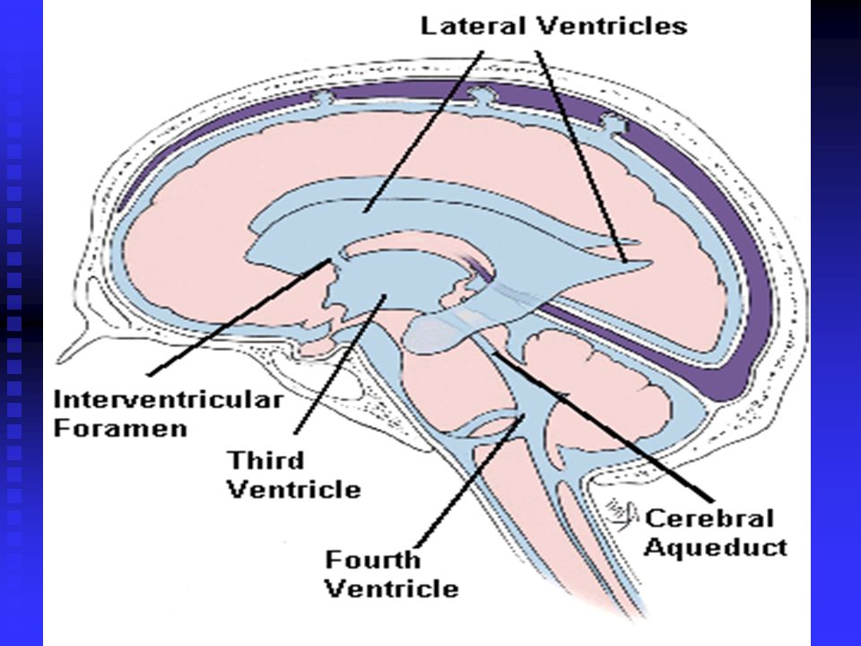
Ventricles of the Brain Ventricles are spaces in the brain that arose from the neural tube. Ventricles are spaces in the brain that arose from the neural tube. There are four ventricles, each filled with cerebrospinal fluid (CSF). There are four ventricles, each filled with cerebrospinal fluid (CSF). Lateral ventricles are in the cerebral hemispheres, separated by a thin membrane (septum pellucidum). Lateral ventricles are in the cerebral hemispheres, separated by a thin membrane (septum pellucidum). The third ventricle is connected to the lateral ventricles by the interventricular foramen. The third ventricle is connected to the lateral ventricles by the interventricular foramen. The fourth ventricle is in the brain stem and connects with the central canal of the spinal cord (also connected to the third ventricle by the cerebral aqueduct. The fourth ventricle is in the brain stem and connects with the central canal of the spinal cord (also connected to the third ventricle by the cerebral aqueduct.
. There are four ventricles, each filled with cerebrospinal fluid (CSF). Lateral ventricles are in the cerebral hemispheres, separated by a thin membrane (septum pellucidum). Lateral ventricles are in the cerebral hemispheres, separated by a thin membrane (septum pellucidum). The third ventricle is connected to the lateral ventricles by the interventricular foramen. The third ventricle is connected to the lateral ventricles by the interventricular foramen. The fourth ventricle is in the brain stem and connects with the central canal of the spinal cord (also connected to the third ventricle by the cerebral aqueduct. The fourth ventricle is in the brain stem and connects with the central canal of the spinal cord (also connected to the third ventricle by the cerebral aqueduct..")
13
Hydrocephalus The fluid produced by the ventricles must circulate and drain. If fluid is overproduced, or cannot drain properly, it builds up causing enlarged skull and intracranial pressure. Hydrocephalus can be treated by inserting a straw-like shunt to help drain the CSF.

14
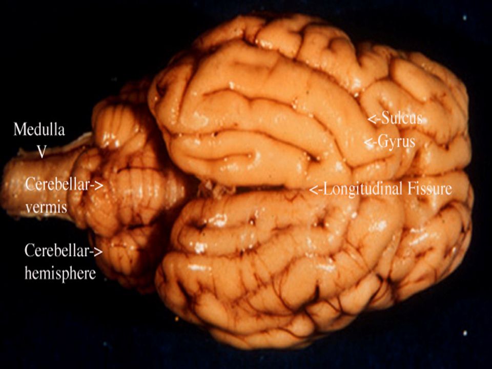
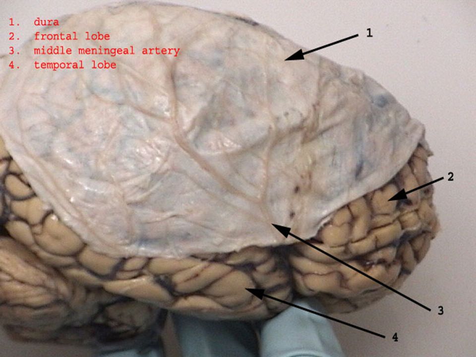
Cerebral Hemispheres Cerebral hemispheres form the superior part of the brain, making up about 83% of total brain mass. Cerebral hemispheres form the superior part of the brain, making up about 83% of total brain mass. Surface is marked by elevated ridges of tissue called gyri separated by shallow grooves called sulci (singular forms are gyrus and sulcus). Surface is marked by elevated ridges of tissue called gyri separated by shallow grooves called sulci (singular forms are gyrus and sulcus). Deeper grooves called fissures separate larger regions of the brain; longitudinal fissure separates the cerebral hemispheres, transverse fissure separates the cerebral hemispheres from the cerebellum. Deeper grooves called fissures separate larger regions of the brain; longitudinal fissure separates the cerebral hemispheres, transverse fissure separates the cerebral hemispheres from the cerebellum.
. Surface is marked by elevated ridges of tissue called gyri separated by shallow grooves called sulci (singular forms are gyrus and sulcus). Deeper grooves called fissures separate larger regions of the brain; longitudinal fissure separates the cerebral hemispheres, transverse fissure separates the cerebral hemispheres from the cerebellum. Deeper grooves called fissures separate larger regions of the brain; longitudinal fissure separates the cerebral hemispheres, transverse fissure separates the cerebral hemispheres from the cerebellum..")
16
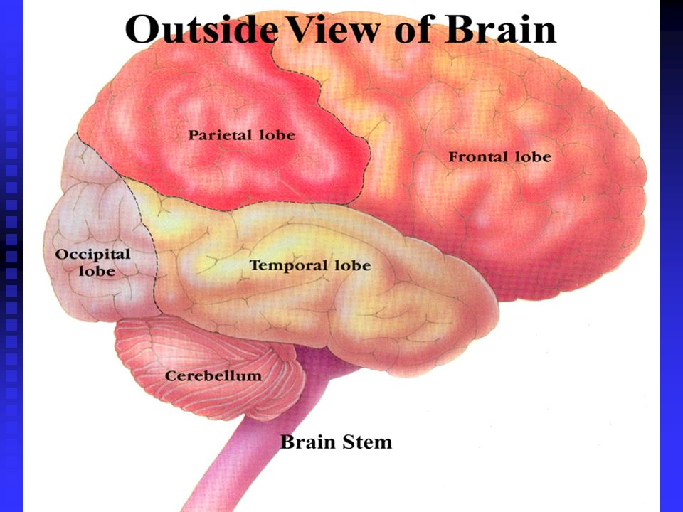
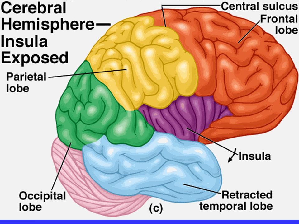
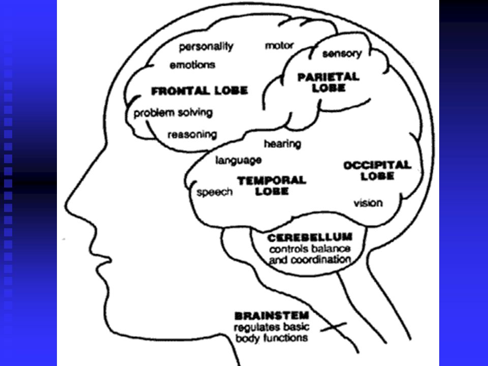
Lobes of the Brain Deep sulci divide each hemisphere into five lobes: frontal, temporal, occipital, and insula– all but the last named after the cranial bones that cover them. Deep sulci divide each hemisphere into five lobes: frontal, temporal, occipital, and insula– all but the last named after the cranial bones that cover them. The central sulcus separates the frontal lobe from the parietal lobe. The central sulcus separates the frontal lobe from the parietal lobe. The parieto-occipital sulcus separates the occipital and parietal lobes. The parieto-occipital sulcus separates the occipital and parietal lobes.

18
Lobes of the brain, cont. The insula is buried deep within the lateral sulcus, and is covered by parts of the temporal, parietal, and frontal lobes. The insula is buried deep within the lateral sulcus, and is covered by parts of the temporal, parietal, and frontal lobes. The lateral sulcus outlines the temporal lobe and separates it from the parietal lobe and frontal lobes. The lateral sulcus outlines the temporal lobe and separates it from the parietal lobe and frontal lobes.

20
Cerebral Cortex The cerebral cortex is the area that carries out the processes of the “conscious mind”. The cerebral cortex is the area that carries out the processes of the “conscious mind”. It enables us to be aware of our sensations, to communicate, remember, understand, and to initiate voluntary movements. It enables us to be aware of our sensations, to communicate, remember, understand, and to initiate voluntary movements. Composed of gray matter (neuron cell bodies, dendrites, and unmyelinated axons). Composed of gray matter (neuron cell bodies, dendrites, and unmyelinated axons). Accounts for about 40% brain mass. Accounts for about 40% brain mass.
. Composed of gray matter (neuron cell bodies, dendrites, and unmyelinated axons). Accounts for about 40% brain mass. Accounts for about 40% brain mass..")
21
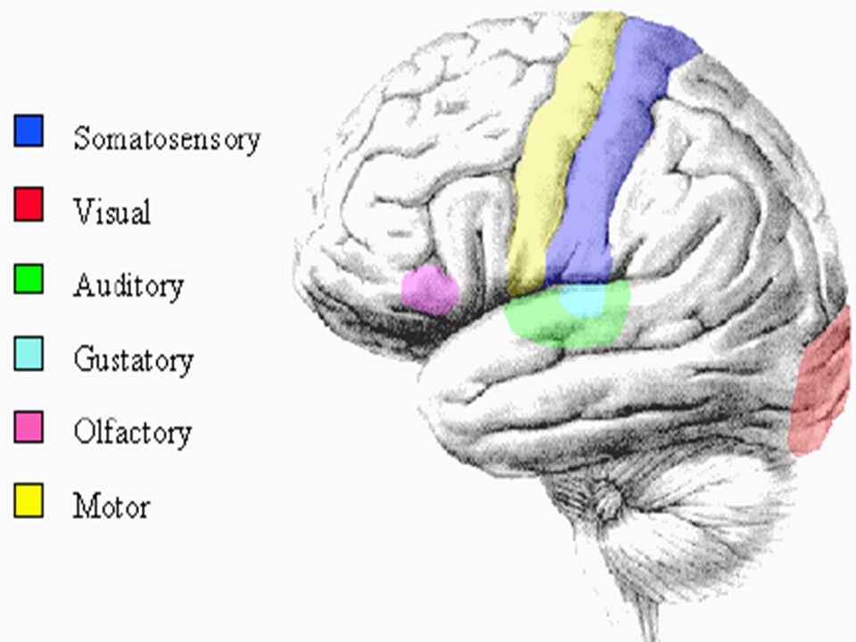
Cerebral cortex, cont. Contains three functional areas: Contains three functional areas: 1) Motor areas that control voluntary motor functions 1) Motor areas that control voluntary motor functions 2) Sensory areas that provide conscious awareness of sensation 2) Sensory areas that provide conscious awareness of sensation 3) Association areas that integrate a wide range of information for purposeful action 3) Association areas that integrate a wide range of information for purposeful action
 Motor areas that control voluntary motor functions 1) Motor areas that control voluntary motor functions 2) Sensory areas that provide conscious awareness of sensation 2) Sensory areas that provide conscious awareness of sensation 3) Association areas that integrate a wide range of information for purposeful action 3) Association areas that integrate a wide range of information for purposeful action.")
22
Cerebral cortex, cont. Each hemisphere is chiefly concerned with the sensory and motor functions of the opposite side of the body. Each hemisphere is chiefly concerned with the sensory and motor functions of the opposite side of the body. The two hemispheres are not equal in function; each is partially specialized The two hemispheres are not equal in function; each is partially specialized Conscious behavior involves the entire cortex; no functional area acts alone Conscious behavior involves the entire cortex; no functional area acts alone

24
Motor areas Cortical areas controlling motor functions lie in the posterior part of the frontal lobes. Cortical areas controlling motor functions lie in the posterior part of the frontal lobes. 1) Primary (somatic) motor cortex- allow conscious control of skeletal muscles, skilled and precise movements. 1) Primary (somatic) motor cortex- allow conscious control of skeletal muscles, skilled and precise movements. 2) Premotor cortex- Controls learned motor skills with patterns, such as playing an instrument. Also involved in planning movements. 2) Premotor cortex- Controls learned motor skills with patterns, such as playing an instrument. Also involved in planning movements.
 Primary (somatic) motor cortex- allow conscious control of skeletal muscles, skilled and precise movements. 1) Primary (somatic) motor cortex- allow conscious control of skeletal muscles, skilled and precise movements. 2) Premotor cortex- Controls learned motor skills with patterns, such as playing an instrument. Also involved in planning movements. 2) Premotor cortex- Controls learned motor skills with patterns, such as playing an instrument. Also involved in planning movements..")
26
Motor areas, cont. Broca’s area- lies anterior to the premotor area. Involved in speech, including motor control of muscles involved in speech production and mental preparation for speaking. Broca’s area- lies anterior to the premotor area. Involved in speech, including motor control of muscles involved in speech production and mental preparation for speaking. Frontal eye field- Controls voluntary movement of the eyes. Frontal eye field- Controls voluntary movement of the eyes.

27
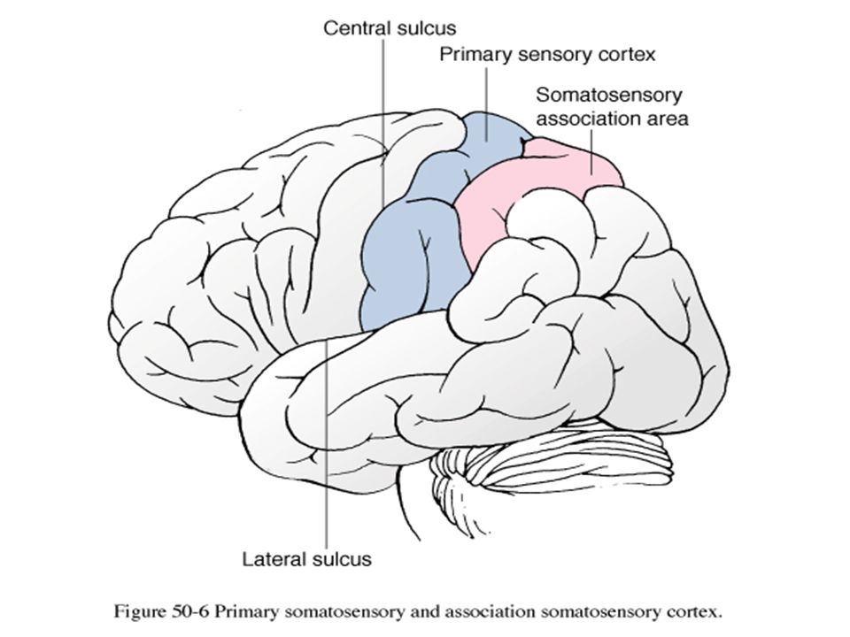
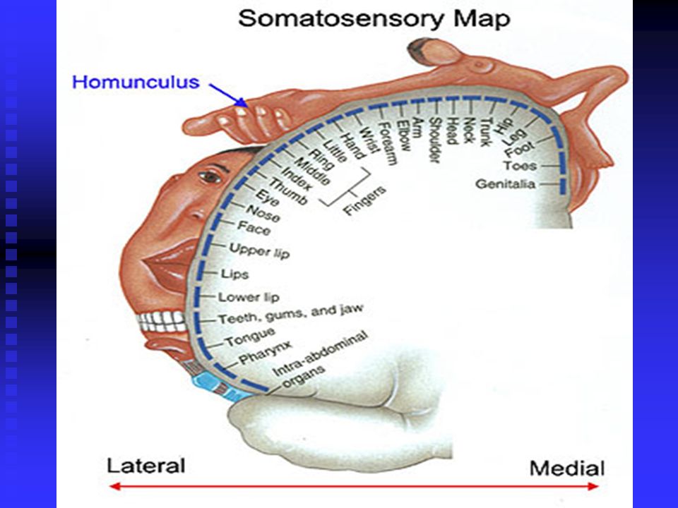
Sensory Areas Unlike the motor areas, which are confined to the frontal lobe cortex, areas concerned with conscious awareness of sensation occur in the parietal, temporal, and occipital lobes. Unlike the motor areas, which are confined to the frontal lobe cortex, areas concerned with conscious awareness of sensation occur in the parietal, temporal, and occipital lobes. 1) Primary somatosensory cortex- located in the postcentral gyrus of the parietal lobe. Receives information from sensory receptors in the skin from proprioceptors in skeletal muscles. Identifies body region being stimulated (spatial discrimination). 1) Primary somatosensory cortex- located in the postcentral gyrus of the parietal lobe. Receives information from sensory receptors in the skin from proprioceptors in skeletal muscles. Identifies body region being stimulated (spatial discrimination).
 Primary somatosensory cortex- located in the postcentral gyrus of the parietal lobe. Receives information from sensory receptors in the skin from proprioceptors in skeletal muscles. Identifies body region being stimulated (spatial discrimination). 1) Primary somatosensory cortex- located in the postcentral gyrus of the parietal lobe. Receives information from sensory receptors in the skin from proprioceptors in skeletal muscles. Identifies body region being stimulated (spatial discrimination)..")
29
Sensory Areas, cont. 2) Somatosensory Association cortex- lies posterior to the primary somatosensory cortex. 2) Somatosensory Association cortex- lies posterior to the primary somatosensory cortex. Function is to integrate different sensory inputs (temperature, pressure, etc.) to produce a comprehensive understanding o an object being felt. Function is to integrate different sensory inputs (temperature, pressure, etc.) to produce a comprehensive understanding o an object being felt. Someone with damages to this area would not be able to recognize objects without looking at them. Someone with damages to this area would not be able to recognize objects without looking at them.
 Somatosensory Association cortex- lies posterior to the primary somatosensory cortex. 2) Somatosensory Association cortex- lies posterior to the primary somatosensory cortex. Function is to integrate different sensory inputs (temperature, pressure, etc.) to produce a comprehensive understanding o an object being felt. Function is to integrate different sensory inputs (temperature, pressure, etc.) to produce a comprehensive understanding o an object being felt. Someone with damages to this area would not be able to recognize objects without looking at them. Someone with damages to this area would not be able to recognize objects without looking at them..")
31
Visual areas Primary visual cortex- located on the posterior tip of the occipital lobe. Primary visual cortex- located on the posterior tip of the occipital lobe. Largest of all cortical sensory areas, the primary visual cortex receives visual information that originates on the retinas of the eyes. Largest of all cortical sensory areas, the primary visual cortex receives visual information that originates on the retinas of the eyes. The visual association area interprets visual stimuli (color, form, movement) based on past visual experiences. The visual association area interprets visual stimuli (color, form, movement) based on past visual experiences.
 based on past visual experiences. The visual association area interprets visual stimuli (color, form, movement) based on past visual experiences..")
33
Auditory areas Primary auditory cortex is located in the superior part of the temporal lobe. Primary auditory cortex is located in the superior part of the temporal lobe. Sound energy stimulating receptors in the inner ear transmits impulses to the primary auditory cortex, where they are related to pitch, rhythm, and loudness. Sound energy stimulating receptors in the inner ear transmits impulses to the primary auditory cortex, where they are related to pitch, rhythm, and loudness. The auditory association area interprets sound stimuli, identifying it based on past experience. The auditory association area interprets sound stimuli, identifying it based on past experience.

34
Olfactory area Olfactory (smell) cortex is found in small areas in the frontal lobe, above the orbits. Olfactory (smell) cortex is found in small areas in the frontal lobe, above the orbits. Afferent fibers from smell receptors in the superior nasal cavities send impulses along olfactory tracts that are relayed to the olfactory cortex. Afferent fibers from smell receptors in the superior nasal cavities send impulses along olfactory tracts that are relayed to the olfactory cortex. Results in conscious awareness of odors. Results in conscious awareness of odors.
 cortex is found in small areas in the frontal lobe, above the orbits. Afferent fibers from smell receptors in the superior nasal cavities send impulses along olfactory tracts that are relayed to the olfactory cortex. Afferent fibers from smell receptors in the superior nasal cavities send impulses along olfactory tracts that are relayed to the olfactory cortex. Results in conscious awareness of odors. Results in conscious awareness of odors..")
36
Gustatory area/Vestibular area The gustatory (taste) cortex is involved in the perception of taste stimuli. The gustatory (taste) cortex is involved in the perception of taste stimuli. Located in the parietal lobe just deep to the temporal lobe. Located in the parietal lobe just deep to the temporal lobe. The vestibular (equilibrium) cortex is located deep to the temporal lobe (insula). Responsible for conscious awareness of balance. The vestibular (equilibrium) cortex is located deep to the temporal lobe (insula). Responsible for conscious awareness of balance.
 cortex is involved in the perception of taste stimuli. Located in the parietal lobe just deep to the temporal lobe. Located in the parietal lobe just deep to the temporal lobe. The vestibular (equilibrium) cortex is located deep to the temporal lobe (insula). Responsible for conscious awareness of balance. The vestibular (equilibrium) cortex is located deep to the temporal lobe (insula). Responsible for conscious awareness of balance..")
37
The Diencephalon Consists of three paired structures: the thalamus, hypothalamus, and epithalamus. Consists of three paired structures: the thalamus, hypothalamus, and epithalamus. Each is surrounded by cerebral hemispheres. Each is surrounded by cerebral hemispheres. These are gray matter areas enclosing the third ventricle. These are gray matter areas enclosing the third ventricle.

39
Thalamus Makes up 80% of the diencephalon. Contains about a dozen nuclei, named for their relative locations. Makes up 80% of the diencephalon. Contains about a dozen nuclei, named for their relative locations. Major relay station for sensory impulses ascending to the sensory cortex. Major relay station for sensory impulses ascending to the sensory cortex. Transmits inputs from subcortical motor nuclei and the cerebellum traveling to the cerebral motor cortex. Transmits inputs from subcortical motor nuclei and the cerebellum traveling to the cerebral motor cortex. Transmits impulses traveling to association cortices from lower centers. Transmits impulses traveling to association cortices from lower centers.

40
Hypothalamus Positioned below the thalamus, caps the top of the brainstem Positioned below the thalamus, caps the top of the brainstem Important autonomic nervous system control center and part of the limbic system. Important autonomic nervous system control center and part of the limbic system. Maintains water balance and regulates thirst, eating behavior, gastrointestinal activity, body temperature, and activity of the anterior pituitary gland. Maintains water balance and regulates thirst, eating behavior, gastrointestinal activity, body temperature, and activity of the anterior pituitary gland. Mammilary bodies that bulge anteriorly from the hypothalamus are relay stations in the olfactory pathways. Mammilary bodies that bulge anteriorly from the hypothalamus are relay stations in the olfactory pathways.

41
Epithalamus Most dorsal portion of the diencephalon, forming the roof of the third ventricle. Most dorsal portion of the diencephalon, forming the roof of the third ventricle. Consists of the pineal gland and choroid plexus of the third ventricle. Consists of the pineal gland and choroid plexus of the third ventricle. The pineal gland secretes a hormone, melatonin, which is thought to help regulate sleep-wake cycles and some aspects of mood. The pineal gland secretes a hormone, melatonin, which is thought to help regulate sleep-wake cycles and some aspects of mood.

42
Homeostatic Imbalance: Hypothalamus A disturbance in the hypothalamus can cause a number of disorders in body homeostasis: A disturbance in the hypothalamus can cause a number of disorders in body homeostasis: Severe body wasting or obesity Severe body wasting or obesity Sleep disturbances Sleep disturbances Dehydration Dehydration Emotional disturbances Emotional disturbances

Similar presentations


>")


 CNS = Brain + spinal cord Surface anatomy includes.>")


 Brain and spinal cord Peripheral Nervous System (PNS) ◦ nerves.>")
 CNS –brain –spinal cord.>")


 – grooves on surface of cerebrum. 1) Sensory areas 2) Association areas 3) Motor areas Three kinds of cerebral functional area: Gyri.>")