Download presentation
Presentation is loading. Please wait.

1
Muscular system

3
Types of the muscle Skeletal:striated, and voluntary. Skeletal:striated, and voluntary. Smooth:nonstiated, and involuntary. Smooth:nonstiated, and involuntary. Cardiac:striated, and involuntary. Cardiac:striated, and involuntary.

4
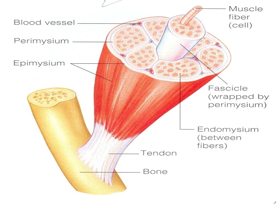
Types of connective tissues wrappings of skeletal muscle

5
Endomysium : a delicate connective tissue sheath surrounds muscle fibre. Endomysium : a delicate connective tissue sheath surrounds muscle fibre. Perimysium : a coarser fibrous membrane surrounds several sheathed muscle fibers to form bundle of fibers called a fascicle. Perimysium : a coarser fibrous membrane surrounds several sheathed muscle fibers to form bundle of fibers called a fascicle. Epimysium : a tougher overcoat of connective tissue surrounds many fascicles. Epimysium : a tougher overcoat of connective tissue surrounds many fascicles.

7
The epimysia blend into the strong, cord- like tendons, or into sheet-like aponeurosis. The epimysia blend into the strong, cord- like tendons, or into sheet-like aponeurosis. Function:attaching the muscle to bone…. Function:attaching the muscle to bone…. Providing durability Providing durability Conserving space Conserving space

8
Muscle function Providing movement Providing movement Maintaining posture Maintaining posture Stabilizing joint Stabilizing joint Generating heat Generating heat

10
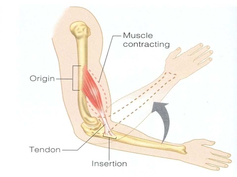
Origin : is the point where the muscle is attached to immovable or less movable bone. Origin : is the point where the muscle is attached to immovable or less movable bone. Insertion : is the point where the muscle attached to movable bone. Insertion : is the point where the muscle attached to movable bone.

11
Prime mover : the muscle that has the major responsibility for causing a movement. Prime mover : the muscle that has the major responsibility for causing a movement. Antagonist : the muscle that reverse a movement. Antagonist : the muscle that reverse a movement.

12
Naming Skeletal muscles Direction of the muscle fibers Direction of the muscle fibers Relative size of the muscle Relative size of the muscle Location of the muscle Location of the muscle Number of the origin Number of the origin Location of the muscle’s origin and insertion Location of the muscle’s origin and insertion Shape of the muscle Shape of the muscle Action of the muscle Action of the muscle

13
Types of body movements Flexion &extension: Flexion &extension: movement in sagittal plane movement in sagittal plane

15
Rotation: Rotation: It is the movement of a bone around its long axis It is the movement of a bone around its long axis atlas around the dens of axis atlas around the dens of axis

16
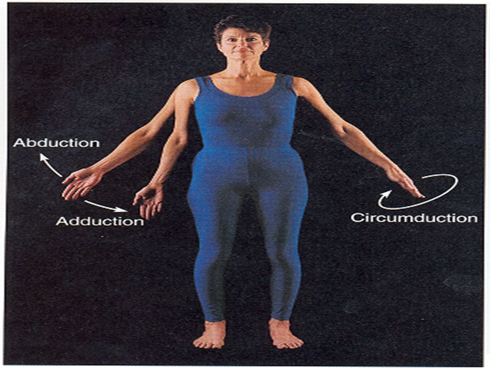
Abduction &adduction: Abduction &adduction: moving the limb away or toward the midline of the body moving the limb away or toward the midline of the body

17
Circumduction : Circumduction : is a combination of is a combination of flexion, extension, abduction and adduction flexion, extension, abduction and adduction

19
Special movement Dosiflexion&planter flexion Dosiflexion&planter flexion Inversion and eversion Inversion and eversion Supination and pronation Supination and pronation opposition opposition

20
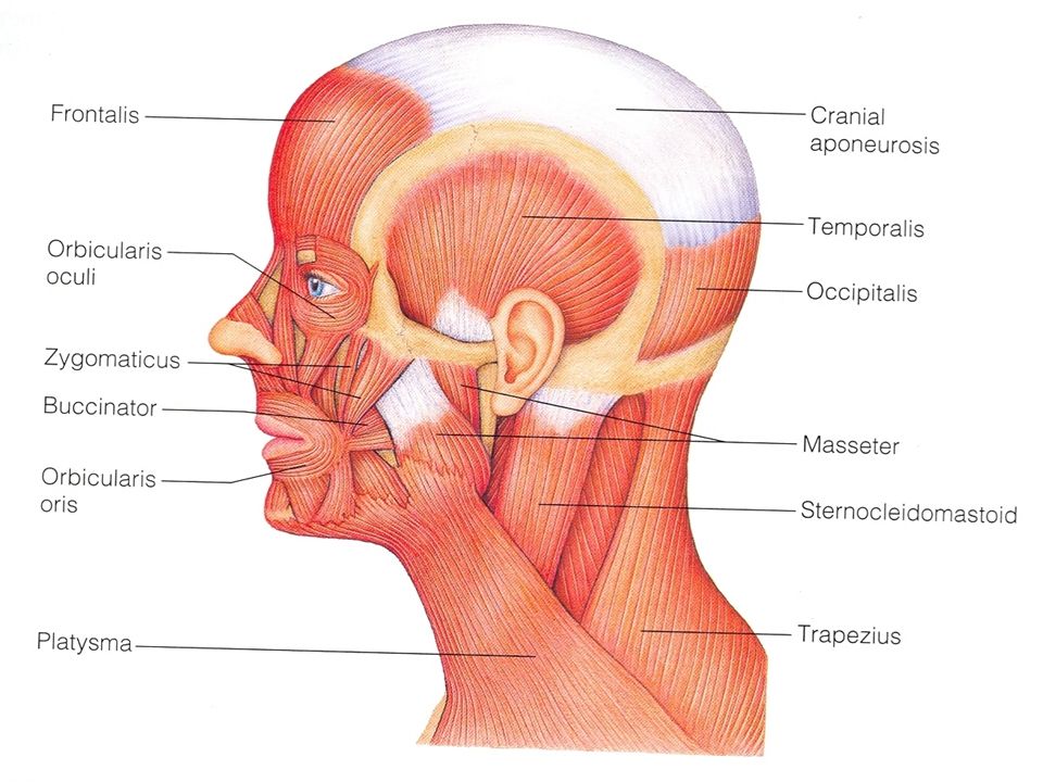
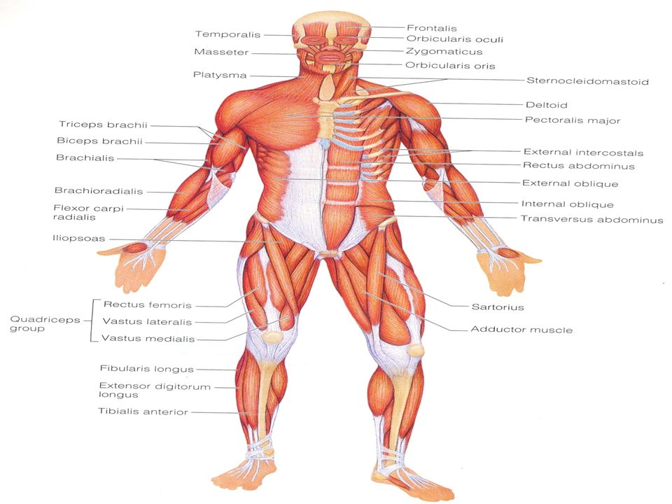
Head muscles Facial Muscles (Frontalis, Orbicularis oculi Orbicularis oris, Buccinator,Zygomaticus). Facial Muscles (Frontalis, Orbicularis oculi Orbicularis oris, Buccinator,Zygomaticus). Chewing Muscle (Buccinator,Masseter, Temporalis). Chewing Muscle (Buccinator,Masseter, Temporalis).
. Chewing Muscle (Buccinator,Masseter, Temporalis). Chewing Muscle (Buccinator,Masseter, Temporalis)..")
22
Facial muscles Frontalis : Frontalis : It raise the eyebrows It raise the eyebrows It wrinkle the forehead It wrinkle the forehead Orbicularis oculi : Orbicularis oculi : It closes the eyes, blink. It closes the eyes, blink.

23
Orbicularis oris: Orbicularis oris: It closes the mouth It closes the mouth Buccinator : Buccinator : It flatten the cheek. It flatten the cheek.

25
Chewing Muscle Buccinator Buccinator Masseter : Masseter : It closes the jaw by elevating the mandible It closes the jaw by elevating the mandible Temporalis: Temporalis: It is synergist of the masseter in closing the jaw. It is synergist of the masseter in closing the jaw.

27
Neck muscles Sternocleidomastoid : it is a paired muscles with two headed muscles, one from each side of the neck. Sternocleidomastoid : it is a paired muscles with two headed muscles, one from each side of the neck. platysma platysma

28
Trunk muscles Anterior muscles Anterior muscles Muscles of the abdominal wall Muscles of the abdominal wall Posterior muscles Posterior muscles

30
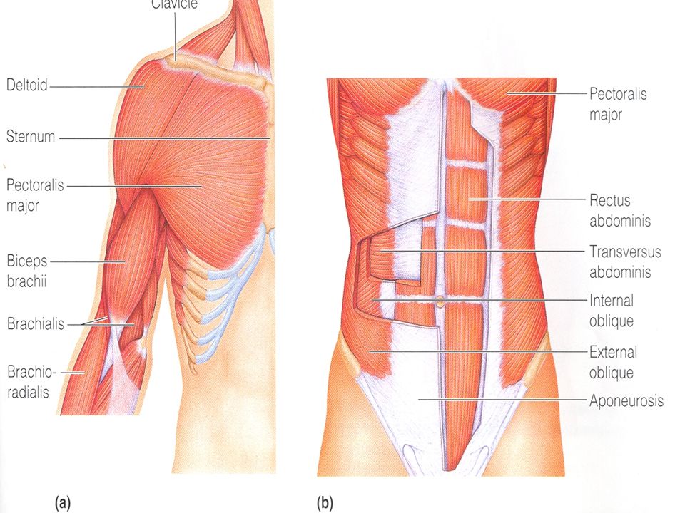
Anterior muscles : Anterior muscles : Pectoralis major : a large fan shaped muscle covering the upper part of the chest. It adduct and flex the arm. Pectoralis major : a large fan shaped muscle covering the upper part of the chest. It adduct and flex the arm. Intercostal muscles: Intercostal muscles: External intercostal External intercostal Internal intercostal Internal intercostal innermost intercostal innermost intercostal

32
Muscles of the abdominal wall : Muscles of the abdominal wall : Rectus abdominis Rectus abdominis External oblique External oblique Internal oblique Internal oblique Transversus abdominis Transversus abdominis

33
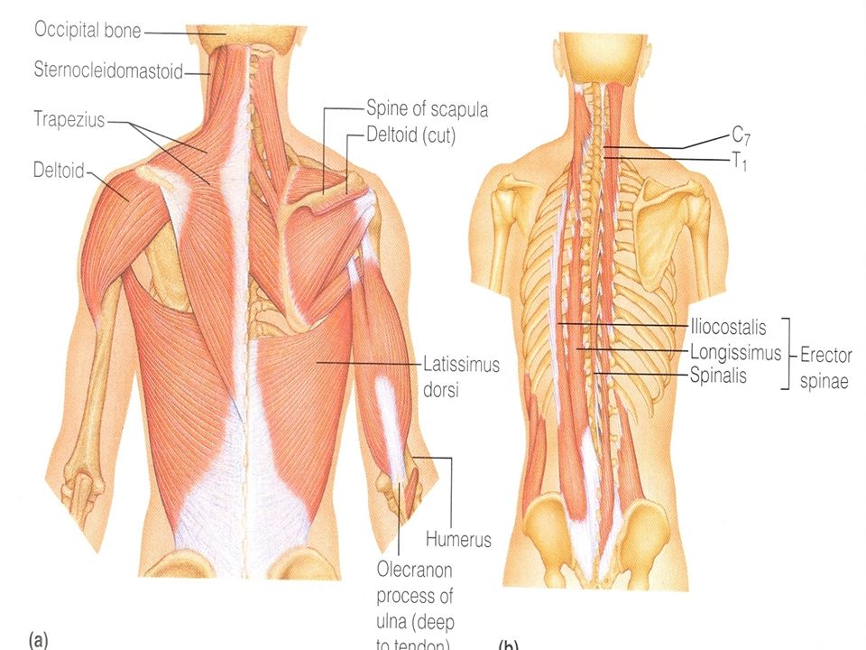
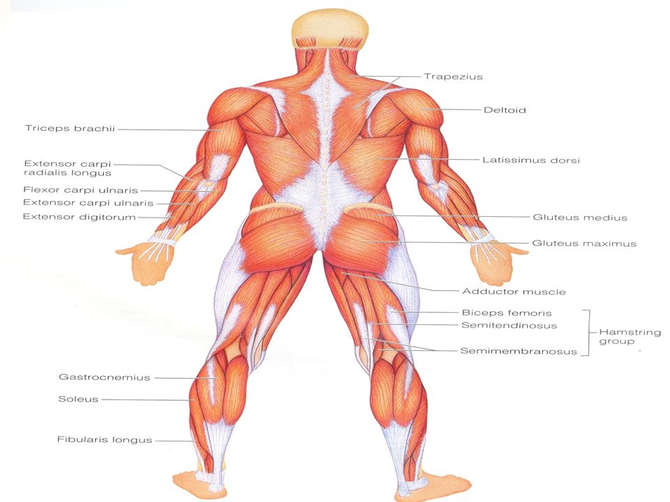
Posterior muscles : Posterior muscles : Trapezius : It is the most superficial muscles of posterior neck and upper trunk. It extend the head &antagonist sternocleidomastoid. Trapezius : It is the most superficial muscles of posterior neck and upper trunk. It extend the head &antagonist sternocleidomastoid.

34
Latissmus Dorsi: Latissmus Dorsi: Large, flat muscle pair that cover the lower back. Large, flat muscle pair that cover the lower back. Extends and adducts the humerus. Extends and adducts the humerus. Erector spinae: Erector spinae: Deep muscles of the back.It extend the back. Deep muscles of the back.It extend the back.

35
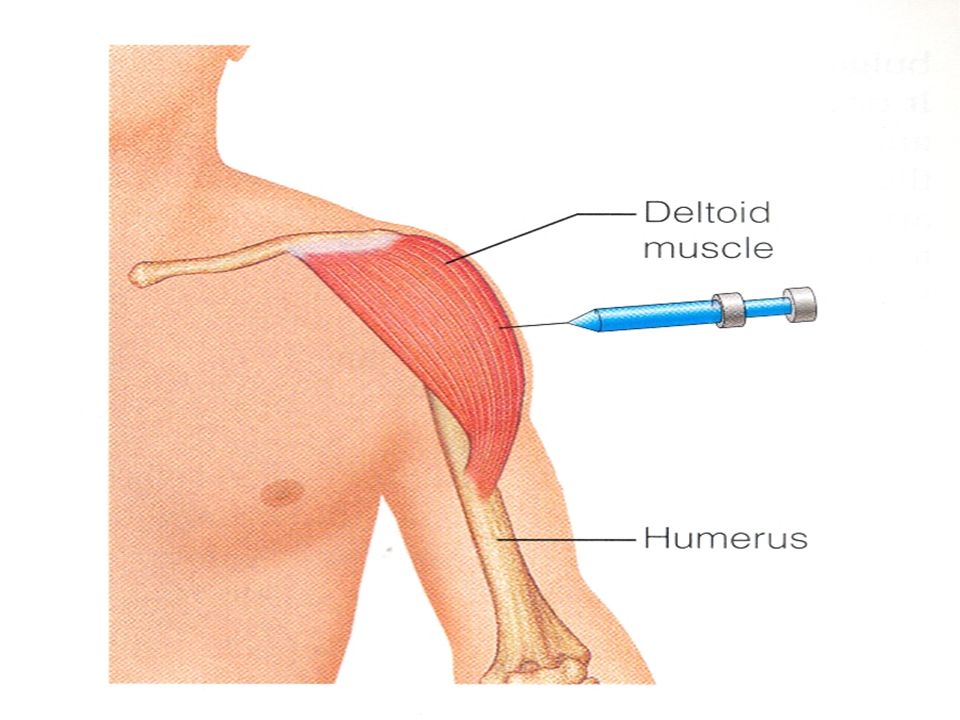
Detoid Detoid Favorite injection Favorite injection Prime movers of arm abduction. Prime movers of arm abduction.

37
Muscle of the upper limb The first group include muscles that arise from the shoulder girdle and cross the shoulder joint to insert into the humerus (pectoralis major, latissimus dorsi, and deltoid). The first group include muscles that arise from the shoulder girdle and cross the shoulder joint to insert into the humerus (pectoralis major, latissimus dorsi, and deltoid).
..")
39
The second group causes movement at the elbow joint (Biceps brachii,brachialis,brachioradialis, and triceps brachii) The second group causes movement at the elbow joint (Biceps brachii,brachialis,brachioradialis, and triceps brachii)
 The second group causes movement at the elbow joint (Biceps brachii,brachialis,brachioradialis, and triceps brachii)")
40
The third group causes movement at the wrist joint (flexor carpi and flexor digitorum) The third group causes movement at the wrist joint (flexor carpi and flexor digitorum) (extensor carpi and extensor digitorum) (extensor carpi and extensor digitorum)
 The third group causes movement at the wrist joint (flexor carpi and flexor digitorum) (extensor carpi and extensor digitorum) (extensor carpi and extensor digitorum)")
41
Muscles of lower limb Muscles causing movement at the hip joint Muscles causing movement at the hip joint Muscles causing movement at the knee joint Muscles causing movement at the knee joint Muscles causing movement at the ankle and foot joint Muscles causing movement at the ankle and foot joint

43
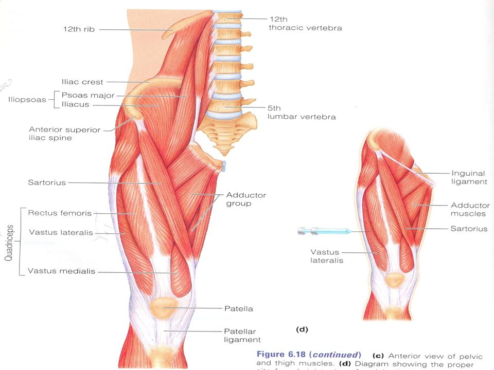
Muscles causing movement at the hip joint (gluteus maximus,gluteus medius,illiopsoas,adductor muscles) Muscles causing movement at the hip joint (gluteus maximus,gluteus medius,illiopsoas,adductor muscles)
 Muscles causing movement at the hip joint (gluteus maximus,gluteus medius,illiopsoas,adductor muscles)")
45
Muscles causing movement at the knee joint (hamstring group,sartorius,quadriceps group) Muscles causing movement at the knee joint (hamstring group,sartorius,quadriceps group)
 Muscles causing movement at the knee joint (hamstring group,sartorius,quadriceps group)")
46
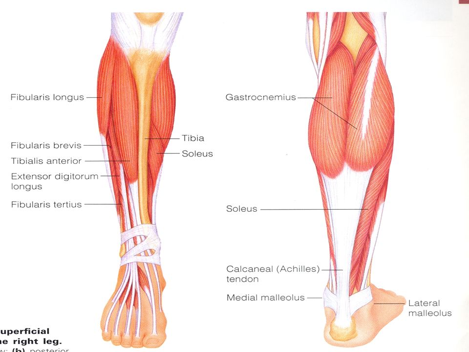
Muscles causing movement at the ankle and foot joint (tibialis anterior,extensor digitorum longus,fibularis muscles,gastrocnemius,soles) Muscles causing movement at the ankle and foot joint (tibialis anterior,extensor digitorum longus,fibularis muscles,gastrocnemius,soles)
 Muscles causing movement at the ankle and foot joint (tibialis anterior,extensor digitorum longus,fibularis muscles,gastrocnemius,soles)")
50
Flexion Motion of a joint which reduces the joint's angle from its anatomical position. Extension Motion of a joint which increases the joint's angle from its anatomical position

51
Abduction moving away from the midline moving toward the midline Adduction

52
Circumduction Combination of flexion, abduction, extension, & adduction

53
supination Motion that rotates the palm of the hand toward the anatomical position. pronation Motion that rotates the palm of the hand away from the anatomical position.

54
Lateral (external) rotation when the bone rotates away from the midline Medial (internal) rotation when the bone rotates towards the midline
 rotation when the bone rotates away from the midline Medial (internal) rotation when the bone rotates towards the midline")
55
Inversion Medial movement of the plantar surface of the foot. Eversion Lateral movement of the plantar surface of the foot.

56
Dorsi flexion Flexion of the ankle joint (toes move superiorly from the anatomical position). Planter flexion Extension of the ankle joint (toes move inferiorly from the anatomical position).
..")
Similar presentations


. There are three types of muscle found in the.>")

