Download presentation
Presentation is loading. Please wait.

1
GENERAL PRINCIPLES OF FRACTURES II

2
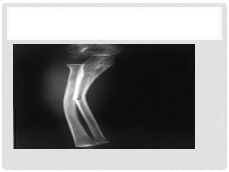
CLINICAL FEATURES History There is usually a history of 1. injury, 2. followed by inability to use the injured limb. 3. But beware the fracture is not always at the site of the injury a blow to the knee may fracture the patella, the femoral condyles, the shaft of the femur or even the acetabulum.

3
4. The patient's age and the mechanism of injury are important. If a fracture occurs with trivial trauma, suspect a pathological lesion 5. Pain, bruising and swelling are common symptoms, but they do not distinguish a fracture from a soft-tissue injury. 6. Deformity is much more suggestive.

7
Always ask about symptoms of associate injuries; 1. numbness or loss of movement 2.skin pallor or cyanosis, blood in the urine. 3. Abdominal pain, 4. transient loss of consciousness. 5. Finally a general medical history is important, in preparation for anesthesia or operation.

8
GENERAL SIGNS A broken bone is part of a patient. It is important to look for evidence of: (1) Shock or hemorrhage. (2) Associated damage to brain, spinal cord or viscera. (3) A predisposing cause (such as tumour or malignancy).
 Shock or hemorrhage. (2) Associated damage to brain, spinal cord or viscera. (3) A predisposing cause (such as tumour or malignancy)..")
9
LOCAL SIGN Injured tissue must handled gently. To elicit crepitus or abnormal movement is unnecessarily painful; x-ray diagnosis is more reliable. The familiar headings of clinical examination should always considered and damage to arteries and nerves may be overlooked.

10
LOOK 1.Swelling. 2.Bruising. 3.deformity may be obvious. 4. the important point is whether the skin is intact; if the skin is broken and the wound communicates with the fracture, the injury is 'open' ('compound').
..")
15
FEEL There is localized tenderness, but it is necessary also to examine distal to the fracture in order to feel the pulse and to test sensation. A vascular injury is a surgical emergency.

17
MOVE Crepitus and abnormal movement may be present, but it is more important to ask if the patient can move the joints distal to the injury.

18
X-RAY X-ray exanimation is mandatory. Certain pitfalls must be avoided by using role of twos: 1. Two views: fracture or a dislocation may not be seen on a single x-ray film, and at least two views (anteroposterior and lateral) must be taken.
 must be taken..")
20
2. Two joints in the forearm or leg, one bone may be fractured and angulated. Angulation, however, is impossible unless the other bone also is broken, or a joint dislocated. The joints above and below the fracture must both be included on the x-ray films.

24
3. Two limbs in x-ray of children's bones, normal epiphysis may confuse the diagnosis of a fracture and films of uninjured limb are helpful.

26
4. Two injuries severe force often causes injuries at more than one level. Thus, with fractures of the calcaneum or femur it is important also to x-ray the pelvis and spine.

28
5. Two occasions Soon after injury, a fracture (e.g. of the carpal scaphoid) may be difficult to see. If doubt exists, further examination 10-14 days later may as a result of bone resorption, make diagnosis easier.
 may be difficult to see. If doubt exists, further examination days later may as a result of bone resorption, make diagnosis easier..")
30
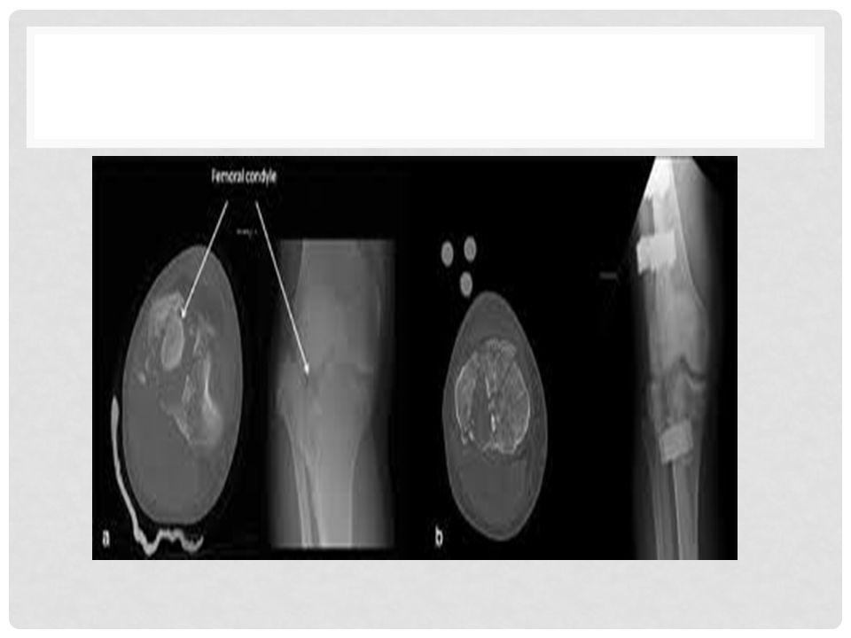
Special imaging Sometimes the fracture - or the full details of the fracture is not apparent on the plain x-ray. Tomography may be helpful in lesions of the spine or fractures of the tibial condyles; CT or MRI may be the only way of showing whether a fractured vertebra is threatening to compress the spinal cord; indeed, transectional images are essential for accurate visualization of fractures in 'difficult' sites such as the calcaneum or acetabulum, and three dimensional reconstructed images are even better.

36
Radioisotope scanning is helpful in diagnosing a suspected stress fracture or other undisplaced fractures.

38
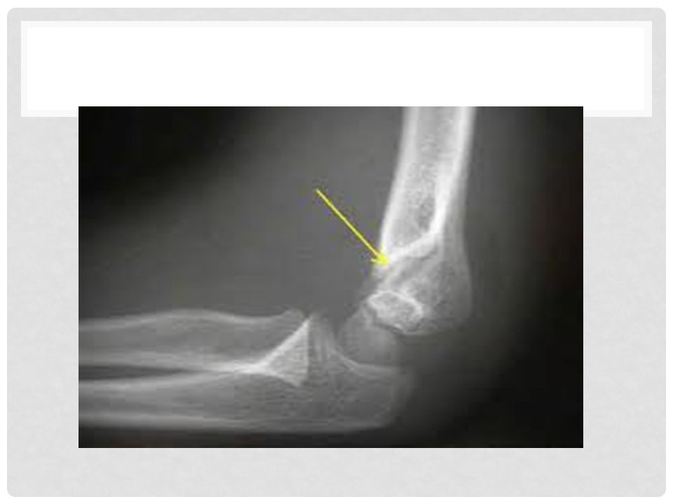
Description Diagnosing a fracture is not enough; the surgeon should picture it (and describe it) in all it complexity. (1) Is it open or closed? (2) Which bone is broken, and where? (3) Has it involved a joint surface? (4) What is the shape of the break?
 Is it open or closed. (2) Which bone is broken, and where. (3) Has it involved a joint surface. (4) What is the shape of the break .")
39
How to read x-ray of fracture 1.Define the image e.g. plain x-ray or C-T scan. 2.Label for the name of the patient, the date and the side. 3.The region and the view.

40
4. The word fracture should be followed by the questions a.Which bone? b.Which site in the bone? c.What is the type and the shape of fracture? d.Does it displaced? e.Which type of displacement? f.Does the fracture recent or old? g.Does it treated or not? h.Does it pathological?

49
SECONDARY INJURIES Certain fractures may cause secondary injuries and these should always be assumed to have occurred until proved otherwise. 1.Spinal cord and nerve root injury may associate with fracture of the spine. 2.Urethral, bladder, rectal, vaginal and neurovascular injuries may associate with fracture pelvis.

52
3. Lung and heart injuries may associate with fracture of the sternum and the ribs. 4. Brachial plexus injuries may associate with fracture and dislocation of the pectoral girdle.

54
Treatment of closed fractures General treatment is the first consideration: to treat the patient, not simply the part. The sequence is:

55
(1) First aid. (2) Transport. (3) The treatment of shock, hemorrhage and associated injuries.
 First aid. (2) Transport. (3) The treatment of shock, hemorrhage and associated injuries.")
56
In principle the treatment of fractures consists of manipulation to improve the position of the fragments, followed by splintage to hold them together until they unite; meanwhile, joint movement and function must be preserved

57
. Fracture healing is promoted by physiological loading of the bone. So muscle action and early weight bearing are encouraged. These objectives are covered by three simple actions: REDUCE, HOLD, and EXERCISE.

58
REDUCE Although general treatment and resuscitation must always take the priority, treatment of fracture should not be delayed because the soft tissue swelling associated with the fracture within first 12 hours make the reduction difficult.

59
There are certain situations in which reduction is not necessary like 1.When there is little or no displacement. 2.When displacement does not matter like in fracture clavicle. 3.When reduction is unlikely to succeed like in compression fracture of the spine.

63
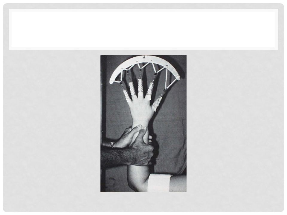
Methods of reduction 1. Closed reduction a.Manipulation under anesthesia ( GA, RA, or LA) b.Traction (skin, skeletal, or gravity). 2. Open reduction.
 b.Traction (skin, skeletal, or gravity). 2. Open reduction..")
64
MANIPULATION Under proper anesthesia and muscle relaxation, fracture reduced by three fold manoeuver: 1)The distal part of the limb is pulled in the line of the bone. 2)As the fragments disengage, they are repositioned by reversing the original direction of the force. 3)Alignment is adjusted in each plane.
As the fragments disengage, they are repositioned by reversing the original direction of the force. 3)Alignment is adjusted in each plane..")
71
Generally manipulation is used for all minimally displaced fractures, for most of fractures in children and for stable fractures after reduction.

72
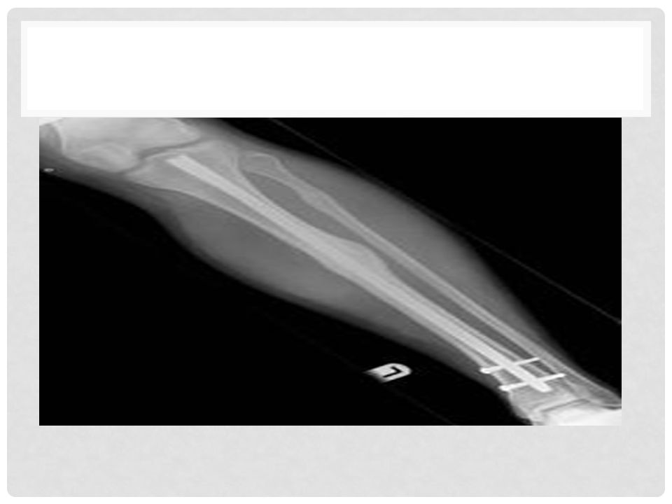
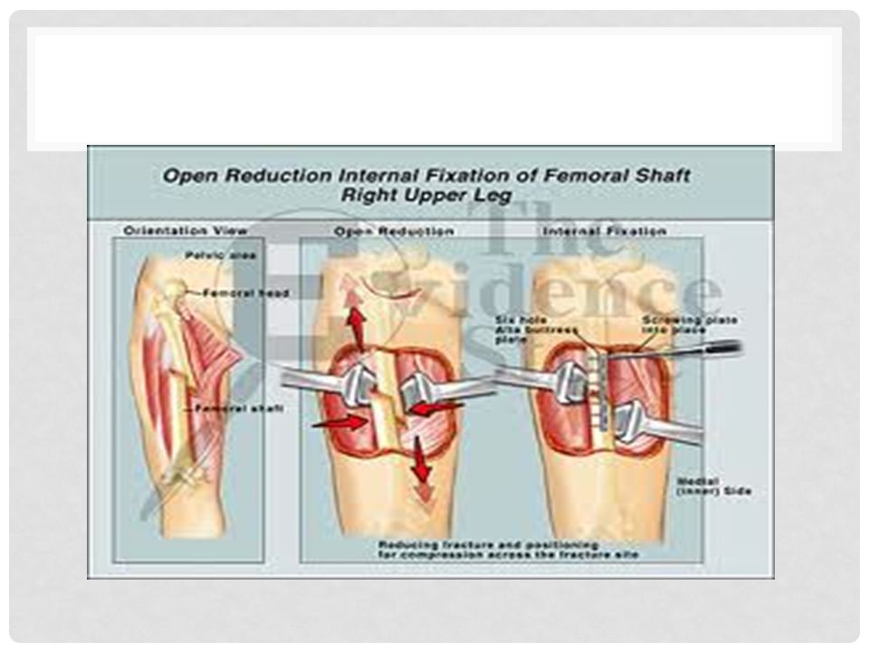
OPEN REDUCTION Opened reduction of the fracture under direct vision is indicated: (1) When closed reduction fails, either because of difficulty in controlling the fragments or because soft tissues are interposed between them. (2) When there is a large articular fragment that needs accurate positioning. (3) For traction fractures in which the fragments are held apart. As a rule, open reduction is merely the first step to internal fixation.
 When there is a large articular fragment that needs accurate positioning. (3) For traction fractures in which the fragments are held apart. As a rule, open reduction is merely the first step to internal fixation..")
Similar presentations


, F.R.C.S.(C )>")


 - Joints - Cartilages - Ligaments.>")


